Peritoneum Flashcards

(54 cards)

1
Q

Gross appearance of endosalpingosis?

A

prominent cyst formation

2
Q

Gross apearance of endocervicosis?

A

mass in outer cervix/bladder

3
Q

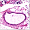
Histological features of endocervicosis?

A

Glands lined by endocervical-type epithelium

Hyalinized, fibroblastic, or edematous stroma

± mucin extravasation

4
Q

IHC for endometrial type stroma?

A

CD10 positive, p16 patchy positive

5
Q

IHC for endometrial epithelium?

A

pax-8, ER, PR positive

6
Q

IHC for ectopic decidua?

A

ER, vimentin, desmin positive

CD10 and inhibin-α may be positive

7
Q

Molecular abnormalities in endometriosis?

A

± KRAS mutations (endometriosis)

8
Q

Features of atypical endosalpingosis?

A

Cytologic atypia that falls short of carcinoma

9
Q

Features of Serous Borderline Tumour (vs. atypical endosalpingosis)?

A

May form mass lesion

Architectural complexity with cellular budding and tufting

Eosinophilic cells with abundant cytoplasm are common

10
Q

Features of Endometriosis with Hyperplasia (vs Endometriosis)?

A

Glandular crowding and complexity + cytologic atypia

11
Q

Features of Metastaic Adenocarcinoma (vs. Endometriosis/Endocervicosis/Mullerianosis)?

A

Prior history &/or concomitant primary

Complex glandular architecture, at least focally

Irregularly shaped and haphazardly infiltrating glands with cytologic atypia

Desmoplastic stroma

12
Q

Features of Stromal Sarcoma (vs Stromal Endometriosis)?

A

Prior history or concomitant mass

13
Q

Features of Low-Grade Müllerian Adenosarcoma (vs. Polypoid Endometriosis)?

A

Well-developed, leaf-like architecture

Pronounced stromal condensation or “cuffing”

± sex cord-like differentiation or sarcomatous overgrowth

± infiltration of underlying tissues

Stromal cytologic atypia with variable mitotic activity

14
Q

Features of Deciduoid Mesothelioma (vs ectopic decidua)?

A

Large, often confluent nodules may be seen

Infiltration of underlying tissues common

May show wide variation in nuclear size and shape

Mitoses may be seen

Numerous, long, slender microvilli by electron microscopy

Keratin (AE1/3, pan keratin, CK7, CK5/6), EMA, D2-40, WT1, mesothelin positive

ER and inhibin negative

15
Q

Features of Metastatic SCC (vs Ectopic Deccidua)?

A

Prior history or concomitant ovarian/uterine tumor

Keratin formation and intercellular bridges (i.e., true squamous differentiation)

Cytologic atypia and mitoses

Keratin and p63 positive

p16 diffusely positive (if cervical origin)

16
Q

Features of Peritoneal Tuberculosis (vs. Necrotic Pseudoxanthomatous Nodules)?

A

Prior history of tuberculosis, positive tuberculosis test, or immunosuppression

Variably sized, sometimes confluent granulomas with central necrosis

Peripheral palisading of histiocytes and giant cells

Positive AFB stain &/or culture

17
Q

Gross features of florid mesothelial hyperplasia?

A

Rough, dull, ± thickened and whitish surfaces

18
Q

IHC for florrid mesothelial hyperplasia?

A
  • Vimentin, AE1/AE3, calretinin, CK5/6, WT1, D2-40 (podoplanin), thrombomodulin positive
  • BAP1 expression preserved
19
Q

Features of Malignant mesothelioma?

A

Widespread peritoneal involvement, often with hemorrhagic ascites

Sarcomatoid/storiform component may be present

Invasion of underlying tissues

Necrosis

Marked uniform cytologic atypia

Homozygous p16 deletion (chromosome 9p21) specific (but not sensitive)

BAP1 loss of expression specific (but not sensitive)

20
Q

Features of well differentiated papillary mesothelioma?

A

Relatively abundant eosinophilic to amphophilic cytoplasm

Papillae covered by single layer of cuboidal cells

No inflammation, granulation tissue, or hemorrhage

21
Q

IHC for Peritoneal inclusion cysts?

A

Vimentin, AE1/AE3, CK5/6, calretinin, WT1, D2-40 (podoplanin), caldesmon, desmin positive

22
Q

DDx for peritoneal inclusion cysts?

A

cystic malignant mesothelioma

cystic lymphangioma

23
Q

Microscopic features of serous borderline tumours of the peritoneum?

A

Identical to noninvasive implants of ovarian serous borderline tumor (SBT)

Papillae, nests (< 20 cells across), glands, clusters of cells, or single cells

Tufting, budding, and epithelial pseudostratification

Stromal desmoplasia (if stroma rich)

Psammoma bodies common

Uniform cells with round nuclei and small nucleoli

Abundant and brightly eosinophilic cytoplasm and mitotic activity, if pregnant

24
Q

IHC of serous borderline tumour of the peritoneum?

A

ER, PR, WT1, MOC-31, BER-EP4, B72.3, pax-8 positive

Calretinin, cytokeratin 5/6, p16 can be positive

p53 wild-type staining

25
Microscopic features of Low-grade serous carcinoma?
Invasion of underlying structures &/or confluent growth (more frequently identified in omentum) Small, cellular papillae or small, solid nests (usually \< 20-30 cells across) with cribriform or slit-like spaces typically free floating in spaces (stroma with retraction artifact) \< 3x variation in nuclear size Mitotic rate ≤ 12/10 HPF Psammocarcinoma: Tumor cell nests \< 15 cells across, psammomatous calcification in \> 75% of tumor cell nests
26
Microscopic features of High-grade serous carcinoma?
## Footnote Complex papillae, pseudoglands, and solid growth Slit-like spaces If papillary, cellular tufting and budding common \> 3x variation in nuclear size Mitotic rate \> 12/10 HPF
27
IHC for Low-grade serous carcinoma?
CK7, WT1, BER-EP4, MOC-31, B72.3, ER, PR, pax-8, pax-2, Claudin-4 positive p53 and p16 heterogeneous positivity Ki-67 usually \< 30% Calretinin may be positive (but often focal) HNF-1-β negative
28
IHC for high-grade serous carcinoma?
CK7, WT1, BER-EP4, MOC-31, B72.3, ER, PR, pax-8, pax-2, Claudin-4 positive p53 either diffusely positive or completely negative (null mutation) p16 diffusely positive Ki-67 positive, usually \> 75% of cells Calretinin may be positive (but often focal) HNF-1-β negative
29
Features and DDX of meatastaic carcinoma vs High grade serous carcinoma?
Serous carcinoma from other locations in female genital tract \> \> primary peritoneal HGSC Extragenital sitesBreast * Clinical history * Pattern of dissemination of disease * Other characteristic histologic patterns (tubular, single file, cribriform) * Mammaglobin (50% of tumors), GATA3 positive * pax-8 negative Urinary tract (micropapillary urothelial carcinoma) * Prior or concomitant urinary tract primary * Other histologic patterns * Uroplakin, thrombomodulin, GATA3 positive * pax-8 negative
30
IHC of Well-differentiated Papillary mesothlioma?
* **_Calretinin, mesothelin, D2-40, and WT1 positive_** * **_Preserved BAP-1 nuclear expression_** * Cytokeratin 5/6 often positive * pax-8 may be positive * MOC-31, B72.3, and BER-EP4 usually negative
31
Micoscopic Features of Solitary Fibrous Tumour?
Patternless growth with hyper- and hypocellular areas Often hyalinized collagenous stroma with perivascular accentuation Thin-walled, branching, staghorn-like vessels Bland, oval, spindled cells with scant cytoplasm Features associated with malignancy: Increased cellularity, cytologic atypia, \> 4 mitoses/10 HPF, and necrosis
32
IHC of Solitary fibrous Tumour?
* CD34, Bcl-2, STAT6, GRIA2, and vimentin typically strongly positive * B-catenin positive in ~ 40% * CD99 (~ 70%) and PR variably positive * NAB2-STAT6 fusion
33
Pathogenesis of malignant mesothelioma?
* Association with asbestos exposure (not as frequent as pleura) * BAP1 tumor predisposition syndrome (BAP1-TPDS) associated with increased risk of Spitz nevus, uveal and cutaneous melanoma, malignant mesothelioma, clear cell carcinoma, and basal cell carcinoma
34
Microscopic features of Malignant Mesothelioma?
* Growth along peritoneal surfaces with invasion of underlying tissues * Epithelioid (3 main patterns): Tubular \> papillary (including micropapillae) \> solid (often admixed) * Irregular, broad papillae with no hierarchical branching and no cell budding * Biphasic: Admixture of epithelial and sarcomatous areas in various proportions (may merge) * Sarcomatoid: Diffuse, fascicular, or storiform growth * Mild to moderate and uniform atypia in most
35
IHC of Malignant Mesothelioma?
* WT1, calretinin, mesothelin, D2-40 (podoplanin), thrombomodulin, CD56, HBME-1 positive * Caldesmon, p16 (~ 30%), pax-8, PR may be positive; BAP-1 often lost * ER, BER-EP-4, pax-2, MOC-31, TTF-1, B72.3 (TAG72), CD15 (Leu-M1), CA19.9, Napsin-A typically negative
36
Microscopic features of Disseminated Peritoneal Leiomyomatosis?
Well circumscribed ± confluent, rounded nodules Intersecting fascicles of bland spindle cells
37
IHC features of Disseminated Peritoneal Leiomyomatosis?
Smooth muscle actin, desmin, caldesmon, ER, PR positive C-kit and rarely DOG1 (focally) positive
38
DDx of Disseminated Peritoneal Leiomyomatosis?
Metastatic leiomyosarcoma Gastrointestinal stromal tumor Pelvic PEComatosis
39
Features of Peritoneal PEComatosis?
Often associated with primary gynecologic PEComa May be associated with tuberous sclerosis ± lymphangioleiomyomatosis Diffuse &/or nested growth Often epithelioid cells ± clear cytoplasm HMB-45, Melan-A focally positive
40
Microscopic features of EGIST?
Epithelioid: Sheets, nests, or trabeculae of uniform round cells with indistinct cell borders Spindle: Short fascicles or whorls of uniform cells with tapered nuclei ± nuclear palisading Delicate vessels may be prominent &/or hyalinized Skeinoid fibers (extracellular eosinophilic deposits of PAS-positive abnormal collagen) rare
41
IHC of EGIST?
C-kit and DOG1 positive; nestin, WT1 (cytoplasmic) positive KIT and PDGFRA mutations
42
Ddx for EGIST?
* Leiomyoma(tosis) * Leiomyosarcoma * Metastatic endometrial stromal sarcoma, high grade * Schwannoma * Fibromatosis (desmoid tumor)
43
Features of Leiomyoma(tosis)?
Long, intersecting fascicles Dense eosinophilic cytoplasm and distinct cell borders Plump spindled cells with blunt-ended nuclei; no perinuclear halos ER, PR, WT1 (nuclear) positive (if gynecologic)
44
Features of Leiomyosarcoma?
Long, intersecting fascicles Plump spindled cells with blunt-ended nuclei and dense eosinophilic cytoplasm Often features significant nuclear pleomorphism and mitotic activity ER, PR, WT1 (nuclear) positive (if gynecologic)
45
Features of Metastatic Endometrial Stromal Sarcoma, High Grade
Prior history or concomitant intrauterine mass Prominent nested architecture and lack of paranuclear vacuoles Presence of low-grade fibromyxoid component Cyclin-D1 positive (\> 50% nuclei) (in high-grade areas) DOG1 negative No KIT mutation despite C-kit positivity (in high-grade areas)
46
Features of a Schwannoma?
Short fascicles of spindled cells Antoni A or B zones Hyalinized, typically medium-sized, blood vessels S100 positive (diffuse, strong) C-kit and DOG1 negative
47
Features of Fibromatosis (desmoid tumour)?
Long, sweeping fascicles Uniformly collagenous background Elongated spindled cells with tapered nuclei β-catenin (nuclear) positivity
48
Microscopic features of Intra-abdominal Desmoplastic Small Round Cell Tumour
Irregular nests and sheets of small round blue cells Desmoplastic fibrous stroma Occasionally, rosette-like formation, trabeculae, tubules, and glandular structures may occur Small, round cells with scant cytoplasm and hyperchromatic, round to oval nuclei ± inconspicuous nucleoli
49
IHC features of intra-abdominal Desmoplastic Small Round Cell Tumour?
Keratin/EMA/desmin positive CD99, WT1 (C-terminal) positive Translocation t(11;22)(p13;q12) RT-PCR or FISH: EWSR1-WT1 fusion
50
Ddx of intra-abdominal Desmoplastic Small Round Cell Tumour?
Ewing sarcoma/primitive neuroectodermal tumor Rhabdomyosarcoma Small cell carcinoma, hypercalcemic type Small cell carcinoma, neuroendocrine (pulmonary) type Non-Hodgkin lymphoma
51
Site of pseudomyxoma peritonei?
Appendix most common primary site (95%) Ovary, pancreaticobiliary, urachus (5%)
52
Microscopic features of pseudomyxoma peritonei?
Peritoneal mucin deposits * Pools of mucin lacking neoplastic mucinous epithelial cells (organizing mucin) Disseminated peritoneal adenomucinosis * Mucin pools containing strips and clusters of cuboidal to columnar mucinous epithelium with only occasional papillary tufting; small glands uncommon * Volume of mucin typically greatly exceeds that of mucinous epithelium * Intestinal-type mucinous epithelium with mild cytologic atypia and rare mitoses Peritoneal mucinous carcinoma * Mucin pools with moderate to abundant strips and clusters of mucinous epithelium often with complex cribriform and micropapillary growth; small glands common * Invasion into underlying structures often with associated desmoplasia * Intestinal-type mucinous epithelium with moderate to severe cytologic atypia
53
IHCof pseudomyxoma peritonei?
Appendix primary * SATB2, CK20, CDX-2, mCEA, pCEA positive * CK7 usually negative (~ 2/3) Colon primary * Typically SATB2, CK20, CDX-2 positive * CK7 negative (except right-sided tumors) Ovary primary (intestinal type associated with teratoma) * CK20 and CDX-2 positive * ~ 22% SATB2 positive * CK7 negative MUC2 often positive at peritoneal sites regardless of primary organ Immunohistochemistry is usually not helpful in distinguishing between potential primary sites * SATB2/CK20 dual stain shows 80% sensitivity and 100% specificity for appendiceal vs. ovarian origin
54