Physiology/Pathophysiology Flashcards
(123 cards)
Draw the simple cascade model of clotting.

Explain the importance of membrane glutamic acid residues in coagulation.
- Gla residues allow for binding of the protein to a membrane surface via interaction between calcium and membrane phospholipids
- The Gla residues must be fully carboxylated via the vitamin-K cycle in the liver or they cannot bind the calcium, which prevents binding to the activated cell membrane surface
- Therefore, vitamin K antagonists prevent carboxylation of the Gla residues and interfere with coagulation
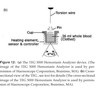
What is the normal distribution of PC, PS, PE on the resting cell membrane? What occurs to these phospholipids with cellular injury?
- In a resting cell, PC is expressed on the external leaflet (being neutral) and PS and PE are on the inner leaflet
- To maintain this resting state:
- Flippase actively transports PS (and sometimes PE) from the external to the internal leaflet
- Floppase moves PC from the internal to the external leaflet
- With injury/activation of the cell:
- Scramblase shuffles the phospholipids between the two membranes
- Occurs in response to increased cytosolic calcium
- Results in appearance of PS/PE on the EXTERNAL membrane

What role does the shuffling of the PS/PE and PC play in promotion of coagulation?
- The expression of PS on the outer membrane of a cell surface essentially causes the membrane to become pro-coagulable
- Cells that do not have PS on their surface are essentially incapable of supporting the coagulation cascade,, generation of enzymes is very slow and cannot lead to thrombin or fibrin generation
- With PS expressed (and PE, which makes coagulation occur even faster) following injury, the cell surface is a procoagulant surface
In addition to the membrane composition/distribution of PC/PS, resting endothelial cells also have a number of other anticoagulant properties. List 3 of these properties.
- Endothelial cells produce HSPGs
- Binding site for anti-thrombin, which can inactivate any thrombin produced in the area
- Endothelial cells express thrombomodulin
- After thrombin binds to TM, it becomes an anti-coagulant molecule through interaction of the thrombin-TM complex with protein C
- Protein C (+protein S) irreversibly cleaves factors Va and VIIIa, which prevents their participation in generation of any additional thrombin
- Endothelial cells express TFPI
- TFPI acts as an upstream inhibitor of FXa and FVIIa
What 2 cell types must be present for cell-based coagulation to occur?
- A cell bearing TF
- Platelets
What is the sole relevant initiator of coagulation?
Tissue factor
**Cells expressing tissue factor are generally localized outside the vasculature; with an injury to the endothelium, flowing blood is exposed to these tissue factor bearing cells**
What are the 3 phases of cell based coagulation?
Initiation
Amplification
Propagation
Describe what occurs during the initiation phase of cell based coagulation.
- TF on the TF bearing cell interacts with FVIIa (which is the ONLY protein floating already active in the circulation)
- The TF-FVIIa complex can interact/activate with Factor X:
- Factor Xa can either interact with it’s cofactor, FV, and thereby generate small amounts of prothrombin and thrombin OR
- Any factor Xa that dissociates free is inactived by TFPI or AT in the region
- The TF-FVIIa complex can interact/activate with factor IX
- Factor IXa can freely dissociate and interact with the surface of platelets and other cells
- The TF-FVIIa complex can interact/activate with Factor X:
**Coagulation ONLY progresses beyond the small amount of thrombin generated during initation IF the injury allows platelets and other proteins to leave the vascular space and adhere to the TF bearing cells!!**

Describe what occurs during the amplification phase of cell based coagulation.
- After the thrombin has diffused away from the TF-bearing cell during initiation, the thrombin can now be used to activate platelets that have leaked from the vasculature to the site of injury
- Thrombin:
- Binds to factor V on the platelet surface, activating it to factor Va
- Binds to factor XI on the platelet surface, activating it to factor XIa
- Cleaves the factor XIII+vWF factor complex, resulting in factor XIIIa and free vWF which promotes platelet adhesion
- Activates the platelet by triggering shuffling of the membrane phospholipids, creating a pro-coagulant surface and release granule contents

Describe what happens during the propagation phase of cell based coagulation.
- After a few platelets are activated in the amplification phase, release of granule contents results in recruitment of additional platelets to the site of injury–this is where propagation occurs.
- Factor IXa
- Is either present due to the release of IXa during initiation OR
- Can be created by interaction of XIa (from amplification) with FIX on the platelet surface
- Factor IXa and factor VIIIa (from amplification) bind together to form the “tenase” complex
- The tenase complex activates factor X to Xa
- The majority of Xa must be generated directly on the platelet surface (as the Xa that was generated during initiation is inhibited if it diffuses away from the TF bearing cell)
- Factor Xa along with FVa cleaves prothrombin to thrombin
- Prothrombinase (FXa+FVa) leads to a burst of thrombin generation, cleaving fibrinogen into fibrin
- When there is a critical mass of fibrin, the soluble molecules will polymerize into fibrin strands and create a fibrin matrix

Draw the complete pathway of cell based coagulation.

Describe the additional roles of thrombin in clot formation/structure (aside from formation of fibrin from fibrinogen).
- Thrombin activates factor XIII to XIIIa, which promotes cross-linking between fibrin strands
- Some thrombin will bind to thrombomodulin on the endothelial cell surface
- TM bound thrombin activates thrombin activatable fibrinolysis inhibitor (TAFI)
- TAFI modifies fibrin molecules by removal of terminal lysine residues
- This makes fibrin markedly more resistant to fibrinolysis
- TM bound thrombin will also activate protein C
- aPC complexes with protein S
- aPC+proS cleave factors Va and VIIIa, which prevents further cofactor activity of either protein
- Consequently shuts down generation of any new thrombin!!!
- TM bound thrombin activates thrombin activatable fibrinolysis inhibitor (TAFI)
How long is the average canine platelet lifespan
6 days
What substances are contained in platelet alpha granules?
Dense granules?
- Alpha granules
- Platelet derived growth factor
- Fibronectin
- TGFBeta
- Fibrinogen
- Factors V and VIII
- VWF
- Dense granules
- ADP, ATP
- Histamine
- Epinephrine
- Serotonin
- Calcium
Briefly describe platelet adhesion and aggregation under high shear conditions.
- Under high shear conditions, adhesion to exposed subendothelium is primarily mediated by collagen and vWF
- vWF is produced by megakaryocytes and endothelial cells
- Stored in Weibel-Palade bodies in endothelial cells and in alpha granules
- After endothelial damage, vWF attaches to the exposed collagen, releasing factor XIII
- Platelets can then roll along the endothelium, mediated by the platelet GP1b-alpha receptor attaching to the vWF on the subendothelium
- After platelets attach to the endothelium via vWF and collagen, undergo a conformational change, exposing the integrin alphaIIbBeta3 receptor
- Causes platelets to expose and assemble membrane glycoproteins which can bind fibrinogen and vWF, promiting platelet aggregation
At high shear rates, vWF mediates platelet aggregation!!!
Briefly describe platelet adhesion and aggregation under low shear conditions.
- Adherence occurs via collagen, fibronectin and laminin
- Fibrinogen is the primary ligand of thrombus growth!!
What are the 2 major roles of vWF?
- Carrier protein for factor VIII, protecting it from proteolysis by protein C
- Mediates adhesion of platelets to damaged endothelium
What are the 3 forms of vWD?
- Type I vWD
- All multimers of vWF are present, but in decreased numbers
- Type II vWD
- The large multimers are absent
- Type III vWD
- All multimeres are absent
List clinical signs seen with primary hemostatic disorders.
List clinical signs seen with secondary hemostatic disorders.

Describe clot retraction as a test for platelet function.
- Influenced mainly by the number and function of platelets and the fibrinogen concentration in plasma
- Other influences may change it–i.e. reduced in anemia, prolonged in polycythemia
- Place 5ml whole blood into a nonadditive tube, insert a wooden applicator and incubated
- The assessment of clot formation and clot retraction is noted over 8-24 hours
- Within 2-4 hours, a normal clot should retract markedly
- To measure % clot retraction, 1ml of whole blood is placed into tubes and incubated
- At 1 hour, the accumulated serum from around the clot can be removed and measured
- Volume multipled by 50 to obtain the percent clot retraction (normal is 25-60%)
Describe the BMBT as a test for platelet function.
- Measure the time for a stable platelet plug to form following a standardized incision on the upper lip
- Normal BMBT is less than 3 minutes in dogs
- A prolonged result is suggestive of thrombocytopenia, thrombocytopathia or vWD
- Best as a screening tool for further more detailed assays
Describe the utility of the PFA100 as a test for platelet function.
- Simulates primary hemostasis by aspirating citrate-anticoagulated whole blood under a high shear rate through a small apearture in a collagen membrane coated with platelet agonists (ADP or epinephrine)
- Provides a “closure time”–CT, which is the time it takes for a platelet plug to form and occlude flow
- CT is highly sensitive to qualitative and quantitative defects in platelet receptors that mediate adhesion (GPIb-V-IX) and aggregation (GPIIbIIIa)
- May be inaccurate in anemic patients, patients with high hematocrits, or platelet counts
- Doesn’t tell specifically WHY/what specific platelet function defect is present, just identifies that there is one