Pleural Diseases Flashcards
(80 cards)
What are the 2 layers of the pleura?
visceral pleura and parietal pleura
What is the pleura?
an elastic serous membrane with a smooth lubricating surface and is divided into 2 layers: visceral and parietal pleura
What is the visceral pleura?
covers the lung parenchyma and extends b/w the lobes (lining of the lung)-Contains NO pain fibers-Drained by the pulmonary venous system
What is the parietal pleura?
covers the inner surface of the thoracic cavity, diaphragm and mediastinum (lining of the inner chest wall)-Contains sensory nerves-Drained by the lymphatic system in the upper abdomen
What is the pleural cavity?
potential space b/c in normal people you can’t see the space -It’s not until air or fluid gets in b/w the 2 layers that you start seeing the 2 separate linings
What nerves supply the costal pleural & the peripheral portion of the diaphragm?
intercostal nerves
What nerve supplies the central portion of the diaphragm?
phrenic nerve E.g. If putting in a chest tube and it hits the diaphragm, pt get referred pain up to the shoulder
What is the primary function of the pleura?
to provide a smooth surface, which reduces friction as the pleurae move against each other As you breathe in and out, want very minimal friction b/w the surfaces
under normal conditions, what is in the pleural cavity?
Under normal conditions, there is a small amount of fluid in the pleural cavity; approx. 1-10mLs (0.1-0.2 ml/kg)
what are the characteristics of normal pleural fluid?
-Clear ultrafiltrate of plasma – looks light beer color -A pH of 7.6-7.64 -Protein count of less than 2% (1-2 g/dL)-Fewer than 1000 WBC’s per cubic millimeter -Glucose content similar to that of plasma -LDH less than 50% of plasma
how is pleural fluid formed?
Starling’s law of trans-capillary exchange -2 forces: hydrostatic pressure & oncotic pressure -> push & pull fluid back & forth in b/w the membranes
what is hydrostatic pressure?
Within the capillaries, think of hydrostatic pressure as the “pushing force” -Pushing fluid out of the capillaries, into the pleural space
what is oncotic pressure?
Think of oncotic pressures as the “pulling force;” -Pulling fluid from the surrounding tissues into the capillaries from the interstitium back into the vasculature -Water gets pulled from pleural space back to vessels
what happens when hydrostatic pressure is greater than oncotic pressure?
fluid will leave the capillaries
what happens when oncotic pressure is greater than hydrostatic pressure?
fluid will enter the capillaries
what is the hypothesis of how pleural fluid gets absorbed?
lymphatic stomata of the parietal pleura
what are pleural effusions?
abnormal accumulation of fluid in the pleural cavity
what are pleural effusions an indicator of?
pathologic process Manifestation of an underlying illness -primary pulmonary origin, or an origin related to another organ system, or to systemic disease
what is the most common etiology of pleural effusions?
increased hydrostatic pressure caused by CHF -back flow into pulmonary vasculature and fluid gets pushed out
what are etiologies of pleural effusions?
-increased capillary permeability (Pneumonia) -***increased hydrostatic pressure (CHF-most common)*** -increased (-) intrapleural pressure (atelectasis) -decreased oncotic pressure (nephrotic/hypoalbuminemia) -decreased visceral pleural drainage (Lymphoma) -decreased lymphatic drainage (Mediastinal node)
what is the clinical presentation of pleural effusions?
-Dyspnea (on exertion) -Cough - as fluid accumulates in confined space, it starts compressing in the lungs & lungs have less space to expand into -Chest pain Others: -Lower extremity edema (CHF) -Night sweats, fevers, weight loss (TB, malignancy)
what are the 4 physical exam signs for moderate to large pleural effusions?
-Dullness to percussion -Decreased tactile fremitus (pt says 99) -Diminished or inaudible breath sounds -Egophony (E to A transition) ***Little physical findings for effusions smaller than 250 cc*** If effusion is small, probably won’t have these
how do you diagnose a pleural effusion?
Pleural effusions of >150ml are usually seen on upright chest radiographs as blunting of the costophrenic angle -CT scans can detect very small effusions that can easily be missed by chest radiographs

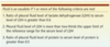
When do you do a thoracentesis?
Effusion of unknown cause