Pneumonics Flashcards
(9 cards)
ABCDE
- Airway control is paramount.
◦ Look – Externally examine for swelling of tongue, lips, or neck. Look inside the mouth for foreign bodies, loose teeth, vomitus or secretions. A word of caution – look but don’t touch! Blind finger sweeps are more likely to worsen than alleviate airway obstruction.
◦ Listen – Noisy breathing is obstructed breathing, so listen with the naked ear to the sound of respiration. Snoring or gurgling are signs of mild-moderate upper airway obstruction, usually with soft tissue or secretions. Stridor, a high-pitched inspiratory sound, suggests high-grade upper airway obstruction – this is always an emergency.
◦ Feel – Palpate the neck and upper chest for crepitus, which can be a sign of pneumothorax or tracheolaryngeal injury. Feel for swelling or masses in the neck as well, as these may lead to airway compromise in the future, even if the airway is patent now.
Check pulse-oxymetry and provide supplemental oxygen if needed.
intubation should be strongly considered for airway protection, oxygenation, decreasing metabolic load by removing the work of breathing and optimization of respiratory status. Keep in mind, though, that many drugs used in intubation, as well as positive pressure ventilation itself, can have negative hemodynamic effects. Arterial oxygen saturation should be maximized, and central venous oxygenation levels targeted above 70%.
◦ ALWAYS MAINTAIN C-SPINE IMMOBILIZATION
◦ Consider performing jaw thrust to establish patency of the airway.
◦ Consider use of a naso or oro-pharyngeal airway during bag-valve mask ventilations (BVM)
◦ Rapid Sequence intubation if needed for airway stabilization or protection (e.g. for GCS of 9 or less)
◦ Evaluate neck for landmarks associated with cricothyroidotomy and to assess the patient for subcutaneous emphysema or tracheal deviation.
Some conditions that can lead to progressive airway compromise:
◦ Anaphylaxis
◦ Angioedema
◦ Foreign body aspiration
◦ Epiglottitis
◦ Retropharyngeal abscess
◦ Ludwig’s angina
◦ Bacterial tracheitis
◦ Neck mass/hematoma
◦ Facial fractures
The following are indications for intubation:
◦ High-grade airway obstruction of any etiology
◦ Suspected imminent airway obstruction of any etiology
◦ Inability to maintain or protect the airway over time
◦ Severe hypoxia refractory to less invasive treatment
◦ Severe hypercarbia refractory to less invasive treatment
- Assess Breathing.
◦ Look: Assess the patient’s respiratory effort – are they breathing slowly or quickly? Deeply or shallowly? Using accessory muscles? Sitting up in the “tripod” position? More importantly, develop your gestalt sense of the patient’s work of breathing – does it look easy or effortful? Remember that normal respiration is almost imperceptible, so anytime you can really see a patient breathing, their work of breathing is increased.◦
Look for cyanosis, JVD (tension pneumothorax or cardiac tamponade), asymmetric movement of the chest (flail chest), accessory muscle use (tension pneumothorax) or open chest wounds (open pneumothroax).
Inadequate ventilation will lead to elevated levels of CO2 (respiratory acidosis) and can cause AMS. Bag-valve-mask ventilation should be provided until adequate ventilation can be restored. In a patient with AMS and a depressed respiratory status, consider narcotic overdose as a possible cause.
Listen to make sure that there are breath sounds – a lot of respiratory effort that produces minimal air exchange is a sign of imminent respiratory failure. Then make sure you hear breath sounds on both sides – unilaterally absent breath sounds may suggest a tension pneumothorax, which requires immediate treatment. Finally the quality of the breath sounds may provide a clue as to the underlying diagnosis.
◦ Ausculate: listen for stridor (upper airway injury), lung breath sounds (pneumo or hemothorax)
◦ Percuss: feel for hyper-resonance (pneumothorax) or dullness (hemothorax), subcutaneous emphysema (airway injury), paradoxical movements (flail chest) crepitence & point tendnerness (rib fractures) or bruising (pulmonary contusion).
◦ Count: There is no substitute for a respiratory rate that you count yourself. Documented respiratory rates are commonly inaccurate, and this is an critical vital sign for a patient in respiratory distress.
◦ Monitor: The oxygen saturation is really the “bottom line” on respiration, at least in the short term. The whole mission of the lungs is to deliver oxygen to hemoglobin, which will in turn deliver it to organs and tissues. If the oxygen saturation is low, some significant pathology is present which is causing the lungs to fail in their mission. Anything less than 96% is abnormal, and anything less than 90% is potentially life-threatening.
The goal of your respiratory assessment is to determine whether your patient has respiratory distress or failure. Patients with respiratory distress are attempting to compensate for underlying pathology that has deranged their pulmonary function.
Management:
The first priority is to make sure your patient IS breathing, and to provide assisted ventilations if not. Assisted ventilations are given using a bag-valve-mask (BVM) device, attached to 10-15L/min of oxygen. BVM ventilation is ONLY performed on patients with agonal or absent respirations. Do not attempt to perform BVM ventilation on a spontaneously breathing patient – this is poorly tolerated and
tends to worsen hypoxia.
◦ Use an oropharyngeal airway: This device hooks over the root of the tongue, pulling it forward out of the hypopharynx. It is so helpful for maintaining airway patency during BVM ventilation that it should feel wrong to use a BVM device without one!
Every patient with respiratory distress should receive some form of oxygen therapy. Oxygen delivery options include:
Noninvasive positive pressure ventilation (NIPPV) is a helpful treatment for some common causes of respiratory distress, including pulmonary edema, asthma, and chronic obstructive pulmonary disease (COPD). Modes of NIPPV include continuous positive airway pressure (CPAP) and bilevel positive airway pressure (BiPAP). Physiologic benefits of NIPPV include improved oxygenation, reduction in work of breathing, recruitment of collapsed alveoli, and redistribution of fluid from alveoli back into the vasculature. NIPPV has been shown to reduce need for intubation for patients with COPD and pulmonary edema, and is considered the standard of care for moderate-severe cases of these conditions. NIPPV requires use of a tight-fitting mask, which is poorly tolerated by some patients, and it is absolutely contraindicated in patients who are unable to maintain or protect their airways.
- Assess Circulatory status.
Obtain IV access through large bore peripheral lines or a central venous
catheter (which can help for rapid fluid and medication delivery, as well as provide invasive monitoring). Vascular access: Every patient with shock must have adequate vascular access. For an adult, this means two “large-bore” peripheral IVs (18g or bigger), or an intraosseous line, or a central line. Remember that intraosseous lines are safe and simple, and are preferred over central lines for patients who need rapid access. Also remember that central lines are long and skinny, and therefore poorly suited for large-volume resuscitation – if your patient needs a lot of fluid or blood, use peripheral or intraosseous access, or place a large-caliber “trauma line.”
Can you feel good distal pulses? Is the blood pressure very high or low? What is the cardiac rhythm? Hypoperfusion starves the brain of oxygen and glucose and leads to AMS. Nonperfusing rhythms require immediate CPR and ACLS. Hypotension should prompt IV fluid bolus and an immediate search for the cause.
First feel for pulses. If a radial pulse is palpable, it suggests a systolic blood pressure of at least 80 mm Hg. If the femoral or carotid are palpable, these suggest a systolic blood pressure of at least 60 mm Hg.
Note if they are thready versus bounding.
Many patients may not mount a tachycardic response:
◦ Neurogenic shock to sympathetic cord disruption
◦ Beta blockade, Calcium channel blockade
◦ Elderly
◦ Children and young adults
◦ Conditioned athletes start with a lower basal level. Doubling their resting heart rate of 45-50 shows a falsely reassuring heart rate of 90-100.
Careful monitoring of fluid status is encouraged, using a urinary catheter, intraarterial blood pressure measurements, and central venous pressure monitoring.
- Check for neurologic disability.
Use Glasgow Coma Score (GCS) or Alert Verbal painful unresponsive (AVPU) scale for a quick assessment of level of consciousness. Look for seizure activity. Are the pupils equal and reactive? Pay attention to spontaneous movements. Lack of movement on one side of the body night indicate stroke while lack of movement below a certain level of the body could indicate spinal cord injury. If there is any suspicion of trauma the cervical spine should be stabilized.
The “D” for disability represents a quick check to assess neurologic status. You can quickly assess mental status via the AVPU scale:
◦ Alert – a fully awake patient.
◦ Voice – the patient responds when verbally addressed. Response to voice can be verbal, motor, or with eyes.
◦ Pain – the patient makes a response on any of the three component measures only when pain stimulus is delivered.
◦ Unresponsive – If the patient does not give any Eye, Voice or Motor response to voice or painful stimuli.
Assess pupils for size, symmetry and reactivity. Uncal herniation will present as a “blown pupil.” This results from the paralysis of parasympathetic fibers of pupillary constrictors of CN III. You will see a dilated pupil due to unopposed sympathetic activity.
- Expose (fully undress) and perform a rapid head to toe look for signs of trauma, transdermal drug patches, dialysis access, infectious sources (such as catheters) or petechiae.
AMS = AEIOU TIPS
- *A** Alcohol
- *E** Epilepsy, Electrolytes, and Encephalopathy
- *I** Insulin
- *O** Opiates and Oxygen
- *U** Uremia
- *T** Trauma and Temperature
- *I** Infection
- *P** Poisons and Psychogenic
- *S** Shock, Stroke, Subarachnoid Hemorrhage and Space-Occupying Lesion
AMPLE
◦ Allergies
◦ Medications
◦ Past illnesses
◦ Last meal
◦ Events / Environment / Mechanism of injury
eFAST
EFAST incorporates use of thoracic ultrasound into a standard FAST examination to detect pneumothorax. In most cases, a high-frequency linear transducer is applied to the anterior chest wall in the midclavicular line at the level of the second intercostal space, and the clinician is looking for the absence of normal lung “sliding” and lack of comet-tail artifact, indicating pneumothorax. Additional views are often obtained in the midaxillary line, and alternative probes may be used. EFAST has been found to be more sensitive than chest radiography for the detection of occult pneumothoraces in patients experiencing trauma (48.8% versus 20.9%), although both have a very high specificity (99.6% for EFAST and 98.7% for chest radiography).
Hints
HINTS exam is composed of three separate tests:
the horizontal Head Impulse test, a test of Nystagmus, and a
Test of Skew.
If vertical or bidirectional nystagmus is present
at rest, or if vertical skew is present at baseline, a central cause
of vertigo should be suspected and a full HINTS examination
is not indicated.
Head Impulse: a positive (abnormal) test is suggestive of a peripheral lesion,
whereas a negative (normal) test is consistent with a more
concerning central lesion.
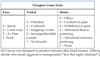
Parkland…
Rule of 9s; 50-50, 8-16
LEMON
“LEMON” is a helpful mnemonic for remembering signs of a potentially
difficult airway:
◦ Look: visual inspection of the face and neck can reveal anatomic or cosmetic factors that make intubation difficult: facial dysmporphism (particularly micrognathia), obesity, dental abnormalities, short or thick neck, facial/neck trauma or swelling
◦ Evaluate 3/3/2: this rule refers to the normal facial and neck anatomy in terms of the patient’s finger breadths – if any of these measurements are less than the amount specified, expect trouble:
◦ 3 finger breadths between the incisors when the mouth is opened
◦ 3 finger breadths from the mental vertex to the hyoid bone
◦ 2 finger breadths from the hyoid bone to the thyroid notch
◦ Mallimpati: this is a great tool for cooperative patients in the preoperative setting, but is less useful for critically ill patients in the ED. It requires that the patient sit upright and voluntarily open the mouth as widely as possible. The score is based on how much of the posterior pharynx can be visualized.
◦ Obstruction: check for foreign body, listen for stridor as above
◦ Neck mobility: assess range of motion of the neck, as limitations can make positioning for intubation difficult. Note that trauma patients have limited neck mobility by definition, as they are typically immobilized for protection of the cervical spine, though this does not usually present a significant obstacle to airway management.
DONT
Coma cocktail:
Dextrose
Oxygen
Nalaxone
Thiamine
ELEVATION
Electrolytes
LBBB
Early repolarizations
Ventricular hypertrophy
Aneurysm
Thiland (brugada)
Inflammation
Osborne “J waves” hypothermia
Non Ischemic Vasospasm
