Polyps, Esophagitis, Surgical Abdomen Flashcards

(104 cards)

1
Q

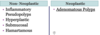
define polyp classifications

non-neo

neo

A
2
Q

Smooth surface “cloud-like” appearance polyp

tx?

A

sessile serrated

Complete excision is recommended - due to their sessile nature and indistinct borders, special care is needed to ensure their complete removal endoscopically.

3
Q

adenomas ≥10 mm in size or with villous components or high-grade dysplasia.

A

Advanced Adenomatous Polyps

4
Q

Most common non-neoplastic polyp

•Normal cellular components but may be indistinguishable from adenomatous polyps

A

Hyperplastic Polyps

Look similar to tubular adenomas

5
Q

Irregularly shaped islands of intact mucosa that forms as a result of mucosal ulceration and regeneration

•Seen in UC and Crohn’s Disease

A

Inflammatory Pseudopolyps

6
Q

define high greade vs low grade dyplasia

A

Low-grade dysplasia – some cells are abnormal, but unlikely to spread

High-grade dysplasia- represents a step in the progression from a low-grade dysplasia to cancer - unlikely to metastasize

•applied to lesions that are confined to the epithelial layer and lack invasion into the lamina propria.

7
Q

Recommend that CRC screening begin at

A
  • age 45 in African Americans, and that colonoscopy is the preferred test - More likely to develop right sided CRC
  • Age 50 for other races
8
Q

CRC screening recc in

lynch syndrome

FAP

A

lynch

  • Age 20-25 or 10 years younger then youngest affected relative
  • Colonoscopy 1-2 years
  • Then yearly at age 40
  • Genetic testing recommended

FAP

  • Age 10-12 sigmoidoscopy yearly
  • Colonoscopy yearly after polyp discovered genetic testing and counseling
9
Q

CRC screening reccomendations

hx of CRC

hx of adenoma

IBD

A

Personal Hx of CRC

  • Total colon exam w/in 1 yr, repeat at 3 yrs
  • Repeat 5 yrs if normal

Personal Hx of Adenoma- Repeat colonoscopy every 3-5 yrs

IBD

  • Begin 8 yrs after diagnosis
  • Colonoscopy every 1-2 yrs
10
Q

differnetiate b/w cancer prevention vs cancer detection tests

A
11
Q

Fecal Occult Blood Test(Stool Guaiac)

A

Testing stool for the presence of blood – 3 separate stools

Lowest specificity / sensitivity

•Detects ANY blood – could be from nose bleed etc

12
Q

Fecal Immunochemical test (FIT)

A

More sensitive for colonic blood loss -Higher CRC detection rates compared to FOBT

Detection of advanced adenomas is VERY low

13
Q

Fecal DNA (FIT-DNA

A

Looks for evidence of mutations associated molecular changes leading to malignancies – KRAS mutations, methylation biomarkers associated with neoplasia, and hemoglobin

•Full stool sample in a special collection kit

Higher specificity for CRC –Still LOW detection od adenomas

Convenient, no sedation

14
Q

The definitive test for detection of precancerous adenomas and CRC

A

colonoscopy

Avg risk – 10 yrs

May be shorter for higher risk (3-5 yrs)

15
Q

Patients who cannot take colonoscopy or who are sick –

why BAD for AAs or younger population ??

A

Sigmoidoscopy

•BAD for AAs or younger population as they are most likely affected by Right sided colon cancer

41-45% of CRC are on the right side and will be missed

16
Q

Most colorectal cancers, regardless of etiology, arise from ____ polyps polyps

A

adenomatous

17
Q

Currently Influence CRC Screening

A
  • Personal or Family History of CRC or polyps
  • Age
  • Hereditary CRC Syndromes
18
Q

Carcionembryonic antigen (CEA) level

A

but used to monitor progression pre-post surgery, of CRC

• indicator of Recurrence. Expect to normalize after surgery

19
Q

si/sx of CRC

A

Change in bowel habits – 74%

Rectal bleeding/bloody stool/black stool – 51%

Anemia

20
Q

tx of CRC

A

Surgery – Resection of primary colonic or rectal cancer is the treatment of choice in all stages (I+II ONLY surgery)

  • Poorly differentiated histology
  • Lymphovascular invasion
  • T2 lesion, cancer at stalk margin

Chemotherapy –Stages III&IV Colon cancer

Radiation + Chemotherapy – Rectal Cancer stages II-IV

21
Q

differntiate b/w iron deficency pattern vs anemia of chronic dz

A

iron deficency

Transferrin/TIBC – Increased

  • Transferrin Sat% - low
  • Ferritin* - low

anemia of chronic dz

•Transferrin/TIBC – low

  • Transferrin Sat% - normal or low-normal
  • Ferritin* – normal or elevated
22
Q

chemo for CRC is reccomended at what stage

A

III

23
Q

Indications to Consider Hereditary Intestinal Polyposis Syndrome

A
  1. Patient with family history of CRC affecting more than one family member.
  2. Personal or family history of colorectal cancer developing early age <50 years
  3. Personal or Family History of multiple polyps >20 cumulative!)
  4. Personal or family history of multiple extracolonic malignancies.
24
Q

Intestinal Polyposis Syndromes

A

Lynch Syndrome / HNPCC

Familial Adenomatous Polyposis (FAP)

25
Lynch Syndrome / HNPCC si/sx
Poorly differentiated tumors in the right colon Presenting at young age \*\* History of rectal bleeding, bowel obstruction, perforation Family history
26
Extra-colonic malignancies: Lynch Syndrome / HNPCC
**Endometrial** , uterine, ovarian, stomach, small bowel, hepatobiliary, urinary tract, brain and skin cancers
27
Familial Adenomatous Polyposis (FAP) Extracolonic Manifestations
Gardner Syndrome – FAP patient with extracolonic manifestations •Desmoid tumors – most common
28
define FAP
characterized by the presence of multiple colorectal adenomatous polyps (typically more than 100) •First polyp age 16 Germline mutation in the APC gene located on chromosome 5q21-q22 •AD
29
dx FAP
Diagnosis should be suspected in anyone with \>10 cumulative colorectal adenomas
30
tx ## Footnote Lynch Syndrome / HNPCC Familial Adenomatous Polyposis (FAP)
Lynch Syndrome / HNPCC - colectomy Familial Adenomatous Polyposis (FAP) Prophylactic Colectomy * Remaining rectum or ileal pouch will need to be screen q6mo-2 yos * Age 20-25 EGD q1-3 years Chemoprophylaxis NSAID and COX2
31
screening guidelines in lynch FAP
lynch - Yearly colonoscopy 1-2 years starting age 20-25 years of age FAP - Age 10-12 yearly flexible sigmoidoscopy Yearly colonoscopy once polyps detected CRC in 100% of pts by 39
32
small bowel cancer etiologies
**Adenocarcinomas -** Lynch syndrome, FAP CF Crohn’s or UC **Neuro -endocrine Tumors (Carcinoid Tumors)** -Assoc w/ multiple endocrine neoplasia type-1 **Lymphoma -** Crohn’s disease Celiac disease Chronic immunodeficiency **Leiomyosarcoma**- Assoc w/ Meckel’s Diverticulum
33
noninvasive imaging modality of choice\*\* for small bowel Cancer
CT scan – IV/PO contrast ## Footnote Angiography if active bleeding Surgical exploration – most sensitive diagnostic modality
34
anal cancer is most commonly assoc w/ etiologies?
HPV, anal warts ## Footnote Small Cell Carcinoma (SCC)- Most common\* Adenocarcinoma Melanoma Sarcoma
35
tx of anal cancer
Systemic chemo and radiation •Cisplatin +mitomycin C Biopsy-proven disappearance in \>80% of patients •lesions \<3 cm
36
Post Treatment Surveillance: anal cancer
Every 3-6 months for five years: * Digital Rectal Examination (DRE) * Anoscopy * Inguinal node palpation * +/-CT chest/abdomen/pelvis annually for three years
37
staging anal cancer O-II III IV
Stage O-II : Node negative Stage III: Node positive Stage IV: Metastatic disease
38
name 2 types of SCC anal cancers
* Non Keratinizing SCC – above the Dentate line * Keratinizing SCC –distal to Dentate line
39
define surgical adbomen
•an acute intra-abdominal condition of abrupt onset, usually associated with pain due to inflammation, perforation, obstruction, infarction or rupture of abdominal organs, and usually requiring emergency intervention
40
Periumbilical pain -\> right iliac fossa pain Colicky -\> dull, constant dx?
apendicitis
41
si/sx of apendicitis in elderly children preganant
**Elderly: \>65** * Diminished inflammatory response → \< findings H&P * Increased rate of perforation at presentation **Children:** * Clinical exam before imaging * Classic presentation with \> WBC, CRP → Surgical Consult before imaging * Atypical or equivocal presentation → _Ultrasound 1ST_ * Possible need for contrast CT or MRI **Pregnant:** * Many present ‘_non-classically’→ heartburn, bowel irregularity, diarrhea, malaise_ * Elevated WBC’s can be normal in pregnancy * _US is imaging of choice_ → non-compressible, \> 6mm diameter
42
US findings in apendicitis
appendiceal diameter of \>6 mm) * Non-compressible tubular structure in RLQ * Wall thickness \>2 mm * Overall diameter \>6 mm * Free fluid in the right lower quadrant * Thickening of the mesentery * Localized tenderness with graded compression * Presence of a calcified ‘appendicolith’
43
Exceptions NO Appe
: Stable patients with perforated appendicitis who have symptoms localized to the RLQ * Treated initially with antibiotics, intravenous fluids, and bowel rest, rather than immediate surgery * These patients will often have a palpable mass; (CT) scan may reveal or abscess
44
pre-op abx in apendicitis
Peri-op IV Abx – broad spectrum to cover aerobic and anaerobic bacteria •3 rd generation Cephalosporin or Gentamycin + Metronidazole (Flagyl)
45
tx of prolonges ileus
NGT ## Footnote * improves patient comfort * Minimizes/prevents recurrent vomiting * Serves as a means to monitor the progress or resolution of these conditions
46
most common causs of SB obstruction
•Post-operative adhesions - most common 70%
47
si/sx of SB obstruction
Dehydration \*\*\* (hallmark) - Tachycardia, orthostasis, decreased urine out +/- Fever High-pitched ‘tinkling’ sounds on auscultation (acute), tympanic on
48
imaging of choice for SB oobstruction
abdominal CT - •Finding the transition point between dilated and non-dilated bowel can identify site and cause of obstruction plain XRay - "string of pearls’
49
Blood gas, lactate in SB obstruction
(marker for mesenteric ischemia), and blood cultures
50
tx of SB obstruction
NPO - bowel rest IVF – fluid resuscitation is integral to treatment Bladder catheter – measure urine output NGT – if emesis - Decreases nausea, vomiting, distention, aspiration •Sx improver!!
51
SB Obstruction 80% of cases w/ _____ intestinal obstruction
mechanical
52
ileus vs SB obstruction bowel sounds pain fever, tachy
**bowel sounds** ileus - quiet SB - high-pitched **pain** ileus - mild and diffuse SB - moderate - severe, colicky **fever, tachy** ileus - absent SB - should raise suspicion
53
ischemia of the small bowel, usually secondary to an acute cause involving the SMA or SMV
Mesenteric Ischemia
54
etiology of Mesenteric Ischemia
**SMA Occlusion (~70% of cases)** * Embolism: MI, Afib, Endocarditis, Valve disease * Thrombosis: Atherosclerosis – plaque rupture **Non-occlusive Mesenteric Ischemia (NOMI)** * Hypoperfusion + vasoconstriction (Transient/partial) * ‘Watershed’ areas of colon with limited collateral circulation **Mesenteric Venous Thrombosis (MVT)** * \*\*\*younger patients w/o CV disease * Primary clotting disorder
55
si/sx of mesenteric ischemia
Rapid onset Severe, unrelenting periumbilical pain Pain out of proportion to clinical exam \*\*\*\*
56
Mesenteric Ischemia test of choice
Mesenteric Angiography
57
Plain X-ray non-specific (normal in 25%) Distended loops of bowel, ‘Thumbprinting"
Mesenteric Ischemia
58
ischemic bowel dz includes
Mesenteric Ischemia Ischemic Colitis
59
tx of ischemic bowel dz
Mild: supportive care , bowel rest, IV fluids, observation, ? NGT Moderate: Empiric ABX-broad spectrum Severe: Surgical exploration - Surgical Laparotomy with resection
60
The presence of four or more risk factors: 100% predictive of colonic ischemia - ischemic colitis
* \> 60 years * Hemodialysis * Hypertension * Hypoalbuminemia * Diabetes mellitus * Constipation-inducing medications
61
si/sx of ischemic colitis
Rapid onset of mild cramping and tenderness over affected bowel -\> Associated with the urgent desire to defecate
62
imaging for ischemic colitis
CT (oral/IV) Colonoscopy -Confirmatory, Not in patients with peritonitis (MRA) – individuals w/ compromised renal function
63
ischemic bowel dz effects what areas of the colon
* Effects are typically prominent at the “watershed” areas of the colon à Collateral blood flow is limited * Splenic flexure * Rectosigmoid junction
64
Potentially lethal complication of inflammatory bowel disease (IBD) or infectious colitis
toxic megacolon
65
define toxic megacolon
Characterized by total or segmental non-obstructive colonic dilatation + systemic toxicity * Marked dilatation of the colon * Thinning of the bowel wall * Deep ulcers
66
imaging in toxic megacolon
X-Ray - Marked dilation CT scan - Thinning of wall lining
67
infectious causes of toxic megacolon
c. diff ## Footnote Stool WBC and cultures including C. diff
68
dx criteria for toxic megacolon
Radiographic evidence of colonic distension _With at least three of the following:_ * Fever \>38ºC * Heart rate \>120 beats/min * WBC \>10,500/microL * Anemia _At least one of the following:_ * Dehydration * Altered sensorium * Electrolyte disturbances * Hypotension
69
tx of toxic megacolon
_Non operative therapy is the first line of treatment_ * Antibiotics broad spectrum for IBD (Vanco and Flagyl for C. Diff) * Intravenous corticosteroids (IBD) * Bowel decompression with a NGT if needed **_Surger_**y reserved for patients who do not improve on non-op management --\> subtotal colectomy with end-ileostomy (up to 50% mortality)
70
Peptic Ulcer Disease PUD RF types
Risk Factors: **H. Pylori**, NSAIDs, Etoh, Bile salts, etc Gastric Ulcer -Pain shortly after or during eating Duodenal Ulcer -Pain hours after eating —Pain wakes pt @ night
71
dx of PUD
H. Pylori testing - Urea breath test ## Footnote Endoscopy – modality of choice\*
72
Helicobacter Pylori: triple therapy
* Omeprazole, lansoprazole or Esomeprazole * AND Clarithromycin 500mg bid * AND Amoxicillin or Flagyl 10-14 days Alternative (PPI, Bismuth, Tetracycline, Flagyl)
73
Epigastric pain \*- Gnawing/Burning s/p meals Chest pain/Heartburn Hematemesis
PUD
74
Dysmotility Disorders etiology (4)
**Achalasia** - Relative obstruction and proximal dilation of esophagus w/ food bolus stasis d/t loss of ganglion cells from esophagus wall causing LES to fail to completely relax •Botox **Diffuse Esophageal Spasm** - Functional imbalance btwn excitatory and inhibitory pathway thus disrupted peristalsis and manometry w/ \>20% simultaneous contraction **Nutcracker esophagus** - Distal esophagus mmhg @ peristalsis \>220mmhg • **Hypertensive LES -** Resting LES \>45mmgh Scleroderma esophagus - Smooth muscle atrophy and fibrosis •Smooth muscle replaced by scar tissue thus lose peristalsis and LES tone
75
Barium Esophagram\* - Birdbeak or Corkscrew appearance Manometry\* -Esophageal motor pattern/intensity w/ LES mmhg & function DX?
Dysmotility Disorders
76
TX OF DYSMOTILIOTY DISORDERS
**Nitrates & CCB** – SMC relaxants •Isosorbide dinitrate OR Nifedipine OR Diltiazem **TCA** - Modify neuropathic pain pathway •Imipramine OR Amitriptyline **Botox in LES**- Esp achalasia and HTN LES **Endoscopy therapy** - Dilation (pneumatic) 50-93% response rate LES pathology) •No response s/p x2 dilations consider surgery **Surgery - Heller Myotomy**
77
name 2 types of esoph striuctures
Distal stricture - Peptic stricture, GERD. Adenocarcinoma Proximal/Mid Stricture - Caustic ingestion, Malignancy, esophagitis
78
si/sx of eso stricture
Dysphagia\* - food impaction
79
tx of eso stricture
**Medication - PPI** **Diet - GERD restrictions** * Weight loss * Small meals & eat slowly and deliberately * Avoid meds that cause pill esophagitis **Esophageal dilation\*** - repeat at 1 yr , tx of choice!!
80
dx eso strictures
Barium esophagram\*Location, length, diameter of the stricture **Endoscopy\*** CT - Stage malignancy
81
White people w/ GERD THINK -
eso stricture
82
si/sx of ## Footnote Mallory Weiss Tears Boerhaave syndrome
**Mallory Weiss Tears** - Hematemesis 85% - Vomit/wretch then _hematemesis classic_ **Boerhaave syndrome** -Repetitive wretching/vomiting then s_udden chest pain_ * ?radiates to back/shoulder * no hematemesis
83
dx ## Footnote Mallory Weiss Tears - Boerhaave syndrome -
Mallory Weiss Tears - EGD - Most have stopped bleeding and risk of rebleed low Boerhaave syndrome - CT (imaging of choice +/- esophagram)
84
tx of mallory weiss
* PPI +/- Sucralfate 1-2 weeks * Anti-emetic
85
tx of Boerhaave syndrome
IVF, antibiotics Surgical consult w/ Thoracotomy w/ direct repair of rupture and mediastinal/pleural cavity drainage
86
esophagitis etiology
reflux infectious medication induced radiography systemic dz
87
Heartburn - Worst w/ large meal, tight clothes, supine or bending over Dysphagia\*
esophagitis
88
dx of esopahagitis
EGD (esophagastroduodenoscopy)
89
tx of reflux esophagitis
Pain- Narcotics , H2 blockade •Liquid antacid therapy or magic mouthwash PPI - Omeprazole, Lansoprazole Sucralfate - Esophageal coating agent
90
2 types of gastritis
•**Erosive** (reactive gastritis d/t exposure & gravity usually @ greater curvature of stomach most often w/ _NSAIDs_ - Superficial, deep, hemorrhagic **•Non-Erosive** - _H. Pylor_i most common cause of gastritis
91
si/sx of gastritis
Epigastric pain, burning, gnawing Nausea/Vomiting +/- w/ eating Melena/hematemesis/hematochezia/coffee ground emesis
92
dx gastritis
. Pylori Tests (same as PUD) EGD •Appearance: Thick, edema, erosions, erythematous gastric folds
93
tx gastritis
H. Pylori+ then tx w/ triple/quad therapy D/C offending agents (NSAIDs, Etoh) Antacid -Aluminum & magnesium hydroxide INC pH and neutralizes gastric acidity Sucralfate/Carafate - Mucosal protectant H2 blocker - Inhibits gastric acid secretion PPI - Inhibit proton pump and INC pH
94
common causes of GERD
**LES transient relaxation\*** * Foods- Coffee, Etoh, chocolate, fatty meal, mint/peppermint * Medications -CCB, B-Blocker, Nitrates, Hormones, anti-cholinergics * Nicotine **Hiatal hernia** * LES migrates into chest thus lose HPZ (high mmhg zone) à Length of HPZ may decrease * Gastric contents trapped in hernia sac and reflux during LES relaxation
95
Heartburn\* -Retrosternal sensation of burning esp s/p •Eating, supine, bending over Dysphagia\* Regurgitation\*
GERD
96
dx GERD
24 hour esophageal pH monitoring\*\*\* Gold standard for GERD dx as quantifies amt reflux w/ sxs
97
tx of GERD
Minimize gastric acid secretion à Antacid - S/P each meal and @ bedtime H2 Blocker -Ranitidine, famotidine, cimetidine •Before meal ie. 30 min PPI\* - Best for GERD * Omeprazole, esomeprazole, lansoprazole * Few side effects and good long term Corrective anti-reflux surgery - Nissen Fundlopication
98
complications of GERD
Stricture Barrett Esophagus
99
types of esophageal cancer
**Small cell carcinoma (Eastern europe, Asia)** * Upper half of esophagus * Smoking & Etoh **Adenocarcinom**a (North America, Western europe) * Lower half esophagus * GERD/Barretts metaplasia
100
definitive dx of esoph cancer
endoscopy and bx
101
tx of esophageal cancer
Esophagectomy- Transthoracic OR Transhiatal •Type of surgery does not influence survival but staging @ time surgery does
102
strongest RF for gastric cancer
h. pylori
103
gastri cancer labs and imaging
CEA : carcinoembryonic antigen INCREASED 45-50% CA 19-9 INC 20% EGD\* - Definitive diagnosis w/ Bx
104
tx gastric cancer
_Surgery_ * Total gastrectomy * Esophagogastrectomy - Tumor @ GEJ and Cardia * Subtotal gastrectomy - Tumors of distal stomach _Neoadjuvant chemoradiotherapy_ * Preoperatively = std of care in US * First line = epirubicin/cisplatin/5-FU or docetaxel/cisplatin/5-FU