Procedures Flashcards
(26 cards)
Differentiate between TPN and EN
Enteral nutrition = given in to the GI tract
- Mouth if no risk of aspiration/choking
- NG allows disease-specific liquid nutrotion eg high amino acids for liver disease
Parenteral nutrition = bypass GI tract, directly into vein
- given through central venous line
- only given if GI tract malfunctioning so would become malnourished without it
Summarise the indications for enteral feeding
- Increased nutritional requirements e.g. sepsis, surgery
- Increases nutritional losses e.g. malabsorption
- Decreased intake e.g. dysphagia, nausea, sedation, coma
- Effect of treatment, e.g. nausea, diarrhoea
- Enforced starvation e.g. prolonged NBM period
- Difficulty with feeding
- Unappetising food
Summarise the indications for parenteral feeding
- GI tract not functioning e.g. bowel obstruction
- Poor absorption e.g. short bowel syndrome or active Crohn’s
- High risk of malnutrition
Identify the possible complications of TPN
- Sepsis
- Thrombosis of central vein leading to pulmonary embolism or superior vena caval obstruction
- Metabolic imbalance, refeeding syndrome
- Mechanical issues such as pneumothorax, embolism of IV line tip
Summarise the indications for NG tube
- To decompress the stomach/GI tract especially when there is obstruction e.g. ileus
- For gastric lavage
- To administer feed/drugs
Identify the possible complications of NG tube
- Pain
- Rare: loss of electrolytes, oesophagitis, tracheal/duodenal intubation, necrosis (retro/nasopharyngeal), stomach perforation
Describe the different types of endoscopy based on area examined
- Oesophagus, stomach and duodenum = oesophagogastroduodenoscopy (OGD) aka upper GI endoscopy
- Small intestine = enteroscopy
- Large intestine/colon = colonoscopy, sigmoidoscopy
- Bile duct = ERCP
- Rectum (rectoscopy) and anus (anoscopy) = both is proctoscopy

Summarise the indications for an upper GI endoscopy
- Haematemesis
- New dyspepsia (if >/= 55 y/o)
- Gastric biopsy
- Duodenal biopsy
- Persistent vomiting
- Iron deficiency
Define Colonoscopy
Endoscopic examination of the large bowel and distal part of the small bowel
Sedation and analgesia first given, before a flexible colonoscope is passed and guided around the colon
indications for a colonoscopy
- Rectal bleeding – when settled, if acute
- Iron-deficiency anaemia
- Persistent diarrhoea
- Biopsy of lesion seen on barium enema
- Assessment or suspicion of IBD
- Colon cancer surveillance
Define + explain ERCP
Endoscopic retrograde cholangiopancreatography

combines endoscopy and fluoroscopy to diagnose and treat problems of biliary or pancreatic ductal systems.
A catheter is advanced from a side-viewing duodenoscope via the ampulla into the common bile duct.
Contrast medium is injected and x-rays taken to show lesions in the biliary tree and pancreatic ducts.
Summarise the indications for ERCP
No longer routinely used for diagnosis
Significant therapeutic role
⇒ Common bile duct stones
⇒ Stenting of benign or malignant strictures
⇒ Obtaining brushings to diagnose nature of a stricture
Identify the possible complications of ERCP
- Pancreatitis
- Bleeding
- Cholangitis
- Perforation
Indications for laproscopy
- minimally invasive so reduced pain
- reduced risk of haemorrhaging
- shorter recovery time
- smaller scar
- fewer wound-related infections
Define laparotomy
surgical procedure involving a large incision through the abdominal wall to gain access into the abdominal cavity
Indications for laparotomy
- Rupture of an organ e.g. spleen, aorta, ectopic pregnancy
- Peritonitis (perforation of a peptic ulcer/duodenal ulcer, diverticulum, appendix, bowel, gallbladder)
- CS
Identify the possible complications of open (laparotomy) abdominal surgery
- Adhesions
- Bleeding
- Infection
- Paralytic ileus
- Shock
- Incisional hernia (20%)
Summarise the indications for a cholecystectomy
- Gall bladder stones (symptomatic)
- Acute cholecystitis
- Gallstone pancreatitis
- Choledocholithiasis
- Cholecystoduodenal fistula
Identify the possible complications of a cholecystectomy
- Damage to bile ducts which can cause bile leak
- Post cholecystectomy syndrome – RUQ pain, dyspepsia, nausea/vomiting
- Post site hernia
- Bleeding
- Infection
- Fat intolerance due to inability to secrete a large amount of bile into the intestine as pt no longer has a gall bladder
Summarise the indications for an appendicectomy
Normally performed as an emergency procedure for acute appendicitis
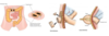
Define stoma
A surgically created opening in the body between the skin and a hollow viscus.
ABDOMINAL STOMAS are used to divert faeces or urine outside the body to be collected in a bag at the skin
A stoma’s position, appearance and contents can point to which type of stoma it is.
Describe these features for each 3 types of stoma
Colostomy- LARGE BOWEL
- Found in the LIF
- Contents – solid (as faeces has had time to travel through colon and undergo water absorption)
- Will be flat against skin
Ileostomy- SMALL BOWEL
- Found in the RIF
- Contents – liquid and lighter (not as much water absorbed)
- As the enzymes in the faeces are toxic and can damage skin, the stoma will not be flat but rather have a spout sticking out from the abdominal wall
Urostomies- post-cystectomy (bladder removal)
- Located in RIF
- Contents – urine (way to distinguish from ileostomy)
- A piece of ileum is resected then attached to the skin with a spout protruding.

Compare permanent vs temporary end-colonostomies
Permanent end colostomies
- done in cases of abdominoperineal resection of large rectal cancers
- when there is removal of entire rectum.
Temporary end colostomies
- done to rest the bowel e.g. diverticulitis
- the rectum and bowel will be re-anastomosed at a later date
- Hartmann’s procedure
Cannot distinguish between these clinically

Describe a loop colostomy
These are done to protect distal anatomoses after recent surgery
A loop of bowel will be brought to the surface and half opened, which allowed the faecal matter to drain into the stoma bag without reaching the distal anastomoses, a supporting rod is used to secure the two parts of the skin.
The two parts are still attached as this is a temporary procedure which will be reversed. As it is a half –opened loop, the healing process is much quicker