RA week 2 Flashcards

1
Q

thoracic cage made up of?

articulations?

seperated by?

girdle of upper limb? part of throacic cage?

A

12 pairs of ribs that form lateral walls

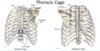
anteriorly = articulates with midline sternum

posteriorly = articulate with 12 throacic vertebrae

seperated by intercostal spaces

girdle of upper limb = scapula and clavicle, not part of thoracic cage

2
Q

space enclosed by thoracic cage?

thoracic apertures?

A

thoracic cavity

3
Q

boundaries of superior thoracic aperture (inlet)?

inferior throacic aperture (outlet)?

A

superior

  • posteriorly = body of T1
  • laterally = 1st pair of ribs + costal cartilages
  • anteriorly = superior border of manubrium

inferior

  • posteriorly = body T12
  • anterolaterally (going from lateral to anterior) = 11th + 12th ribs, costal cartilages of ribs 7-10 (costal margin)
  • anteriorly = xiphisternal joint
4
Q
A

anterior body attached to posterior vertebral arch via pedicle

5
Q

transverse process function?

what forms spinous process?

note long an sliding arrangement of spinous processes

A

attachments for muscles

+ articular surfaces for ribs

lamina extend posteriorly and unite to form spinous processes

6
Q

describe articular surfaces for ribs on T6 and T7

A

on body of T7 (close to pedicle) - can see superior and inferior demi-facets

  • will articulate with head of rib 7 but also rib 8 inferiorly
  • head of rib 7 will also articulate with vertebrae above (T6)

articular surface of transverse process

  • articulates with tubercle of the 7th rib
7
Q

types of ribs

classification of ribs

A
8
Q

atypical ribs?

A

ribs 1, 2, 10, 11 + 12

9
Q
A

head articulates with body of own vertebrae but also with vertebra above (and with intervertebral disc via the crest)

tubercle - non articular part provides attachment for costotransverse ligament

10
Q

most curved part of rib body?

A

costal angle

11
Q

where is cosal groove?

purpose?

A

it is an indentation on the internal surface of the inferior border of the body of the rib

provides protection for intercostal vessels and nerves

12
Q

what makes rib 1 atypical?

what is significant about the bony feature that seperates these grooves?

(pic is superior view)

A

single facet on head - articulates with T1 only

grooves for subclavian vessels on superior surface (seperated by scalene tubercle)

  • scalene tubercle is attachment for anterior scalene muscle of the neck
13
Q

what makes rib 2 atypical?

(pic is superior view)

A

has many typical features but is called atypical because it has a rough area on upper surface called = tubersoity for serratus anterior

  • attachment site for serratus anterior muscle
14
Q

what makes ribs 10, 11 and 12 atypical?

(image superior view)

A

10, 11 + 12 = single articular facet on head, articulates with single vertebra

11 + 12 = very short with no neck or tubercle

15
Q

divisions of the sternum?

connected by?

A

manubrium, body and xiohoid process

  • manubrium + body connect at sternal angle (manubriosternal joint)
  • body and xiphoid process connect at xiphisternal joint
16
Q

bony features of manubrium?

A

jugular notch superiorly

clavicular notche son either side of JN = articular surfaces for the clavicles

17
Q

lateral border of sternum?

articulations?

A

lateral border has a number of indentations called the costal notches

  • 7 costal notches for articulations of costal cartilages of the ribs

1st rib articulates with manubrium adjacent to the clavicular notch

2nd rib articulates at the sternal angle

ribs 3-6 articulate along the side of the body of the sternum

7th rib articulates at xiphisternal joint

18
Q

level of sternal angle?

A

rib 2

19
Q

intervertebral joints?

orientation of articular facets in thoracic region of vertebral coloumn?

A

intervertebral discs between bodies of vertebrae = secondary cartilagenous joints

synovial plane joints between superior and inferior articular facets (NOT costal facets) - zygapophyseal joint

orientation of articular facets in thoracic region allows rotational movement to occur between the vertebrae

20
Q

zygapophyseal joint?

A

synovial plane joints between the articular processes of two adjacent vertebrae

also called facet joints

21
Q

sternocostal joints?

explain

interchondral joints?

A

joints between costal cartilages of the ribs and the sternum

rib 1 = primary cartilagenous joint (synchondrosis)

rib 2-7 = synovial plane joints

interchondral joints (joints between costal cartilages) between 6+7, 7+8, 8+9, 9+10

  • 6+7, 7+8, 8+9 = synovial plane joints
  • 9+10 = fibrous joint
22
Q

costovertebral joints?

type?

movement?

A

found between ribs and vertebrae

in image can see 7th rib with head of 7th rib articulating with body of T6+T7

  • can also see tubercle of 7th rib articulate with transverse process of T7
  • all of these joints are plane synovial joints
    • ​NOTE: movements will occur at joints formed by head and tubercle (rib moves around an axis that runs between both of these joints)
23
Q

movement of ribs during respiration?

A

determined by axis of rotation

  • upper ribs = more lateral xis - greatest degree of movement at anterior aspect of rib “pump handle movement”
    • ribs 1-6
  • lower ribs = more posterior axis - greatest degree of movement at lateral border of rib “bucket handle movement”
    • ribs 7-12
24
Q

rib cage changes in dimension during respiration?

A

on inspiration rib cage expands along 3 different axis

  • sternum + upper ribs move anteriorly and superiorly = increases anterior-posterior dimension
  • lower ribs flare out laterally = increases lateral dimension
  • diaphragm descends = increases vertical dimension
25
Q

primary muscle of inpiration at rest?

A

diaphragm

movements of rib cage are minimal (become more pronounced during forced inspiration and expiration)

26
Q

thoracic wall muscles?

secondary muscles of respiration?

superficial muscles associated with upper limb and anterior abdominal wall?

A
27
Q

upper limb and anterior abdominal wall muscles?

innervation?

A

pectoralis minor deep to pectoralis major

serratus anterior = muscular bands wrap around latral thoracic wall and attach on lower ribs (gets name from serrated appearance)

rectsu abdominus - run vertically on either side of the midline

fibres of external oblique = hands in pockets

28
Q
A

29
Q

secondary muscles of respiration?

attachments?

innervation?

A

2 serratus posterior muscles - superior and inferior

  • superior = medially attached to spinous processes of C7-T3 then extends inferolaterally towards upper borders of ribs 2-4
  • inferior = medially attached to spinous processes T11-L2 then extends superolaterally towards lower borders of ribs 8-12

there are 3 scalene muscles on either side of the neck - anterior, middle and posterior

  • proximal attachments on transverse processes of cervical vertebrae
  • distal attachments - 1st rib for anterior + middle scalene, 2nd rib for posterior scalene
30
Q

scalene muscle function?

innervation?

A

movement of the neck

when neck is fixed, can also act as accessory respiratory muscles

(raise 1st + 2nd ribs)

31
Q

serratus posterior muscle function?

innervation?

A

main function thought to be proprioception

superior muscle may assist with raising upper ribs

+ inferior muscle may assist with lowering the lower ribs

32
Q

which muscles are found in the intercostal spaces?

A

from superficial to deep = external intercostal, internal intercostal, innermost intercostal

33
Q

external intercostal muscle fibres?

found where?

function?

A

pass downwards and forwards (hands in pockets) from lower border of one rib to upper border of the rib below it

they extend from the tubercles of the ribs to the costochondral juction (most obvious posteriorly and laterally on the thoracic wall)

  • beyond this point they are replaced anteriorly by the external intercostal membrane (extends between costal cartilages towards sternum)

raise the ribs in inspiration

34
Q

internal intercostal muscles

found where?

function?

A

fibres pass dowards and backwards from lower border of one rib to upper border of rib below (hands on chest) - at a 90* angle to external intercostal muscles

  • internal intercostal muscle most obvious anteriorly on the thoracic wall
  • extends from sternum to angle of ribs - after which it is replaced posteriorly by internal intercostal membrane

1 - interosseus part (i.e. between ribs) depresses ribs on expiration

2 - interchondral (between costal cartilages) part raises ribs on inspiration

35
Q

innermost intercostal muscles found?

direction?

features

A

deep to lateral part of internal intercostal muscles

fibres pass dowards and backwards (same direction as internal intercostals)

very thin, often incomplete layer (difficult to distinguish from internal intercostal)

36
Q

deepest muscles of thoracic wall?

attachments?

what lies superficial to transversus thoracis?

A

subcostal muscle + transversus thoracis muscle

internal thoracic vessels lie superficial to transversus thoracis (run on either side of the sternum)

37
Q

what is intercostal space?

contains? (2)

A

space between adjacent ribs

contains intercostal muscles

neurovascular plane is found between internal and innermost intercostal muscles:

  • contains intercostal Vein, Artery and Nerve (superior to inferior)
  • run in costal groove at inferior border of rib
  • (also give off branches that can be found near upper border of rib)
38
Q
A

39
Q

intercostal nerve function?

A

within each intercostal space, there is intercostal nerve

this nerve will supply intercostal muscles

  • will also give lateral and anterior branches that pierce through muscle layer into skin where they supply sensory innveration
40
Q

arterial supply to intercostal space?

branches?

A

majority by posterior intercostal arteries (most branch directly from the aorta)

  • posterior intercostal artery will also give cutaneous branch whcih passes through muscle walls to supply the skin

anterior part of intercostal space supplied by anterior intercostal artery (branch directly from internal thoracic artery)

  • will give anterior perforating branch to supply the anterior skin
41
Q

posterior intercostal arteries branch from?

A

1 + 2 from supreme intercostal artery (branch of costocervical trunk from subclavian artery)

3-11 and subcostal artery branch directly from thoracic aorta

(will see in posterior mediastinum dissection)

42
Q

which intercostal spaces also have an anterior intercostal artery?

branch from?

what does internal thoracic artery branch from?

where is it found?

what does it become?

A

intercostal spaces 1-9 have anterior intercostal artery (in addition to posterior)

  • 1-6 direct from internal thoracic artery
  • 7-9 from musculophrenic artery

Internal throacic artery branches from the subclavian artery

internal throacic artery bifurcates to give superior epigastric artery and musculophrenic artery

43
Q

venous drainage of thoracic wall?

A

follows similar pattern to arterial supply

  • anterior intercostal veins drain to –> internal thoracic vein
    • runs alongside internal thoracic artery
  • posterior intercostal veins drain to –> azygos/hemiazygos veins
    • azygos vein on right
    • hemiazygous and accessory hemiazygous on the left
44
Q
A

can see posterior intercostal veins draining into (hemi?)-azygos vein

can also see posterior intercostal arteries coming from the aorta

45
Q
A

46
Q

where is breast located?

A

anterior surface of pectoralis major muscle (also has axillary tail which extends laterally towards axilla)

47
Q

glandular tissue of breast oragnised into?

what happens during lactation?

where do lobules open into?

A

glandular tissue of breast oragnised into lobules

in resting state these lobules are small - during lactation they enlarge and produce milk

lobules of mammary gland open into nipple via lactiferous ducts

48
Q

how is glandular tissue of breast supported?

organised into? - function?

A

supported by fibrous tissue

organised into thick fibrous bands called suspensory ligaments

  • provide support
  • anchors breast at dermis of the skin
49
Q
A

50
Q

blood supply to breast?

venous drainage?

A

medial and lateral mammary arteries

  • lateral mammary arteries branch from lateral thoracic artery (whcih branches from axillary artery)
  • medial mammary artery branches from internal throacic artery

venous drainage follows similar pattern

  • lateral mammary vein
  • medial mammary vein
51
Q

lymphatic drainage of breast?

A

within breast there is subareolar lymphatic plexus

  • from here, most (>75%) lymph drains to axillary nodes
  • remainder will drain to parasternal lymph nodes and some to abdominal nodes
52
Q

layers of thoracic wall dissection

A

1st layer = skin

2nd layer = superficial fascia (carries superficial blood vessels and nerves that supply the skin and fascia), will probabably see subcutaneous fat in this layer

3rd layer = muscles

  • pectoralis major, serratus anterior, external oblique muscles
  • in image you can see the nerve that supplies serratus anterior (long thoracic nerve) on the scissors
53
Q
A

note muscular bands that give serratus anterior its serrated appearance

54
Q
A

The intercostal nerves form branches that supply motor innervation to the intercostal muscles but also carry sensory fibresfrom the skin. Photograph A shows some of these nerves as they pierce through the muscle of the thoracic wall and enter the superficial fascia

Pectoralis minor lies inferior to pectoralis major on the anterior thoracic wall. Photograph B shows pectoralis major reflected away from the ribs and pectoralis minor passing to ribs 3-5

55
Q
A

The pectoral muscles are innervated by the medial and lateral pectoral nerves. The nerves are named for the cords of the brachial plexus from which they originate, not for their position on the thoracic wall

Photograph A shows the lateral pectoral nerve (raised by the forceps) piercing through the posterior surface of pectoralis major. This muscle is also innervated by the medial pectoral nerve

Photograph B show part of the medial pectoral nerve piercing the posterior surface of pectoralis minor and pectoralis major (raised by the forceps in the photograph)

56
Q
A

Photograph B shows serratus anterior reflected away from the thoracic wall to reveal one of the external intercostal muscles lying between the ribs. External intercostal is more muscular posteriorly and laterally

It becomes membranous anteriorly, as shown by the blue arrows on the photograph

57
Q
A

Pic A shows some of the muscle fibres of internal intercostal visible through the membranous part of external intercostal (indicated by the arrows). Internal intercostal is more muscular anteriorly and laterally and becomes membranous posteriorly

Pic B shows the initial stages of removal of the anterior thoracic wall. The ribs have been sectioned and the left lung is just visible. You can also see the internal thoracic vessels that lie either side of the sternum

58
Q
A

Once the ribs have been sectioned the thoracic shield can be lifted away from the heart and lungs, this is shown in photograph A. Note the presence of transversus thoracis muscles radiating from the sternum to the ribs.

Photograph B shows the innermost intercostal muscles visible through the costal parietal pleura (shown by arrows on the photograph)

59
Q

function of non-articular part of tubercle?

A

non articular part provides attachment for costotransverse ligament

60
Q

Divisions of the thoracic cavity? (3)

What do they contain?

A

pulmonary cavities contain lungs

between pulmonary cavities there is the mediastinum (all other organs of throax including heart contained here)

61
Q

lining of pulmonary cavities?

purpose?

A

lined by serous membrane called pleura

produces fluid to lubricate movement of organ in the cavity

62
Q

pleura divided into?

are they completely separate layers?

A

parietal = lines pulmonary cavity, has cervical, costal, diaphragmatic and mediastinal parts

visceral = covers surface of lung

no - continuous at lung root/hilum

63
Q

sleeve of pleura hanging down below lung root? purpose?

layer of connective tissue that attaches… to …?

A

sleeve of pleura hanging down below lung root = pulmonary ligament

  • allows expansion of vessels in the hilum e.g. during exercise

layer of connective tissue that attaches costal parietal pleura to thoracic wall = endothoracic fascia

64
Q

endothroacic fascia?

continuous with?

attachments?

A

endothoracic fascia = layer of connective tissue that attaches costal parietal pleura to thoracic wall

superiorly (yellow part of image) - it is continuous with fibrous suprapleural membrane

suprapleural membrane has attachments on 1st rib and transverse process of C7

65
Q

what is purpose of suprapleural membrane?

A

prevents expansion of lungs up into the neck

66
Q

surface markings of pleura and lungs described when?

where do lungs/pleura extend to at mid-isnpiration?

A

usually described at mid-inspiration

at mid inspiration lungs/visceral pleura:

  • extend in mid-clavicular line to level of 6th rib
  • mid-axillary line to 8th rib
  • scapular line to 10th rib
67
Q

where does parietal pleura extend to at mid-inspiration?

A

attachments of parietal pleura will not change with inspiration and are usually 2 ribs below the level of the lungs

  • mid clavicular line = rib 8
  • mix-axillary line = rib 10
  • scapular line = rib 12
68
Q

parietal pleura extensions create?

A

extensions of parietal pleura create costodiaphragmatic recesses

  • costomediastinal recesses are found posterior to the sternum (larger on left due to cardiac impression)
69
Q
A

70
Q

Features of lungs?

surfaces?

borders?

lobes?

A
71
Q

lobes of left lung?

right lung?

A

left lung = superior + inferior lobe (divided by oblique fissure)

right lung = oblique fissure separates superior and inferior lobe (+ inferior and middle lobe), horizontal fissure separates superior lobe from middle lobe

72
Q
A

73
Q

impressions on right lung?

A

largest impression created by right atrium of the heart (still small compared to cardiac impression on left lung)

SVC leaves impression as it runs towards right atrium of the heart - upper part of this impression is created by right brachiocephalic vein

(the 2 brachiocephalic veins unite to form the SVC)

other impressions by azygos vein + oesophagus

74
Q

arrangement of structures in hilum of right lung?

largest of these structures?

A

largest = pulmonary veins (anteriorly and inferiorly)

pulmonary arteries = more superiorly

right main bronchus = posteriorly

75
Q
A

76
Q

where is lingula located?

what is it?

A

inferior anterior border of left upper lung lobe

it is analogous to the middle lobe of the right lung

76
Q

where is lingula located?

what is it?

A

inferior anterior border of left upper lung lobe

it is analogous to the middle lobe of the right lung

77
Q

impressions of left lung on mediastinal surface?

A

very pronounced cardaic impression on left lung (left ventricle of heart)

arch of aorta above lung hilum and descending aorta posterior to the hilum

impression extending off arch of aorta = left subclavian artery

78
Q

arrangement of structures in hilum of left lung?

A

pulmonary veins = anteriorly and inferiorly

pulmonary artery = superiorly

left main bronchus = posteriorly

78
Q

arrangement of structures in hilum of left lung?

A

pulmonary veins = anteriorly and inferiorly

pulmonary artery = superiorly

left main bronchus = posteriorly

79
Q

how to orient yourself at lung hilum?

A

pulmonary veins = always most anterior and inferior structures (always at least 2 pulmonary veins present)

pulmonary arteries = located superiorly in the hilum

bronchus = located posteriorly in the hilum (very easy to identify because can feel for cartilage in its wall)

80
Q

relations of the right lung hilum?

A

azygos vein posteriorly and arching over top of the hilum to empty into SVC (anteriorly)

oesophagus posteriorly

2 important nerves

  • vagus nerve will always travel posteriorly to hilum
  • phrenic nerve will always travel anteriorly to hilum
81
Q

relations of right lung hilum?

A

azygos vein posteriorly and arching over top of the hilum to empty into SVC (anteriorly)

oesophagus posteriorly

2 important nerves

  • vagus nerve will always travel posteriorly to hilum
  • phrenic nerve will always travel anteriorly to hilum
82
Q

relations of left lung hilum?

A

arch of aorta above left lung hilum, and descending thoracic aorta posteriorly

vagus (posterior) + phrenic nerves (anterior)

on left side, vagus nerve will also give recurrent laryngeal nerve which loops underneath arch of the aorta

83
Q

bronchial tree?

A

trachea will bifurcate at level of sternal angle into right and left main bronchus

  • RMB will further subdivide into 3 lobar bronchi (superior, middle, inferior)
  • LMB will subdivide into 2 (superior + inferior)

lobar bronchi will then further subdivide into many segmental bronchi (multicoloured branches) = supply segments of lung lobes

84
Q

bronchopulmonary segmentation?

what is contained in each segment?

A

lobes of lungs divided into number of segments

  • on left = 5 segments associated with superior lobe + 5 with inferior lobe
  • on right = 3 segments with superior, 2 with middle, 5 with lower lobe

each segment will have: single segmental bronchus + single branch of pulmonary artery + single pulmonary vein

85
Q

does respiratory tree end with segmental bronchi?

A

no - segmental bronchi will subdivide into bronchioles

86
Q

types of bronchioles?

A

conducting

terminal

respiratory → connect to alveolar sac via alevolar duct

87
Q

what is an alveolar sac?

function?

A

an air sac that has a profuse capillary network associated with it

gas exchange

88
Q

lymphatic drainage of lungs?

A

within tissue of lungs, there are pulmonary nodes

as move towards the lung hilum, there are bronchopulmonary (hilar) nodes

surrounding point of bifurcation of trachea = superior + inferior tracheobrachial (carinal) nodes

either side of trachea = paratracheal nodes

89
Q
A

90
Q
A

Photograph C = blue paper has been used to highlight position of pulmonary artery and purple markers have been used to show positions of pulmonary veins

note at the hilum the bronchus is posterior and the pulmonary veins are anterior and inferior in position

on the left lung the pulmonary artery lies above the bronchus

91
Q
A

Pic B = blue paper indicates position of pulmonary artery and purple markers indicate position of pulmonary veins

the bronchus is the most posterior structure at the hilum

the pulmonary veins are anterior and inferior

the pulmonary artery usually lies next to the bronchus in the right lung

92
Q

mediastinum divided into?

separated at what point?

A

superior mediastinum

inferior mediastinum:

  • anterior, middle + posterior mediastinum

separated at transverse thoracic plane - from sternal angle to IV disc between T4 + T5

93
Q

middle mediastinum bounded by?

anterior and posterior?

A

fibrous pericardium of the heart

everything anterior to fibrous pericardium = anterior mediastinum

posterior is the same

94
Q

anterior mediastinum boundaries?

contents?

A

boundaries

  • body of sternum (anteriorly)
  • fibrous pericardium (posteriorly)
  • transverse thoracic plane (superiorly)
  • diaphragm (inferiorly)

contents

  • loose connective tissue
  • fat
  • lymphatics
  • in children = thymus
    • usually in superior mediastinum but as it is larger in children, it often extends below transverse thoracic plane into anterior mediastinum
95
Q

middle mediastinum contents?

boundaries?

adjacent structures?

A

contents

  • heart + pericardium
  • roots of great vessels

bounded on all sides by pericardial sac

adjacent structures

  • great vessels (pulm. a + v, aorta + SVC)
  • phrenic nerves
  • lung root structures
96
Q
A

97
Q

heart located?

relations?

A

heart is a midline structure located posteriorly to the body of the sternum (located slightly more to the left than to the right)

(⅓ right of the midline, ⅔ left of the midline)

right and left phrenic nerves

  • right phrenic nerve associated with pericardium overlying RA
  • left phrenic nerve associated with pericardium overlying LA and LV
98
Q

fibrous pericardium continuous with?

A

superiorly = blends with tunica adventitia of great vessels

inferiorly = continuous with central tendon of diaphragm

99
Q

what will you see if you open up fibrous pericardial sac?

A

sac itself is lined with parietal serous pericardium

this will be reflected onto surface of heart to form visceral serous pericardium

(from images you can see that there is a significant amount of fat deep to the serous pericardium on the surface of the heart)

100
Q

arrangement of pericardium?

A

most superficially = tough fibrous pericardium (forming pericardial sac)

just deep to this = parietal serous pericardium

reflected over roots of great vessels onto surface of heart to become = visceral serous pericardium (also called epicardium)

between the 2 layers of serous pericardium = pericardial cavity (serous pericardium produces fluid that lubricates movements of the heart)

deep to epicardium = myocardium

lining chambers of the heart = endocardium

101
Q

arrangement of serous pericardium creates?

how does T arise?

dissection?

A

arrangement of serous pericardium creates 2 pericardial sinuses = transverse pericardial sinus + oblique pericardial sinus

transverse pericardial sinus arises due to development of heart from simple heart tube with an inflow end + outflow end

  • as heart develops it will close on itself, bringing 2 ends closer together + creating narrow channel between inflow and outflow vessels of heart

during dissection slide finger posterior to pulm. trunk and ascending aorta - ur finger is now in transverse pericardial sinus with outflow vessels in front of your finger and inflow vessels behind

102
Q

what is the oblique pericardial sinus?

dissection?

A

space posterior to the heart

if you slide hand underneath and behind heart (fingers posterior to left atrium), fingers now within oblique pericardial sinus with pulmonary veins on either side

103
Q

shape of heart?

borders?

base?

A

heart is trapezoidal shape

base of the heart is posterior aspect of the heart + made up of left atrium

104
Q

anterior view of heart?

A

most of what we see is right side of heart

105
Q

what separates right atrium from right ventricle?

what is found here?

what separates left and right ventricles anteriorly?

what is found here?

A

atrioventricular/coronary groove

groove contains the right coronary artery

anterior interventriclar groove

groove contains anterior interventricular artery (left anterior descending artery)

106
Q

what enters right atrium?

what leaves left ventricle?

A

SVC + IVC enter right atrium

(IVC pierces through diaphragm and immediately enters inferior part of RA)

ascending aorta leaves left ventricle and then continues as aortic arch (arching over pulm. trunk as it divides into left + right pulmonary arteries)

107
Q

what leaves right ventricle?

what enters left atrium?

A

pulmonary trunk leaves RV

right and left pulmonary veins enter LA

108
Q

branches of the aortic arch?

what unite to form the SVC?

A

brachiocephalic trunk, left common carotid and left subclavian

SVC = brachiocephalic veins

109
Q

posteroinferior view of the heart?

A

most of what we see is left side of heart

base of heart can be seen (mainly LA)

110
Q

left pulmonary surface of heart?

diaphragmatic surface of heart?

A

seen posteroinferiorly

mainly composed of left ventricle

diaphragmatic surface = left + right ventricles

111
Q

what separates left and right ventricles posteriorly?

where is it found?

A

posterior interventricular groove

runs along position of interventricular septum between left and right ventricles

112
Q
A

shiny surface caused by serous visceral pericardium

deep to this layer is significant amount of epicardial fat

113
Q

serous pericardium + epicardial fat cleared away

A

113
Q

serous pericardium + epicardial fat cleared away

A

114
Q

right coronary artery found?

left coronary artery divides into?

A

atrioventricular/coronary groove (between RA and RV)

left anterior descending artery (between right and left ventricle) + left circumflex artery (posteriorly)

115
Q

ligamentum arteriosum?

function?

significance?

A

fibrous remnant of ductus arteriosus

DA allows communication between aorta and pulmonary trunk during foetal life (i.e. allows blood to bypass the lungs)

useful landmark - left recurrent laryngeal nerve runs posterior to it in the adult

(ligamentum arteriosum shown by blue arrow)

116
Q

where is ligamentum arteriosum found?

A

between pulmonary trunk and arch of aorta (remnant of ductus arteriosus)

117
Q

arteries that supply the heart arise from?

names?

A

arteries that supply the heart arise from ascending aorta

right and left coronary artery

118
Q

right coronary artery branches?

A

first branch is small SA nodal branch = supplies SA node in wall of right atrium

right coronary will then continue in atrioventricular/coronary groove (will give off numerous branches to supply walls of RA and RV)

before turning into inferioposterior surface of heart, will give off right marginal branch (runs along inferior border of RV)

will then continue to inferoposterior surface of heart in AV groove and will give off AV nodal branch (located in wall of RA)

right coronary artery will then terminate by giving posterior interventricular artery which runs in posterior interventricular groove (between RV and LV)

119
Q

left coronary artery branches?

A

by comparison to RCA, LCA is very short vessel quickly terminating as circumflex artery and anterior interventricular artery

anterior interventricular artery/LAD in anterior interventricular groove (between RV + LV)

anterior interventricular artery will give diagonal artery

circumflex artery will continue to posterior aspect of the heart (runs in left atrioventricular groove between LA + LV)

  • before it passes to posterior surface it will give left marginal branch that runs along left border of heart
  • it will then continue in AV groove ultimately anastomosing with RCA
120
Q

variation in arterial supply of heart?

A

SA nodal branch + posterior interventricular artery can sometimes arise form circumflex artery

diagonal artery not always present in all individuals

121
Q

areas of heart supplied by RCA?

A

basically right side of heart:

right atrium + most of right ventricle

SA (usually) and AV nodes

posterior ⅓rd of interventricular septum (via posterior interventricular artery)

part of left ventricle (again via posterior interventricular artery)

122
Q

areas of heart supplied by left coronary artery?

A

left side of heart:

  • left atrium + most of left ventricle
  • anterior ⅔rd of interventricular septum (anterior interventriclar artery)
  • AV bundle (part of the electrical component of the heart found within IV septum - so supplied by anterior interventricular artery)
  • part of right ventricle via anterior interventricular artery
123
Q
A

last pic can also see diagonal artery coming off of anterior interventicular artery

124
Q

venous drainage of heart?

A

via cardiac veins

all veins drain to coronary sinus except anterior cardiac veins which drain directly to right atrium

125
Q

what is coronary sinus?

A

coronary sinus = swollen venous structure on posterior surface of heart between LA + LV

all cardiac veins drain here except anterior cardiac veins (drain wall of RA and drain directly into RA)

126
Q

cardiac veins?

found?

A

anterior cadiac veins (drain into RA)

great cardiac vein

  • found in anterior IV groove alongside anterior interventricular artery
  • will then curve around left border of heart to reach posterior surface where it empties into coronary sinus

middle cardiac vein

  • posterior interventricular groove alongside posterior interventricular artery

small cardiac vein

  • wall of RV alongside right marginal artery
  • will curve around inferior border of heart to reach posterior surface where it empties into coronary sinus
127
Q

main tributary to coronary sinus?

A

great cardiac vein

128
Q
A

129
Q

other venous structures on the heart?

A

(posterior surface of heart)

left marginal vein runs alongside left marginal artery

left posterior ventricular vein drains posterior wall of left ventricle

oblique vein of left atrium drains wall of left atrium

130
Q

coronary sinus drains into?

A

coronary sinus opens directly into right atrium

131
Q
A

132
Q

internal wall of right atrium?

smooth area?

A

internal wall of RA can be divided into 2 distinct areas

  • area with muscular ridges = musculi pectinati
  • smooth area = sinus venarum

sinus venarum will connect entrance of SVC with entrance of IVC below

133
Q

what separates sinus venarum and muscular wall of RA?

A

ridge called crista terminalis

134
Q

interatrial wall (between RA and LA)?

what is this?

what can also be seen within right atrium?

A

there is a small thumb-shaped impression = fossa ovalis

fossa obvalis is a remnant of foramen ovale (allowed commonuication between RA and LA during development)

opening of coronary sinus can also be seen within right atrium

135
Q

blood flow from RA to RV? found?

what is found along its margins?

function?

what else is found in right ventricle?

A

blood flows from RA to RV through tricuspid valve (can be found at right atrioventricular canal)

along margins of tricuspid valve there are string-like structures called chordae tendineae (found in right ventricle)

anchor valve at muscular projections of right ventricular wall called papillary muscles

a structure unique to right ventricle = moderator band

  • extends from IV septum to anterior papillary muscle
  • will carry part of conducting system of heart
136
Q

muscular ridges in atrial wall?

muscular ridges in ventricular wall?

A

muscular ridges in atrial wall = musculi pectinati

muscular ridges in ventricular wall = trabeculae carneae

137
Q

wall of right ventricle?

blood flow?

A

muscular ridges called trabeculae carneae

RA → RV → lungs via pulmonary trunk

138
Q
A

139
Q

internal wall left atrium?

however…

blood flow?

A

smooth and featureless

however, left atrium has left auricle as an expansion - auricle has muscular ridges called musculi pectinati

blood flows from LA to LV via bicuspid (mitral) valve

140
Q

cusps of mitral valve connected to?

A

the 2 cusps of mitral valve connected at their edges by papillary muscles by chorda tendineae in left ventricle (anchors them to left ventricular wall)

141
Q

features of left ventricular wall?

blood from LV?

A

much thicker than wall of right ventricle

will have numerous muscular ridges = trabeculae carneae

LV → aorta via aortic valve

142
Q

blood flow through heart

A
  • deoxygenated venous blood returned to RA via SVC + IVC
  • pass through tricuspid valve to RV
  • pumped into pulmonary trunk → lungs via pulmonary arteries
    • pulmonary arteries only arteries in body that carry deoxygenated blood
  • blood oxygenated in lungs + returned to heart via pulm. veins
    • pulmonary veins only veins that carry oxygenated blood (4 total, 2 form either side)
  • pulmonary veins enter LA
  • LA → LV via mitral (bicuspid valve) → aorta via aortic valve
  • rest of body
143
Q

purpose of heart valves?

types of heart valve?

mechanisms of action?

A

ensures unidirectional flow of blood

2 types

  • semilunar valves = aortic + pulmonary valves
  • cuspid valves = mitral (bicuspid) + tricuspid

different mechanisms of action

  • semilunar valves open in response to high pressure (ventricles contract)
    • valves closed in low pressure
  • cuspid valves open during low pressure
    • close in response to ventricular contraction
144
Q

how to semilunar valves close?

what does this allow?

A

when heart relaxes, blood will flow back towards ventricles

this blood will trickle in between cusp of valve and vessel wall (red dot) filling that space and forcing cusps of valve to close

this backflow also allows heart to receive blood supply through coronary vessels (remember coroanry arteries arise from acending aorta at level of aortic valve)

coronary arteries filled during low pressure via backflow of blood

145
Q

what helps prevent prolapse of cuspid leaflets?

A

prolapse would allow regurgitation of blood back into atria

to prevent this happening chordae tendineae are anchored at papillary muscles and help to hold valve in place

papillary muscles will contract at same time as ventricles holding cusps in place :))

146
Q

which valves are open during ventricular diastole?

systole?

A

diastole = cuspid valves open + semilunar valves closed

systole = cuspid valves closed + semilunar valves open

147
Q
A

148
Q

pulmonary valve cusps?

aortic valve cusps?

A

pulmonary = anterior, left + right

aortic = right, left + posterior

(can see opening of right coronary artery within the valve)

149
Q

what are heart valves supported by?

other function?

A

fibrous skeleton of the heart

supports valves + electrical insulation between atria and ventricles (allows delay of contraction of ventricles until after atrial contraction)

pic = can see rings that support cuspid valves + coronets that support semilunar valves

150
Q

SA node function? located?

signals?

located?

A

SA node (located in upper part of crista terminalis adjacent to entrance of SVC - in wall of RA) allows heart to regulate its own heart rate

signals from SA node pass through wall of right atrium + into wall of LA causing atria to contract at the same time

signals from SA node will also be picked up by AV node (located in lower part of interatrial septum)

151
Q

AV node function?

how does it do this?

RV?

A

transmits signal from SA node into ventricles

does this via AV bundle which divides into right and left bundle branches

these bundle branches will give off many branches into walls of right and left ventricles

within RV - some fibres from RBB will travel via septomarginal trabecula (moderator band)

152
Q

why do some fibres from right bundle branch travel via septomarginal trabecula (moderator band)?

A

ensures anterior papillary muscle + anterior part of wall of right ventricle contract at same time as rest of ventricular walls

153
Q

SA node innervation?

effect?

A

can function without any external input from CNS - however, does receive autonomic supply via cardiac plexus

  • parasympathetic - vagus (X) - intrinsic ganglia
  • sympathetic - T1-T5/6 spinal levels - cervical and superior thoracic paravertebral ganglia (sympathetic chain)

parasympathetic = decrease HR + constrict coronary vessels

sympathetic = increase HR + dilate coronary vessels

154
Q

what does cardiac plexus supply?

A

SA node

155
Q

sakhc

A

pic A = fibrous pericardium, note fat on outer surface. Pericardial sac blends with fascia of diaphragm + great vessels

pic B = right and left phrenic nerves pass over fibrous pericardium on their way to supply innervation to diaphragm

pic C = surface of heart itself is only visible if pericardium is sectioned - parietal serous layer adheres to fibrous pericardium + visceral serous layer covers surface of the heart (visceral serous layer sometimes called epicardium)

156
Q

kkb

A

pic A = anterior surface of heart - RA, RV, LV can be seen. Aorta + pulm/trunk can also be seen (SVC seen lying behind aorta in this image but usually lies to the right of aorta)

pic B = roots of coronary arteries (fat usually surround surface of coronary arteries so have to remove this)

157
Q

qfe

A

coronary arteries

pic A - largest part of arteries as they leave ascending aorta

pic B - branches of right coronary artery on anterior surface of heart

pic C = anterolateral view of the heart, some branches of left coronary artery can be seen

158
Q

efbw

A

pic A + B = posterior surface of heart. LA + LV can be seen

there is fat associated with posterior surface of heart

pic C = shows middle + great cardiac veins

159
Q

vqv

A

pic A = external surface of RA, shows SVC as drains into RA, muscle of wall is deep red/brown colour

pic B = ridged patter of wall of atrium (musculi pectinati i.e. the pectinate muscles) - end/originate at crista terminalis

pic C = can see fossa ovalis

no photo of internal wall of LA as this is a mainly smooth wall except opening of pulm.veins and ridges inside left auricle

160
Q

vqev

A

when wall of RV is opened you can see trcuspid valve

3 valve flaps each attached to chordae tendineae and papillary muscles

moderator band (septomarginal trabecula)

161
Q

ebfiugb

A

inner surface of both ventricles forms muscular network - trabeculae carneae

2 valve flaps in mitral valve - anterior an dposterior papillary muscles associated with them (pic B)

162
Q

qebwrn

A

pic A = semilunar valves + origins of left and right coronary arteries from ascending aorta

pic B = aortic valve - right, left cusps (associated with right and left coronary arteries) and posterior cusp

pic C = pulmonary trunk, right, left + anterior cusps

pic D = fatty plaque formation on flaps of aortic valve