Radiology II Flashcards
(61 cards)
the MRI can be used to image what elements?
which element does it image in medicine?
why?
- can be used to image any element with an odd number of protons
- in medicine, it is tailored to image / manipulate H+ (one proton)
- this is b/c there are so many H+ atoms in the body
hydrogen is in what state when bound to a molecule within body tissues?
which body tissues most dense in hydrogem?
- when bound: H+ goes from having 1 elecron + 1 proton to just 1 proton.
- tissues most abundant with H+: water, fat
what is the significance of a spinning proton?
the spinning positive charge creates a tiny electrical current
how does hydrogen behave organically vs in the presence of an external magnetic field?
why is this important?
- no external magnetic field:
- protons oreinted different directions
- their individual electric signals cancel out
- external magentic field (in an MRI):
- protons align (either parallel or antiparallel)
- this generates magnetic field
how is an image generated from aligned protons in an MRI?
- a radiofrequency (RF) pulse is sent onto, inducing protons to temporal change their alignments
- the new spinning magnetic field produce an electrical signal
- this electrical signal is detected by a antenna (coil) then mapped into an image
what are TE and TR?
- TE (echo time): time between sending RF pulse and measuring the signal
- TR (reptition time): time between successive RF pulses
what is the difference between T1 and T2 weighted signals?
- T1: fat is white
- T2: fat AND fluid (CSF, for example) are white
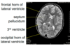
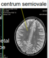
label
T1 vs T2?

cerebellar tonsils
T1 (fluid is dark)
label
T1 vs T2?

cerebellar tonsils
T2 (eye fluid is white)
tonsillar herniation
- definition
- causes
- when tonsils pass inferior to foramen magnum
- causes:
- Chiari I malformation, often associated w/ syrnix - congenital, mild
- intracranial hemorrhage - acquired, life threatning
- tumor - aqcuired, life threatning
identify

tonsilar herniation (inf to foramen magnum)
d/t chiari I malformation
identify
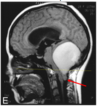
tonsillar herniation (inf to foramen magnum)
d/t posterior fossa tumor
label
T1 vs T2


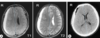
label
T1 vs T2

T2

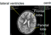
label
T1 vs T2
middle cerebellar peduncle
T2
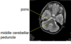
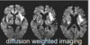
how does multiple sclerosis appear on an MRI
- T2 intense plaques (lesions) within white matter, which if often:
- radiating perpendendicular from lateral ventricles
- within the corpus collosum
- in middle cerebellar peduncle

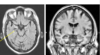
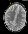
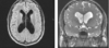
identify
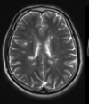
multiple sclerosis
plaque (lesion) perpendicular from lateral ventricle
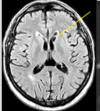
identify

multiple sclerosis
plaque (lesions) perpendicular from lateral ventricles
identify

multiple sclerosis
plaque (lesion) in corpus collosum
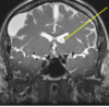
identify

multiple sclerosis
plaque (lesion) in the middle cerebellar peduncle
label

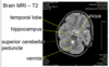
identify

uncal herniation

hippocampus
hippocampus atrophy - cause?
Athzheimers, commonly