Renal Pathophysiology Flashcards

(235 cards)

1
Q

How is GFR measured?

A

clearance of inulin or creatinine

estimates based on serum creatinine

2
Q

azotemia

A

accumulation of nitrogenous waste products in the blood

i.e. urea

any rise in serum BUN or creatinine above normal

3
Q

uremia

A

clinical syndrome or symptom compelx associated with severe impariment of renal function

4
Q

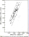
specific gravity of urine

A

lower specific gravity correlated with low osmolarity (more dilute urine)

5
Q
A

This is an example of a urine

1: Here a white cell with red blood cells around it

When you see cells in the urine you do not know if they have come from the kidney or someplace else in the urinary tract a

6
Q
A

WBCs and bacteria in urine

7
Q
A

tubular epithelial cell (not round like WBC)

8
Q
A

squamous epithelial cells - from bladder ureter or urethra NOT kidney

9
Q

casts

A

cylindrical masses of agglutinated material

formed in distal nephron, have to come from kidney

Tamm-Horsfall mucoprotein is the major protein constituent

Hyaline, granular or cellular

10
Q

Where are casts formed?

A

distal nephron

11
Q

Tamm-Horsfall mucoprotein

A

major protein constituent of casts
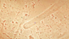
12
Q
A

Hyaline cast

we think the hyaline cast and granular cast are degenerated cellular casts

There is a lot of other amorphous material here

13
Q
A

tubular epithelial cell cast

you can see the shape of the cells here are not perfectly round which you would see in a white blood cell cast

14
Q
A

broad cast

it was formed further down in the nephron, again there are red cells around this cast

15
Q
A

coarse granular cast

notice the granules and degenerating cells

16
Q
A

RBC cast

17
Q
A

WBC cast

18
Q
A

waxy cast (probably has cholesterol)

19
Q
A

triple phosphate crystals

often in people with UTIs

20
Q
A

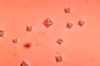
calcium oxalate crystals

21
Q
A

On the left there are stellar and amorphous Ca Phosphate crystals

On the right Ca Oxalate crystals

22
Q
A

cysteine crystals

23
Q
A

uric acid crystals

24
Q

What does dipstick look for?

A

•Dipsticks (mainly picks up albumin, may miss low molecular weight and other nonalbumin proteins)

25
Heat and Acetic Acid for urine test?
take a specimen of urine and heat it up and if there’s protein you will see it form at the bottom of the test tube The test tube has been heated (left) and as it cools you see the protein on the bottom This is with heat and acetic acid but with sulfosalicylic acid it will look very similar We don’t do this often, usually send sample off to the lab and they can measure protein or albumin
26
sulfosalicylic acid test for urine
detects all proteins in the urine
27
Microalbuminuria - how do we test?
dipsticks not positive until rel high to find smaller amounts - use direct measrements of albumin secretion microalbumin - to - creatinine ratio!
28
How do you determine the type of protein in the urine?
protein electrophoresis
29
glomerular proteinuria
increase in permeabilty of glomerular capillary wall leads to increased glomerular filtration of protein
30
tubular proteinuria
impaired reabsorption of normally filtered proteins
31
overflow proteinuria
increased production of smaller proteins in multiple myeloma and ther plasma cell issues
32
nephrotic syndrome
massive loss of normal serum proteins in the urine 1. heavy proteinuria (\>3.5) 2. hypoalbuminemia 3. edema 4. hyperlipidemia 5. sometimes HTN
33
hypoalbuminemia in nephrotic
urinary loss of protein liver is making more but can't keep up with loss
34
Why edema in nephrotic syndrome?
overfill hypothesis glomerular disease/tubular inflammation leads to increased renal sodium retention (reabsorb mostly in collecting tubules
35
Why is there no hypertension in nephrotic syndrome?
Na retention USUALLY results in hypertension but nephrotic patients to not MAY be secondary to hypoalbuminemia
36
Why hyperlipidemia in nephrotic syndrome?
elevated cholesterol, TG, phospholipids low plasma albumin? increased lipoprotein synthesis
37
lipiduria in nephrotic syndrome
oval fat bodies (tubular cells w fat drops) maltese crosses (fat drops under polarized light) on urinalysis
38
oval fat bodies in neprhotic synd urine ## Footnote 1: oval fat body (tubular cell that is filled with fat)
39
maltese crosses - polarized light on fat lipid in urine in nephrotic If you think a patient has nephrotic syndrome it is important to look at the urine for oval fat bodies and then look under polarized light
40
thromboembolic events
in nephrotic syndrome! hypercoagulable state DVT and renal vein thrombosis
41
Minimal Change Clinical Picture
acute onset variable fluid retention HTN infrequent renal function is normal EDEMA and protein in the urine!
42
urinalysis in minimal change disease
proteinuria (ALBUMIN - selective) oval fat bodies few cells
43
treatment for minimal change
high dose steroids - usually remission in 2-4 wks
44
membranous nephropathy presentation
insidious - asymptomatic proteinuria or microscopic hematuria
45
urinalysis in membranous
massive proteinuria (non selective - not just albumin) HTN and azotemia if late
46
acute glomerulaonephritis presentation
follows GSA - pharyngitis or skin gross hematuria and oligouria edema and pulmonary congestion flank pain hypertension
47
when do you see congested circulation?
nephritic!! renal retention of salt and water decreased urine output dyspnea, orthopnea, cardiomegaly, rales, gallop
48
urinalysis in acute glomerulonephritis
GAS rxn hematuria (coca cola) RBCs, RBC casts prteinuria (low) low urine sodium (retaining, vol overload) very concentrated urine
49
treatment of acute glomerulonephritis
treat HTN manage fluids and electrolytes treat renal failure/dialysis
50
Hematuria in which syndromes/
gross - nephritic only microscopic - sometimes nephrotic, always nephritic
51
hypertension in which syndromes
sometimes in nephrotic, always in nephritic
52
decreased GFR in which syndromes?
sometimes nephrotic always nephritic
53
congestion in which syndromes
only nephritic
54
hypoalbuminemia in which syndromes
always nephrotic rarely nephritic
55
urinalysis in UTI
pyuria and WBC casts bacteria
56
urinalysis in pyelonephritis
WBCs and WBC casts
57
Urinalysis in acute interstitial nephritis
eosinophils granular or WBC casts
58
59
systemic glomerulopathies
nephrotic diabetes, amyloid
60
primary glomerulopathies
nephrotic minimal change fsgs membranous
61
systemic nephritis
SLE Endocarditis MPGN ANCA
62
kidney only nephritic
post infectious IgA
63
congenital nephrotic syndrome of the newborn
finnish severe NS at birth - all ESKD need dialysis and transplant because mutation in nephrin (in the podocyte slit diapragm) how we learned about it!
64
secondary causes of minimal change
malignancy (Hodgkin and non-hodgkin lymphoma drugs (NSAID, lithium, rifampin) Infections (syphilis, malaria)
65
clinical presentation of minimal change
mostly children explosive onset - edema, hypoalbuminemia kidney biopsy to make diagnosis
66
treatment of minimal chagne
prednisone - usually dramatic and quick response treat underlying secondary disease
67
urokinase plasminogen activating receptor (suPAR)
role in FSGS
68
Treatment for PRIMARY FSGS
steroids first line most are steroid resistant second = calcineurin inhibitors some targetted thereapy? recur post transplant!
69
secondary FSGS
secondary to other kidney disease and obesity
70
how does primary FSGS present?
NS or asymp prteinuria normal or elevated BP
71
how does secondary FSGS present?
NON-nephrotic preinuria, decreased GFR
72
How do you treat secondary FSGS?
ACEI/ATR blocker
73
collapsing glomeruloathy etiology
variant of FSGS characterized by dedifferentiation and proliferation of podocytes with collapse of glomerular tuft HIV nephropathy (infects podocytes causing proliferation) or infections, meds, malignanc
74
treatment of collpsing glomerulopathy
anti retroviral correct underlying ACEI/ARBs
75
APOL1
worse prognosis in FSGS, more likely to develop kidney failure in african americans 1 risk allele = prptection from trypanosomes 2 = risk for kidney failure
76
histopath of membraous nephropathy
characterized by C3, IgG deposits SUBEPITHELIAL
77
how does membranous nephropathy present
NS or asymptomatic proteinuria
78
outcomes of membranous nephropathy
25% spontaneous remission 50% persistent proteinuria 25% renal failure
79
treatment for membranous nephropathy
ACEI/ARB prednisone/calcineuron?
80
primary membranous nepropathy pathogenesis
IgG antibody to podocyte ag (PLA2R) Ab fixes compliment and C3 is present in renal tissue Here would be the podocyte (brownish stuff) - Expresses the antigen (phospholipase A2 receptor) - Ab is generated to that autoimmune antibody receptor that binds the receptor - That then activates complement à destroys the podocyte and gives you this disease - To remind you, an Ag-Ab complex can activate complement à which ultimately can form this membrane attack complex
81
PLA2R
phospholipase A2 receptor on the membrane of the podocyte IgG ab bonds to it and fixes comp Here would be the podocyte (brownish stuff) - Expresses the antigen (phospholipase A2 receptor) - Ab is generated to that autoimmune antibody receptor that binds the receptor - That then activates complement à destroys the podocyte and gives you this disease - To remind you, an Ag-Ab complex can activate complement à which ultimately can form this membrane attack complex
82
secondary membranous nephropathy pantogenesis
trapping of preformed antibody-angigen complexes leading to fixation of complement and podycte damage SLE syphilis malaria hep B drugs tumor
83
rapidly progressive glomerulonephritis (RPGB)
presentations OF nephritic syndrome that are emergent based on percent of cresecents (not time!!) require urgent treatment
84
post infectious glomerular nephritis
small circulating immune complexes of low-avidity antibody an oligovalent angigen (any infection can cause)
85
clinical presentation of post infections GN
nephritic syndrome 1-2 wks after strep infection (skin, throat)
86
pathology of post infectious GN
subepithelial deposition of immune complexes granular on immunoflorscence
87
clinical course of post infectious gn
most recover in a couple weeks control - BP, diuresis, infection
88
IgA nephropathy pathology
mesangial IC deposits with IgA and usually C3 and IgG - Shown biopsy with immunofluorescence - Slice of kidney à primary Ab against IgA, IgG or IgF à secondary Ab with something that can be detected fluorescently - In this case see IgA deposited in kidney in mesangium and around glomerular capillaries (L piecture) - You also see complement (R picture) - This would be enough to give you a dx of IgA nephropathy
89
pathogenesis of IgA nephropathy
1. incrased levels of galactose deficienct IgA 2. production of unique auto antibodies 3. formation of pathogenic IgA contianing ICs circulating 4. mesangial deposition and glomerular injury Somehow you get this galactose-deficient IgA à produce unique auto antibody à some systemic (maybe driven by a third factor like infection) à deposition of immune complex à activate immune response à inflammation
90
membranoproliferative glomerulonephritis causes
hep B, hep C, malignancy, eds can present w systemic signs of vasculitis and renaly insufficiency and nephritic syndrome skin rash etc treat underlying
91
ANCA associated vasculitis
antibodies to proteins expressed in neutrophil binding of abs to neutrophil plasma membrane leads to neutrophil activation which causes kidney disease get autiantibodies by molecular mimicry - present protein that looks like self
92
treatment of ANCA-associated vasculitis
cancer model inductions (steroids and abs) plasmapheresis if severe renal impairment interfere w immune system
93
anti-gbm mediated glomerularnephritis
i.e. goodpastures (+ pulmonary hemorrhage) auto antibodies against alpha3 chain of collagen IV in renal and lung BM ab activates compliment
94
presentation of anti-GBM mediated GN
oliguria advanced renal failure
95
treatment for anti-gmb mediated gn
recovery of renal function is rare can use aggressive drugs if fewer crescents
96
primary GBM disease
alports! hereditary nephritis
97
renal progression of alport's
1. hematuria 2. proteinuria 3. eskd (30s, 40s)
98
pathophys of alports
genetic mutation in alpha 3/4 (ch 2) or alpha 5 or type IV collagen inflammation
99
treatment of alports
ACEI (before proteinuria) to protect GBM from damage from deposition transplant
100
C3 glomerulopathy
primary deposition of C3 in absence of IgG was referred to MPGN2 or dense deposit disease DDD alternative pathway!!!
101
glomerulonephritis w dominant C3 - what can it be?
C3GN - DDD, C3GN without IC post infectious GN other
102
DDD vs C3GN
DDD - younger, many more ESKD faster, more C3 C3GN - older, less end stage both recur!!
103
therapy for C3GN
monoclonal abs
104
DDD treatment
alternate day steroids abs ACEI/ARB
105
106
role of hypothalamus
sense osmolality control thirst secrete/suppress ADH (post pit)
107
ADH action on collecting duct
prinipal cell ## Footnote AQP2, 3, and 4 are all affected by ADH. ADH attaches to the V2 receptor on the basolateral surface of principle cells in the collecting duct. That activates GTP-associated protein which activates adenylate cyclase. cAMP activates PKA in the cytoplasm and PKA in the nucleus. In the nucleus, PKA phosphorylates various transcription factors and increases the synthesis of AQP2, 3, and 4. Activation of PKA in the cytoplasm phosphorylates AQP2. AQP2 is located in a subapical region (in vesicles) and when it is phosphorylated, the aquaporin 2 moves to the apical membrane, fuses with it, and makes it water permeable. When AQP2 in the membrane is dephosphorylated, it moves back to the subapical region and vesicles accumulate. There is a shuttling of AQP2 between apical membrane and subapical region that is ultimately under the influence of ADH. AQP3 and 4 are located on the basolateral surface and are not thought to shuttle between sub-basolateral membrane and membrane; they tend to stay on the membrane. You can see that water flows in the CD across AQP2 on the apical surface and out the basolateral membrane via AQP3 and 4. Exactly how water transport occurs through the cytoplasm is currently unknown.
108
AQP 2
AQP2 is located in a subapical region (in vesicles) and when it is phosphorylated, the aquaporin 2 moves to the apical membrane, fuses with it, and makes it water permeable. When AQP2 in the membrane is dephosphorylated, it moves back to the subapical region and vesicles accumulate. There is a shuttling of AQP2 between apical membrane and subapical region that is ultimately under the influence of ADH.
109
AQP
AQP2 is located in a subapical region (in vesicles) and when it is phosphorylated, the aquaporin 2 moves to the apical membrane, fuses with it, and makes it water permeable. When AQP2 in the membrane is dephosphorylated, it moves back to the subapical region and vesicles accumulate. There is a shuttling of AQP2 between apical membrane and subapical region that is ultimately under the influence of ADH. AQP3 and 4 are located on the basolateral surface and are not thought to shuttle between sub-basolateral membrane and membrane; they tend to stay on the membrane. You can see that water flows in the CD across AQP2 on the apical surface and out the basolateral membrane via AQP3 and 4. Exactly how water transport occurs through the cytoplasm is currently unknown.
110
causes of hypernaturemia
requires loss of hypotonic body fluid AND Absence of thirst OR inability to get sufficient water
111
management of hypernaturemia
replace water rapidly if acute (less than three days) slowing if greater than 3 days or unknown if there is hypernaturemia + Na deficit - replace sodium first (isotonic fluids) and then replace water
112
hyperglycemia and Na
without insulin - glucose is in blood - shifts water from ICF to ECF lowers Na by 1.6 mEq per 100 mg/dl glucose hyperglycemia can entirely account for hyponaturemia or not
113
water deficit equation
((Na/144) -1) x TBW
114
hyperglycemia and Na equation
what degree hyperglycemia accounts for hyponaturemia 1.6 mEq/L for 100 mg/dL
115
if hyponaturemia and increased Posm?
hyperglycemia!!
116
role of Uosm in presence of hyponaturemia?
indicator of the relative roles of the kidney's ability to dilute and fluid intake ans contributors to the hyponaturemia
117
if Uosm is low, what is the role of fluid intake in hyponaturemia?
high!! kidney is not having trouble - problem is likely intake
118
max dilute urine
50-100 mosm
119
primary polydipsia
hyponaturemia despite max dilution of urine! episodic drinking in excess of normal kidney's ability to excrete water polyuria, low Na in urine
120
hyponaturemia and max dilution of urine
failure of defense of Posm after water intake - hyponautemia primary polydipia tea and toast diet (poor intake of protein and salt drinking beer renal failure (can't dilute) failure to suppress ADH (SIADH) There are also patients that dilute normally but have a limitation in the amount of urine they can excrete. This is because they have a decrease in the number of osmoles in their urine– elderly people that eat tea and toast, very low protein diets or diets that primarily consist of beer— very low in Na, K; lots of sugars and very little or no protein. The number of osmoles in their urine are very low, a total of maybe 100 mOsm/day. These people can only excrete 2 L/day even though the urine has a Uosm of 50. This is a group of patients with dilute urine that becomes hyponatremic. 2 groups: primary polydipisa and decreased urinary solute.
121
low ECFV, low EABV
diarrhea glycosuria diuretics hyponaturemia
122
high ECFV, low EABV
cirrhosis, CHF, nephrotic syndrome
123
high ECFV high EABV
ARF CRF
124
if Uosm \> 100 mOsm/kg
evidence of ADH effect
125
hyponaturmia in decreased EABV
replenishment of decreased ECFV or decreased EABV with relatively more water than Na in the presence of reduced water (and Na) excretion due to failure to filter normal amts of water (decreased GFR) failure to deliver water and solute to TALH (increased proximal Na reabsorption) failure to suppress ADH
126
hyponaturemia in high ECFV and high EABV
intake of more water than Na in presence of reduced water (and Na excretion) due to ARF or CRF failure to filture nomral amts of water due to decreased GFR
127
SIADH
if exclude other causes of euvolemic hyponaturemia - increased hypothalamic release of ADH (pain, stress, pulm disease, drugs) or ectopic release of ADH (tumors) hyponaturemia high Uosm clinically normal ECFV suppression of Na reabsorption by water expansion of ECFV - effect on vol receptors high urine Na! DECREASED serum uric acid!!
128
treatment of hyponaturemia
low ECFV - give NS to turn of ADH secretion high ECFV - water restrict, diuretics, ACEI
129
treatment of symptomatic SIADH
3% saline and sometimes Lasix increase Osm enough t get rid of yptoms desmopressin (DDAVP) to slow rate of correction
130
treatment of asymptomatic SIADH
water restrict Tolvaptam (ADH antagonist) correct slowly!!! brain swelling
131
osmotic demyelination syndrome
increase Na - opens BBB - C3 and cks get in and attack oligodendrocytes - demylination rate of correction of chronic hyponaturemia is a risk
132
Living Donor Kidney Pros
immediate funtion longer lifespan rapidly transplantable/able to plan can avoid dialysis elective scheduling
133
deceased donor kidneys
delayed funtion shorter lifespan waitlist unpredictable
134
kidney graft survival
longer in living donors
135
eligible for transplant?
GFR \< 20 eval safety of immune suppression eval safety ensrure patient will be able to comply w regimen
136
blood type w longest wait for transplant?
O - most common
137
blood type with shortest wait for transplant
Ab
138
heterotopic kidney
the new kidney is placed in the iliac fossa, native kidney remains in place
139
meds post-transplant
1. innunosuppresants (3) 2. anti fungal, anti-viral, antibiotic 3. anti hypertensives
140
diet on dialysis
low sodium low potassium low phosphate limited fluids
141
immune response against transplant
once activated, t cells clonally expand - induce cytotoxic t cell activity, help b cellss make antibodies and help macrophages induce delayed hypersensivity MHC!!
142
143
what can impair autoregulation?
NSAIDs, ACEi, vascular disase, CKD, chronically elevated BP
144
impaired autoregulation threshold
when MAP \<80 mm - autoregulation can't work and GFR falls
145
definition of ARF
ABRUPT fall in GFR by increase creatinine by .5 or 50% over baseline oliguria decline in GFR to require dialysis
146
causes of ARF
pre-renal intrinsic post-renal (obstruction)
147
pre-renal definition
fall in RBF leading to fall P of glomerulus
148
intrinsic ARF definition
fall in K(f) fall in RBF increase in P(t) * Intrinsic acute renal failure, which may result from anything happening in the kidney tissue/parenchyma itself * This may affect the ultrafiltration coefficient, e.g. the permeability or surface area of the membrane It may result in a fall in renal blood flow, or a rise in tubular pressure
149
post renal ARF definition
increase in Pt * Post-renal acute renal failure is typically caused by an obstruction to the flow of urine in the renal pelvis or distal to that * Obstruction initially increases the tubular pressure—the pressure increases proximal to the obstruction When tubular pressure equals the glomerular pressure, filtration will stop
150
Fe Na
percentage of filered Na that is excreted in the urine U(na) x P(cr) / P(na) x U(cr) x100
151
pre renal urine dipstick micro Uosm FeNa
min protein no micro high osmolarity (low water) lower Na
152
interstitial urine dipstick micro Uosm FeNa+
tubular protein, casts, RBC, WBC low Uosm (a lot of water) \>1% Na - getting rid of Na
153
post-renal urine dipstick micro Osm FeNa+
min protein everything varies
154
causes of low perfusion states
hypovolemia low CO distributive (sepsis, vasodilators) renal (renal artery stenosis, drugs)
155
156
treatment of low perfusion states
effective treatment of vol loss, infection, reversible causes of impaired autoregulation avoid additional insults (toxins, infections)
157
Intrinsic ARF
ischemia and toxins
158
sediment in prerenal vs ATN/AKI
pre renal - normal or occ hyaline ATN - epithelial cells, grnular and muddy casts
159
pathology of ATN/AKI
ischemia or toxins damage the tubular cells - leads to sloughing of brush border and cells into lumen w cast formation decrease in GFR is out of proportion to pathological changes
160
mechanisms of decreased GFR in ATN/AKI
hemodynamic (hypoperfusion or vasoconstriction of outer medulla) alteration of tubule cell structure (necrosis and apoptosis) tubular obstruction activation of Tubulo Glomerular feedback
161
ischemic damage to the tubule - where is it the worst?
the outer medulla! relatively hypoxic oxygen demand is high because of transport * The cartoon shows the cortex, medulla and the thick ascending limb * In this portion of the kidney, in the thick ascending limb, there is a high oxygen demand because this is where the NKCC transporter lives * This transport takes up a lot of oxygen * In addition, the vasa recta are shunting blood around the ascending limb * This is how the medullary interstitial gradient is generated, which allows for concentration of the urine * In this segment of the kidney particularly, the balance between oxygen delivery and oxygen demand is pretty precarious * This segment is very sensitive to any decrease in oxygen supply * When oxygen demand exceeds oxygen supply, there is ischemic damage, ischemic reperfusion injury, free radicals, etc. * This is one possibility as to why ischemia causes tubular damage
162
pathophysiology of ATN/AKI
* This is a little more detailed view of the various mechanisms that are proposed to explain why GFR falls so much in tubular injury * Note: the peritubular capillary is shown at left * If you have shock, toxins, you have endothelial damage, an increase in adhesion molecules, and the inflammatory cells can enter the tubular lumen * There are cytokines that damage the tubular cells * In the proximal tubule (to the right of the peritubular capillary), you either lose the integrity of the cells, or lose cells entirely * When this occurs, there is nothing to protect the basement membrane from allowing the filtrate in the lumen to leak back into the circulation * You can filter urea, for example, but if it leaks back, it will not be excreted * These cells lose microvilli, some become necrotic or apoptotic and slough off in the tubular lumen à they no longer function like normal tubular cells * The image at right shows a region later on in the distal portion of the nephron * That image is a picture of tubular cells now contacting Tamm-Horsfall proteins, polymerizing with the protein, adhering to one another and forming a cast that now obstructs the tubular lumen and will prevent urine flow It is a combination of ischemic damage, necrosis, loss of normal polarization of the tubular cells, cast formation, back leak à these all lead to renal failure when you have toxic or ischemic damage to the tubules
163
alterations in tubule cell structure after ischemic AKI
loss of brush border and polarity necrosis and apoptosis sloughing off of cells w lumenal obstruction dedifferentiation of viable cells
164
tubuloglomerular feedback
decreased Na reabsorption in the proximal tubule increases delivery to the MD signalling the glomerulus to reduce GFR
165
treatment of ATN/AKI
treat infection, support BP, avoid insults duiretics to increase urine output bicarb for acidosis if needed limit Na, water, K intake adjust meds for reduced GFR
166
postrenal ARF
GFR falls when tubular pressure rises and gradient favoring filtration falls for renal failure, obst must be bilat or in patient w only one kidney - not ruled out w normal urine output
167
treatment of post-renal ARF
remove obstruction
168
timecourse of glomerular hemodynamics in obstruction
* The kidneys are designed to protect themselves * The first thing that happens in obstruction is the tubular pressure goes up * The kidneys think they may need more blood flow in order to overcome that * Afferent resistance will go down * Glomerular pressure will rise in an attempt to balance tubular pressure * Depending on how severe the obstruction is, the glomerular filtration rate (GFR) may not initially fall * If the tubular pressure stays very high, the kidney now knows any further perfusion will deleterious * Therefore, you have renal vasoconstriction that causes glomerular pressure to be lower and GFR falls still further * After you relieve the obstruction, it takes some time—not too long, but a little while—for the decrease in tubular pressure to be reflected in a decrease in afferent resistance * This is less than 24 hours, but you may not see an immediate increase in GFR * It may take time because the tubular obstruction has set off all of these compensatory mechanisms to try to initially maintain GFR and then prevent further damage * The take-home message in obstruction: tubular pressure goes up, GFR goes down * When you relieve the obstruction, this recovers within 24 hours
169
Definition of chronic kidney disease
GFR \< 60 for 3 months with or without kidney damage OR kidney damage for greater than 3 months with or without decreased GFR manifested by either pathologic abnormalities or markers of kidney damage
170
end stage renal disease definiton
CKD requiring dialysis or transplant
171
stages of CKD
1 - GFR \> 90 2 60-89 3 GFR 30-59 4 15-29 5 less than 15
172
ApoL1
african americans - 1 allele protects against trypanosomes 2 alleles increase the risk for kidney disease, FSGS and HIV nepropathy circulating domain that pokes holes in trypanosimes
173
major cause of death in CKD?
cardiovascular disease
174
lipoprotein abnormalities in CKD
low cholesterol, low LDL, high TG treat with a statin - lowers all cause mortality, CV death, non CV mortality what is uremic toxin leads to heart disease?
175
primary kidney disease
diabetes, GSFS MPGN IgA, membranous. post strep always treat underlying kidney disease!
176
secondary kidney disease
once a critical reduction of renal mass occurs, either by initiating disase or surgical removal of a kidney tissue, progressive rena; failure can ensue by a set of common mechanisms
177
intact nephron hypothesis
lose nephrons as units what remain are intact functional nephrons that must compensate for the loss of function of the lost neprons try to compensate and damage themselves increase single nephron GFR by increasing glomerular capillary P - hypertrophy glomerular plasma low, pressure, filtrtion rate increases
178
TGF beta in secondary ckd
endothelial cells: mechanical stretch activate EC to secrete factors that cause fibrosis mesangial cell - mechanical stretch - narrowing
179
how to decrease glomerular capillary pressure in CKD?
1. control BP 2. use ACEI as first line therapy
180
How do we prevent the progression of CKD
inhibiting RAS
181
how do we treat proteinuria in CKD
RAAS! antiprtoeinuric effects are due to direct effect of ATII on permeability barrier (podocyte) and decreas intraglomerular P
182
how does proteinuria contribute to progression of ckd?
increased filtered albumin is taken up by PT accumulate in cytoplasm - perturbation of cell function - recruit macrphages and T cells, activate TGF beta - bind to cells fibroblast proliferation and matrix depositioon
183
how do you treat proteinuria?
ACI, ARB
184
causes that contribute to progression of renal failure
metabolic acidosis high phosphate smoking obesity hyperlipidemia african american
185
CKD and acedemia
CKD causes acidemia - increase interstitial fibrosis ## Footnote This is just to highlight, because it is really important, that a lot of the things that you do, there’s not good data. There is good data for angiotensin system for progression. There is good data if you can decrease proteinuria. There is also data for treating the acidosis, for preventing progression. I mean, controlled trials that if you treat with bicarbonate and get the serum bicarbonate to normal, you will preserve kidney function. So ammonia can increase renal acid production, can activate complement, can activate a cytokine called endothelin.
186
indicartions for starting dialysis
uremic symotms hyperkalemia and acidosis fluid overload or HTN
187
30-20-10 rule
for GFR 30 - educate, referral for transplant 20 - create fistula 10 - start HD
188
HIF-1
There is a transcription factor regulated by oxygen called HIF-1 (hypoxia inducible factor), which under normal conditions when oxygen is present, it is degraded. When oxygen is very low, the enzyme that causes it to get degraded, a prolyl hydroxylase, gets inhibited, and so HIF-1 is not degraded. It accumulates, it goes to the nucleus. It turns on erythropoietin, which then circulates and goes to the bone marrow, and commits RBCs to differentiate. If you don’t have kidney’s, your erythropoietin levels are low.
189
anemia in CKD
kidney makes erythrpoetin - interstitial fibroblast senses O2 content/RBCs and if it decreases turns on erythropoetin give recombinant erythropoeitin
190
darbepoetin alfa
binds to same receptors with same mechanism as endogenous erythropoeitin carb chains to increase half life guidelines: do NOT go to Hgb\> 12
191
PO4 and PTH
So here is sort of the standard idea. As you lose kidney function, phosphorus goes up, it then will combine with calcium and precipitate in the bone. (Something about lay down in the bone and could be … go other ways? Could not make out sentence/clause). So high phosphorus will eventually lower in the serum the free ionized calcium. That sets (?) by the parathyroid gland and the parathyroid gland will make PTH, which then goes and circulates, and will stimulate and tell the osteoclasts to break down more bone and release calcium.
192
Ca and Vit D
vitamin D needs to be activated by 2 ways, there is a 25 hydroxylase in the liver and then a one (hydroxylase?) in the kidney. So 1,25 (OH)2D is your active vitamin D. If you don’t have kidney function your 1,25 is decreased. That’s important for calcium reabsorption for the gut. So that’s another reason why your calcium could be low. directly suppresses PTH in parathyroid
193
tadeoff hypothesis
decreased nephron mass - decrease PO4 clearance - increased PO4 - decrease C, decreased D - increased PTH also high FGF 23
194
FGF 23
, fibroblast growth factor 23, this is actually one of the major stimuli for the kidney to decrease reabsorption of phosphorus. And it blocks phosphorus reabsorption by the kidney. So what happens is as you lose kidney function, phosphorus goes up, FGF23 is made to try to… is one of the other… maybe more important than PTH. FGF23 then causes increased phosphate loss, but FGF23 does a lot of other things. It can lower 1,25. It blocks the 1 hydroxylase. So it lowers vitamin D, which has its other effects.
195
secondary hyperparathyroidism
increased osteoclast activation and bone resorption
196
osteitis fibrosa cystica
painful bones - increased osteoclast - fractures ## Footnote The disease that you get from hyperparathyroidism is a disease called osteitis fibrosa cystica where the osteoclasts are activated. They resorb bone and you get really painful bones, the patients will tell you their body aches all over. They can develop actual cysts in the bone and it can actually be crippling. That should never happen anymore, we can actually treat it. How do you treat it, really quickly?
197
how do you treat hyperparathyroidism?
restrict phosphorus oral phosphate binders (bind phosphate so not absorbed) give vit d (decreases PTH syntehsiss and serum Ca levels)
198
199
What is removed in dialysis?
creatinine and urea surrigates for uremic toxins
200
mean survival of patients on hemodialysis
2.5 years major COD is cardiovascular - HF, SCD, arrhythmias atherolsclerosis but not by traditional risk factors
201
Organic Anion Transporters
OATs on cell membranes in PT and endothelium transport many small protein bound solutes (? uremic toxins)
202
indoxyl sulfate
candidate for uremic toxins - when administered to nephrectomized rats - accelerated renal scarring and changed the expression of many genes mimics the effect of uremia on gene expression gene dysregulation not corrected by dialysis (leading to cardiac death) may be due to IS which is poorly dialyzable!!!
203
indoxyl sulfate mechanism
signals gene transcription (like a steroid hormone) in nucleus transported by OAT in proximal tubule
204
Anglerfish hypothesis
patients with end stage renal disease who rentain residual renal function (continue to produce urine) have better survival and less CV disease than patients who do not produce urine ? reflects activtiy of PT transporters and the small quantity of urine produced has IS (or another uremic toxin) elim of toxic products could serve survival function
205
Where is indoxyl sulfate made?
in the gut! gut derived binds albumin
206
207
hyperchloremic acidosis
gain of HCl!! RTA (I, II, IV) Diarrhea (loss of HCO3 = gain of HCl)
208
anion gap acidosis
gain of HA, A is not Cl ingestions ketoacidosis lactic acidosis renal failure
209
diarrhea acid-base abnormality
hyperchloemic metabolic acidosis loss of stool (NaCl, KCl, NaHCO3) = gain of H+ decrease serum HCO3, decrease amt filtered Kidney must reabsorb Na (esp bc high renin, low vol) Renal Na reapsorption is accompanied by more Cl becaue no HCO3 is available!
210
lactic acidosis acid-base abnormality
anion gap metabolic acidosis (gain of acid w non-Cl anion) anaerobic metabolism H+lactate- added to the plasma H+ titrates HCO3 - decrease HCO3 - lactate replaces HCO3 in serum
211
anion gap
electroneutrlity requires that cations = anions Na + UC = Cl + HCO3 + UA even thugh everything is measured we pretend it is not
212
Anion gap calculation
Na - Cl - HCO3 = UA-UC = Anion Gap nl = 6-12 (major UA is albumin)
213
causes of anion gap metabolic acidosis
M — Methanol U — Uremia (chronic kidney failure) D — Diabetic ketoacidosis P — Propylene glycol ("P" used to stand for Paraldehyde but this substance is not commonly used today) I — Infection, Iron, Isoniazid, Inborn errors of metabolism L — Lactic acidosis E — Ethylene glycol (Note: Ethanol is sometimes included in this mnemonic as well, although the acidosis caused by ethanol is actually primarily due to the increased production of lactic acid found in such intoxication.) S — Salicylates
214
Distal RTA (Type I)
distal tubular defect in H+ ion secretion (congenital or acquired) decreased rate of H+ ion secretion - can't get rid of daily proton load urine pH \> 5.5 (can't acidify) hyperchloremic metabolic acidosis (like gain of HCl) H+ + Bone --\> hypercalciuria and osteoporosis
215
urine in type I RTA
high urine pH high urine calcium low urine citrate calcium phosphate stones common bc citrate stops it!!
216
Type II RTA
proximal tubular defect in H+ ion secretion impaired HCO3 reabsorption in the PT congenital or acquired - ocular abnormalities still able to max acidify urine (pH \< 5.5) hyperchloremic acidosis but patients are in acid balance! To maintain Na - reabsorb Cl (really low HCO3) No hypercalciuria or stones if give HCO3 - pee it right out bc can't reabsorb
217
Urine in type II RTA
can max acidify urine! hyperchloremic acidosis but still in acid balance (unlike distal) - no hypercalciuria or stones reabsorb Cl with Na because no HCO3
218
type IV RTA
hyporenin/hypoaldo is primary defect conmmon in DM and with spiro/ACEI hypoaldo --\> hyperkalemia hyperkalemia inhibits glutaminase activity and NH4 generation - no urinary buffer so urine is acidic hyperchloremic metabolic acidosis
219
urine in type IV renal failure
lack of urinary buffer (no NH3) leads to acidified urine
220
acidosis of renal failre
GFR \< 30 results from impaired ammonia synthesis because of reduced nephron mass in CKD max acidified urine though patients are not in acid balance!! (remaining intact nephrons acidify urine) lower GFR means less nephrons to get rid of acid anion gap variable - depeneds on retention of SO4, PO4
221
metabolic alkalosis
decrease in H due to increase in HCO3 2 components: generation (cause of increased HCO3) maintenence (reason kidney's can't excrete it) i.e. eat HCO3 - usually NO maintenence (volume expansion - decrease in RAS - rapid excretion of HCO3\_
222
vomiting acid-base disorder
generation: Lose H, Cl usually when secrete H into stomach, HCO3 goes into blood but in balance because HCO3 into duodenum so in balance instead - no H entry into duodenum and no balance high HCO3 in serum - urinary losses of Na and K - filtred HCO3 exceeds threshold of of PT maintenence: since lost Na and K - decrease in ECFV - increase in renin and aldo increase serum HCO3 - Cl is low so reabsorb Na with HCO3 Cl responsive metabilic alkalosis (low Cl)
223
urine in vomiting
low Cl! Cl responsive!!
224
Cl unresponsive metabolic alkalosis
UCl \> 20
225
paradoxic aciduria
during maintenence phase of vomiting, filtered HCO3 is being reabsorbed in proximal tubule - HCO3 is high but none in urine bc reabsorbing all of it
226
Role of K in metabolic alkalosis maintenance
K loss leads to shift K out of cells and H in despite alkalosis stim ammoniagenesis - H is being secreted so HCO3 reabsorption
227
contraction alkalosis
loss of fluid relatively high in Cl leads to a higher [HCO3] does not account for the kidney! diuretics cause
228
diuretics and acid base disorders
Cl responsive metabolic alkalosis 1. generation phase - loss of NaCl in TALK, higher [HCO2] results ue to loss of Cl - contraction alkalosis 2. maintenance phase - decrease EABV - renin and AII and Aldo - Na distally leads to hypokalemia
229
Gitelman syndrome
like taking thiazides hypokalemic metabolic alkalosis - salt losing (hyperrenin/aldo)
230
Bartter syndrome
like taking furosemide hypokalemic hetabolic alkalosis salt losing hypomag NOT hypocalciuria
231
primary hyperaldo
Cl unresoonsive metabilic acidosis continued NaCl delivery to CCD Renin, ATII suppressed - turns off proximal Na reabsorption NOT cl depleted so it' won't help
232
conditions with LOW urine Cl
vomiting diuretics (when not acting) CF low chloride intake
233
conditions with HIGH urine Cl
primary hyperaldo diuretics (acting) alklai admin bartters/gitelmans severe hypokalemia
234
treatment for low UCl
replete ECFV replete Cl (NaCl, KCl)
235