Renal Physiology Flashcards
(30 cards)
Describe the blood flow to the kidneys
Heart → Renal artery → Afferent arteriole → Glomerular capillaries → Peritubular capillaries/Vasa Recta → Efferent arteriole → Venule → Renal Vein → Heart

What are the 7 major functions of the kidneys?
- Regulation of extracellular fluid volume and blood pressure
- Regulation of osmolarity (300 mOsM)
- Maintain ion balance (Cells are just salty bananas)
- Maintain pH (~7.2-7.4)
- Excretion of wastes (i.e. pharmaceutical drugs - Tylenol)
- Production of hormones
- Gluconeogenesis (production of glucose)
Draw a kidney with the following structures:
- Calyces
- Renal Artery
- Renal Vein
- Renal Pelvis
- Medulla
- Nephron
- Ureter
- Cortex

Draw the nephron with the following labels:
- Renal Corpuscle
- Glomerulus
- Bowman’s Capsule
- Tubule
- Proximal tubule
- Loop of Henle
- Descending limb
- Ascending limb
- Distal convoluted tubule
- Collecting duct
- include other nephrons joining
- Arterioles

What are the two types of nephrons? Compare.
1. Cortical nephrons
- corpuscle is in the cortex
- Shorter loop of Henle
-
Peritubular capillaries
- capillaries are tangled; intertwined
2. Juxtamedullary nephrons
- corpuscle is near the medulla
- Longer loop of Henle
-
Vasa Recta
- capillaries run parallel to loop of Henle

Draw the corpuscle
Define Bowman’s capsule (BC) and the Glomerulus
BC:
- fluid-filled, hollow, ball-like structure
- made up of epithelial cells that surround the glomerulus.
- Continuous with the proximal tubule.
Glomerulus:
- fenestrated (“leaky”) capillaries made up of endothelium (type of epithelial cell)

What are the 3 barriers of glomerular filtration?
1. Fenestrated endothelial cell
- contains many pores (fenestrae)
- allows all components of the plasma membrane (except R/WBC’s) to filter
2. Basal Lamina
- composed of negatively charged glycoproteins
- serve as a rough sieve to filter
- exclude plasma membrane proteins from entering BC
3. Podocyte
- long “foot-like” structures that interlace w/ each other and around GC
- Also connected to basal lamina leaving narrow slits around the capillaries to provide further barrier

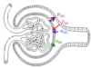
Explain the processes in the nephron:
- Filtration
- Reabsorption
- Secretion
Filtration (blue arrow):
- plasma + substance from GC move through tubule and excreted as urine
Reabsorption (orange arrow):
- Substances from filtrate move through tubule epithelial cells (transcellular reabsorption) back into the blood
Secretion (pink arrow):
- Selected additional molecules from blood move through tubule epithelium to the filtrate in nephron (paracellular reabsorption)
Note: Transcellular does NOT require channels/transporters, but paracellular reabsorption does.

What controls how much plasma is filtered into the nephron?
4 Starling Forces:
Hydrostatic Pressure of Glomerular Capillaries (PGC):
- Pressure of blood flowing inside GC is ~55 mmHg
(very high pressure considering capillaries being so small! —
Recall: ~20% of cardiac output goes to kidneys) - Enables blood to be pushed through the kidney
Colloid Osmotic Pressure of Glomerular Capillaries (πGC):
- Pressure created due to presence of proteins inside GC
- Since most proteins stay in GC, pressure is high: ~30 mmHg
Hydrostatic Pressure of Bowman’s Capsule (PBC):
- Pressure of fluid already in BC creates back pressure: ~15 mmHg
Colloid Osmotic Pressure of Bowman’s Capsule (πBC):
- Pressure created due to presence of proteins in BC: ~0 mmHg
(since almost NO protein should be found in BC, if not ~0 mmHg, kidney disease)
(PGC + πBC) — (PBC + πGC) = Net Filtration Pressure (NFP)
PGC — PBC — πGC = 55 mmHg - 15 mmHg - 30 mmHg
Normal NFP = ~10 mmHg
If NFP > 0, Plasma will filter into BC
If NFP < 0, Plasma will NOT filter out of GC into BC
What is glomerular filtration rate (GFR) and what are the factors affecting GFR?
GFR is the amount of fluid/solutes that are filtered per unit time into BC from GC. Changes in GFR changess the amount of salts and water being excreted from body:
- Higher the GFR, more excretion of solutes;
- Lower the GFR, less excretion of solutes
Factors affecting GFR:
-
Net Filtration Pressure
- Influenced by renal blood flow + blood pressure
-
Filtration Coefficient (“how leaky are GC?”)
- Influenced by…
- Surface area of GCs available for filtration
- Permeability of barriers between GC and BC
- Influenced by…
How is GFR regulated?
1. Myogenic Response:
- Constriction of arterioles in response to increased BP
- Increased BP → Smooth muscle in arterioles stretch → Streth-sensitive ion channels open → Muscle cells depolarize → Voltage-gated Ca2+ channels open → Smooth muscle cells contract, vessels contract → Decreased blood flow in glomerulus
2. Tubuloglomerular Feedback:
-
Macula densa cells are salt detectors
- found at junction of late ascending limb of loop of Henle which passes b/w aff. & eff. arterioles
- increased [NaCl] in distal tubule, cells release chemical signal that stimulates aff. arteriole to constrict ⇒ decrease fluid, decrease BP, therefore decrease GFR
- if eff. arteriole constricts, increase GFR
How is GFR measured?
To determine GFR:
- Excretion = Filtration - Reabsorption + Secretion
- only interested in what is filtered
- measure rate of filtration
- To do so, need a substance that is filtered easily but is not reabsorbed into the body and is fully excreted in the urine:
- **Inulin **(plant product), OR
- Creatinine (breakdown of creatine)
- To do so, need a substance that is filtered easily but is not reabsorbed into the body and is fully excreted in the urine:
- Since no reabsorption & no secretion of inulin/creatinine, then:
- Rate of filtration = Rate of excretion
- Measure how much inulin/creatinine is being excreted in urine, and how much it is in blood. Find the average for % filtration.
Once GFR is calculated and the amount of substance (X) dissolved in the plasma is known, how can we determine how the kidney handled substance X?
By calculating the filtered load:
Filtered Load of X = [X]plasma x GFR
Once filtered load is calculated, measure how much X was excreted
Ex. Individual with plasma concentration of glucose of 1 mg/mL of plasma has GFR of 125mL/min. What is the filtered load of glucose?
Filtered Load of glucose = [Glucose]plasma x GFR
                                      = 1 mg/mL x 125mL/min

                                      = 125 mg glucose/min.
Once the urine is analyzed and find that there is no glucose in the individual’s urine, it is assumed that all of the glucose that was filtered must have been reabsorbed while flowing through the nephron.
What is the average renal handling of the following substances?
- Water
- Sodium
- Glucose
- Urea
- Water = 99%
- Sodium = 99.5 %
- Glucose = 100%
- Urea = 50%
For the following tubules, determine what is reabsorbed and how much (%) is reabsorbed:
- Proximal Tubule
- Descending Limb of Loop of Henle
- Ascending Limb of Loop of Henle
- Distal Convoluted Tubule
- Collecting Duct
Proximal Tubule:
- 65% of filtrate reabsorbed
- Reabsorbs everything (ions, glucose, AA, water)
Descending & Ascending Limb of Loop of Henle:
- 20% of filtrate is reabsorbed
- Descending Limb of Loop of Henle — Reabsorbs water
- Ascending Limb of Loop of Henle — Reabsorb ions
Distal Convoluted Tubule:
- ~14% of filtrate reabsorbed
- Based on daily consumption
- Reabsorbs ions
& Collecting Duct:
- ~1% remaining in collecting duct for excretion
- Fine tunes filtrate
Draw a tubule of nephron cut length-wise and label the following structures:
- Capillaries
- Lumen
- Epithelial cells
- Luminal membrane
- Basolateral membrane
- Interstitium

Describe the mechanisms of tubule transport:
- REABSORPTION & SECRETION:
- Paracellular
- Transcellular
- Channels
- Transporters
- Uniporters
- Symporters
- Antiporters
- Primary Active Transporters
CHANNELS
- Small protein-lined pores that permit specific molecules through simple diffusion (passive), driven by [] or electrochemical gradient
- Ex. Na+ Channel
- Ex. Aquaporins (AQ)
- 4 different types of AQ:
- AQ I: found in the luminal membranes of the proximal tubule + descending limb of the loop of Henle
- AQ II: hormonally regulated by anti-diuretic hormone (ADH). Found in the luminal membrane of the collecting duct
- AQ III & IV: Found in the basolateral membrane of collecting duct
- 4 different types of AQ:
TRANSPORTERS
-
Uniporters
- permit movement of single type of molecule through the membrane by facilitated diffusion
- Involves binding of a molecule to itself in order to pass it through (unlike channel)
- Ex. Glucose uniporters — moves glucose from cytosol of tubule epithelial cells across the basolateral membrane to the interstitium
-
Symporters (co-transport)
- permit the movement of two molecules in the same direction across the membrane
- One molecule must move down its [] gradient to move both molecules across membrane
- For symporters that do not use ATP to move molecules, the use of one molecule’s energy-derived [] gradient is called secondary active transport.
- Ex. Na+/Glucose Symporters — important for reabsorption of glucose & Na+ from filtrate. Driven by Na+ gradient for the transport of glucose out of filtrate
-
Antiporters (exchangers)
- permit the movement of two molecules in opposite directions across the membrane
- One molecules must move down its [] gradient in order for the other molecule to also move (like symporters)
- Ex. Na+/H+ Antiporter — important for exporting protons out of the tubule cell in exchange for Na+ entering the cell; driven by Na+ gradient
PRIMARY ACTIVE TRANSPORTERS
- Require ATP to move molecules against their [] gradients
- Some of these transporters found in kidney are antiporters and some are uniporters
- Ex. Na+/K+ ATPase (Na+/K+ Pump) — important for maintaining a sodium gradient for other transporters to function. Located on the basolateral membrane of tubule epithelial cells. Pumps 3 Na+ out of the cells into the interstitium and 2 K+ into the cell.

How are transporters/channels of the kidney regulated?
Specific transporter/channel is changed in its function in response to a specific type of hormone; regulation can occur at multiple levels:
- Regulation at the level of gene expression **
- When more transporters are needed, the cell increases the amount of mRNA that is transcribed encoding the proteins for that transporter
* *2. Regulation at the level of cellular location** - Cell is capable of adding/removing transporters from the plasma membrane; If transporter is not embedded in the plasma membrane, it cannot move things across the plasma membrane
- Transporters are stored in vesicles when they are not needed
- Regulation at the level of activity**
- Speed at which the transporter functions can be altered
Note: Some channels/transporters are non-regulated & transport occurs at a constant rate
What is diabetes mellitus?
- increased [glucose] in the filtrate
- all glucose is not reabsorbed in the proximal tubule through Na+/glucose symporter
- glucose is excreted in the urine
- resulting in increased urine production (osmotic diuresis)
Draw the Proximal Tubule. Include…
- all channel/transporters
- luminal/basolateral
- regulated?

Draw the Descending Limb of Loop of Henle. Include…
- all channel/transporters
- luminal/basolateral
- regulated?

Draw the Ascending Limb of Loop of Henle. Include…
- all channel/transporters
- luminal/basolateral
- regulated?

What does the Distal Convoluted Tubule reabsorb and secret?
- How are calcium channels regulated?
Note: Don’t need to know how to draw channels/transporters for distal tubule
Distal Convoluted Tubule:
- reabsorbs ions (Ca2+, Na+, K+, Cl-)
- Calcium channels are regulated by parathyroid hormone (PTH)
- Secrets none
Draw the Collecting Duct. Include…
- all channel/transporters
- luminal/basolateral
- regulated?


