Repro Path Flashcards
mature cystic terratoma
from somatic totipotentgerm cell
Most common ovarian tumor of reproductive age group
95% of germ cell tumors are mature teratomas
Tumor differentiation towards all three germ cell layers
Skin is the most common tissue seen
46XX
Rarely malignancy may arise in one of the elements of tumor tissue ( in older women)
what age do embryonal tumors peak in children?
befoer age 5 - very rare after that

what would be the expected endometrial biopsy with unopposed estrogen?
proliferative endometrium with breakdown
- You have estrogen without progesterone.
- Eventually you will have breakdown of the EM, because normally proliferative EM is not meant to be there forever.
- It’s meant to be opposed by progesterone, and undergo secretory differentiation.
- If you don’t have the progesterone, then the proliferative EM grows to a certain point and then starts to break down.
- Usually what you see, you have proliferative EM with breakdown.

serous carcinoma
- Large solid and cystic areas
Necrosis and hemorrhage
fibroadenoma vs phyllodes tumors

fixation of cells in cytopathology
- When you’re taking specimens for histopathology, you always use formalin, but formalin is not a good fixative for cells because it bursts the cells- makes them swell and then you can’t interpret them.
- Most samples can go to the laboratory “fresh”, which means with no fixative in it- you can just take the sample and bring it on over to us. If it’s going to be delayed, you can refrigerate it, and cells will last overnight in refrigeration.
- In some cases you have to fix the cells, and the fixative that we use is ethyl alcohol. One of the problems that we always have in the beginning of July is that ethyl alcohol is typically not available on the hospital wards and rubbing alcohol is, so people put rubbing alcohol in their solutions and it bursts the cells.
- In some of our clinics, they will actually teach you how to make the slides, because in some areas the clinician is actually doing the fine needle aspiration. So they’ll teach you how to smear the slides and then let them dry by air or fix them by ethanol.
stage I ovarian carcinoma
Stage I: Limited to one or both ovaries. Five year survival 90%
what is the functional unit of the breast?
the lobule

•The terminal duct and ducules that come out compose the terminal duct lobular unit and that again is where most carcinomas occru
- AT menarchy these ducts proliferate and give rise to some 30 epithelium lined ductules and acini and all those ducules and acini come together to form the lobule
- The breast is divided in to major duct systems that then divide up into lobules
- It’s really lobules that become functional unit of mammary gland
- It’s where you see most of the benign and malignant lesions that occur in the breast.

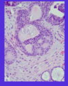
bartholian gland abcess

- The Bartholin glands can get infected and form abscesses.
- Bartholin glands normally are mucinous glands, with a mucinous lining, but with an infection shown here [bottom right], it can undergo a squamous metaplasia.
- Management is with drainage and antibiotics.
placental abruption
painful third trimester bleeding
premature separation of plaenta after 20 wks, secondary to rupture of maternal vessels
if marginal - vaginal bleeding
if central - retroplacental hematoma (accumulation of blood - uterus hard and painful, no bleeding!)
higher incidence of toxemia and HTN
fetal complications from prematurity
risk inversely related to GA at delivery
respiratory distress syndrome (hyaline membrane disease)
necrotizing enterocolitis
retinopothy
sepsis
neuro issues
proliferative fibrocystic change vs proliferative fibrocystic change with atypia

what day of the cycle is ovulation
day 14
what gene has a bad prognosis for neuroblastoma
N-myc amplification
nonprolif fibrocystic change vs proliferative fibrocystic change of the breast
prolif - 1.5-2x increased risk of cancer

Rh mechanism
- Rh negative woman before pregnancy
- pregnancy occurs, fetus is Rh-positive
- after deliver, Rh alloimmunization occurs in the mother and she dvelops antibodies for Rh
- The next positive with an Rh positive fetus - maternal abs can ross the placenta, enter the fetal blood stream, attach to the Rh-positive red cells causing hemolysis

what is the leading cause of death in infants without birth defects?
prematurity
how do we follow neuroblastoma?
▫Elevated urine metabolites (vanillylmandelic acid [VMA] and homovanillic acid [HVA]). Measuring elevated serum catecholamines is not practical. Typically clinicians measure elevated urine metabolites. Looking at VMA and HVA in terms of whether you have overall catecholamine overexpression. Unlike pheochromocytoma which also produces catecholamines, children with neuroblastoma tend not to present with signs of hypertension. Urine catecholamines are important because they set up a baseline. If a child comes in with high urine catecholamine and then after their tumor is resected they’re still high, it suggests incomplete resection or unknown metastatic disease. If they’re low and subsequently become high several months later, it can suggest recurrence.
if twins and embryo splits at second wk of gestation (late)
monochromatic
monoambiotic
only thing that splits is laminar disk!!

3 components of Wilms Tumor
- Triphasic tumor
- (1) small round blue cell component-primitive blastema; [blue arrow]
- (2) epithelial component-immature tubules (most often) and glomeruli [black arrow]
- (3) mesenchymal stromal component [green arrow]


neuroblastoma
The other aspect that we can see in this tumor, parts of this tumor can have an appearance like this. Rather than having a diffuse round blue cell component, they’ll have Schwannian differentiation. Schwannian differentiation suggests that the tumor has fascicles of eosinophilic spindle cells intermixed with ganglion cells. So these are the ganglion cells that we just talked about (black arrow) and these spindle cells with elongated oval nuclei (blue arrow) and this longer eosinophilic cytoplasm. This is an example of Schwannian stromal differentiation.

proliferative fibrocystic change without atypial
1.5-2x risk
mild dysplasia
- This is the surface layer [the top layer].
- In the normal maturation process, the cells get bigger and their nuclei get smaller as they go to the surface.
- The very early terms used to describe these lesions were mild dysplasia, moderate dysplasia, severe dysplasia and in situ carcinoma.
In mild dysplasia, up to 1/3 of the epithelium shows immature cells – they do not mature and they become atypical. But above that, you get maturation of cells.

serum marker for choriocarcinoma
HCG