Reproduction with Hormones Flashcards
(34 cards)
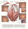
Female Reproductive Structures

- Ovary attached to ligaments but not fallopian tubes just hangs around it w/finger things
- Ovary releases an egg in ovulation into abdominal space and usually ciliated columnar epithelial will beat enough to bring egg down fallopian tube to start path toward uterus
- If egg falls into abd and fertilized there it can be ectopic pregnancy or if egg implants in wall of fallopian tube
- Uteran wall has outer surrounding membrane called parametrian and them myomedrian which is smooth muscle and then inner layer is the endometrian (thicker and more vascularized and then sluffs off again)
- Uterus ends in cervix, extra thick layer of muscle

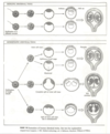
Uterus Types
- Uterus in diff varieties depending on animals
- In some organisms there’s a double uterus (rats, mice, rabbits)
- B. single uterus w/split and found in animals w/litters – dogs, cats, cows – implantation all along uterine horn

Structure of Ovary
- Ovary – @ birth female have primary follicles frozen in prophase I
- In normal cycle: 10-20 follicles begin to grow; follicle cells around primary follicle, replicate and make thicker layer over time
- As follicle matures and makes more follicle cells, it creates spaces in layers of follicle cells, and that space around developing follicle is called the antrum
- Eventually antrum is large and follicle cells are growing begin secreting estrogen
- In each cycle as follicle gets larger and larger there is more estrogen in the blood; midd of cycle before ovulation, antrum and follicle are large and follicle will bulge out wall of uterus
- At ovulation the thin area will break open and oocyte w/associated follicle cells will fall out – attached follicle cells are called corona radiate
- Then its swept into opening and end of fallopian tube in mean time the residual follicle cells reorganize and become a corpus leteum (yellow body), which is an endocrine organ that secretes estrogen and progesterone
- In corpus leteum there is an internal timer, after 10 days after release of ovulated oocyte, it degrades and breaks down to form white scar structure called corpus albicans which stays in ovary for rest of life of woman
- White spots are representing all the previous cycles she went through
- When corpus leteum breaks down it stops producing and releasing progesterone and estrogen

Reproductive Hormones and Organ Changes
•Secretory pathway: Hypothalamus (at base of brain) which secretes a signal molecule called GnRH (gonadotropin releasing hormone) that affects the –> anterior pituitary gland and that is signaled to release reproductive involved hormones called FSH (follicle stimulating hormone) and LH (leuteinizing hormone) –> target organ like the ovary
- FSH and LH from anterior pituitary gland (@ base of brain) and this is levels of them in the blood
- 28 day cycle w/ovulation in middle at 14 days•
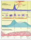
Follicle Phase and then Luteal Phase
First is the follicular phase since follicle is developing and second half of cycle is luteal phase since controlled by action of corpus lituem
•Blood levels of hormones from the overy – estradiol and progesterone
•Then shows what happens in wall of uterus in endometrial layer – at beginning of cycle is onset of menstration phase, wall of uterus is sluffing off to make the period, then new cycle begins
•At beginning phase, the FSH is little higher than LH, and FSH tells primary follicles to begin to grow and the replicating follicle cells secrete more estragen which causes the endometrium of uterus to thicken and become vascularized
•Estrogen gets higher and FSH and LH get lower
•At mature follicle then a surge in LH which causes the ovulation and break open the follicle and amount of estrogen by follicle is < and then becomes corpus leteum and produces estrogen and progesterone which go up are produced by corpus luteum

Luteal Phase
- Oocyte travels down fallopian tube, and lining of uterus is prepped for pregnancy
- In leuteal phase estrogen and progesterone are high and feedsback to pituitary and inhibits release of FSH and LH so inhibits release of additional follicles – same basis for birth control which inhibits release of LH and FSH to prevent new ovulation by secreting progesterone and estrogen
- Timing of corpus leteum runs out and turns itself off and forms corpus albican which ends estrogen and progesterone ; fall off of progesterone causes the start of menstrual phase
- Before end of timing period, i_f fertilization occurs a signal comes to corpus luteum to rescues and won’t progress and degrade_

Regulation by Hormone Feedback
- feedback inhibition here too
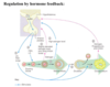
Human Chorionic Gonadotropin (HCG) - rescues corpus leteum
- Estrogen and progesterone both up in second half of cycle - luteal phase
- Near end of cycle and pregnancy has occurred and hormone called HCG (Human Chorionic Gonadotropin) is secreted by the chorion the outerlayer of developing embryo ; acts on corpus leteum and said to rescues the corpus luteum and progesterone and estrogen won’t drop off and wall of uterus will maintain itself and corpus luteum from self-destructing
- Estrogen as preparing the wall of the uterus via cell division and vascularization
- Progesterone hormone – maintains the wall of the uterus so when it falls off you get start of menstration

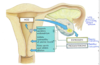
Early in Pregnancy
- Oocyte passes into fallopian tube and fertilized in tube and then travels down as it goes through cleavage, blastulation, and gastrulation and in uterus its passed blastula stage and inner cell mass (w/synchitrophoblast cells dissolve into wall of uterus) and interactions here cause the chorion to secrete HCG and that’s the signal that pregnancy has occurred
- HCG is hormone you detect in home pregnancy test, sticks to antibody there and turns it plus (pregnancy work 10-12 days after fertilization since takes time to implant
- You can buy morning after pills – do diff things; some are intended to prevent ovulation itself if in mid of cycle; some prevent ovulation for 5 days since sperm don’t live that long
- Prevent implantation – irritate wall of uterus so implantation doesn’t occur
- Competitively inhibit binding of progesterone; blocking progesterone signal, then wall will sluff off and menstruation will start even if implanted embryo there
- HCG is similar to LH – use urine sample and if rabbit ovulates then know HCG is in urine
- LH surge to test for ovulation
Menstral/Ovarian Cycle
- Menstral /ovarian cycle – humans and primates
- Estrous Cycle – used by other mammals, shorter luteal phase (2nd part of cycle) – less endometrial build up so no menstral phase ; period of heat which represents period of receptivity; interest and ability to reproduce is only during period of heat – seasonal in many kinds of mammals, ex. Deer in fall once a year; other animals have heat period 3 to 4 times a year
- Deer have monstrous cycle but some other animals are polyestrous like mice and rabbits (every 1-2 weeks have heat period all year long)
- We are placental mammals vs. marsupial mammals
Reproduction in marsupial mammal
- Placental vs. marsupial (pouch) mammals
- Developmental time period of marsupial go from fertilization to period of weaning; shorter gestation and longer lactation
- Fertilization to birth is called gestation and short (8-15 days in marsupial) and then young are born and very undeveloped except forelimbs and drag selves into pouch and then period of lactation in pouch, very long for birth to weaning
- Placental mammal is reversed, period of gestation is longer and lactation is shorter where birth separates the two
- Placental mammals out competed marsupial mammals due to greater developed young of placental mammals
- Teets for young kangeroo and for older young
- Can freeze development – embryonic diapause, until older child is weaned and then small baby gets more developed
- Kangaroos carry 3 babies at 3 stages of life at same time

Twins
- Ways to form twins – less frequent as move down diagram:
- 2 eggs ovulated at same time and both get fertilize and both implant
- Monozygotic/identical twins – fertilized egg in first division is split at 2 cell stage and each forms embryo and each implants in wall of uterus
- Split happening later? Forms blastula w/inner cell mass and before its established there’s a split w/2 inner cell masses in one blastula and so when implants then get 2 amnions
- Cell mass doesn’t split much so 2 embryos in same amnion w/2 umbilical cords w/same placenta - siemes twins
Cleavage Stages
- Fertilize egg, then first division of cleavage which occurs after 30 hours
- 60 hours get 4 cell stage
- Blastula/blastocyst (fluid-filled cavity) have 4-6 days : have inner cell mass and trophoblast cells
- Blastula stage and zona pellucida goes along and then it degenerates around 6 days – 7 days and then the embryo itself hatches and breaks out of zona pellucida layer which allows cells of trophoblast layer to interact w/walls of uterus for implantation

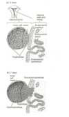
Implantation at about 1 week
- 6-7 days and have inner mass formed and is blastula and sincytiotrophoblast are on side where wall of uterus and digest their way into the wall – embryo moves into the wall here
- Lots of cells and blood vessels
- No zona pellucida at this point
- Receptors between trophoblast cells and uterine wall then starts drilling in w/sincytiotrophoblasts
- Can’t implant unless hatch out of zona pellucida layer, if hatch too early then embryo might implant in fallopian tube, want timing right
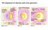
Development of Chorion and Early Placenta
- Inside wall of uterus now not sticking out
- Sincytiotrophoblast cells that ate way in
- Inner cell mass drops down and form amniotic cavity
- Yolk sac and surrounding membrane and chorion and sincytiotrophoblast cells reorganize selves and form a placenta – chorionic villi are formed which > SA between embryo and surrounding uterus wall
- Stalk between developing embryo and outer layer which is the placenta
- Can detect pregnancy around 10-14 days
Weeks of Development
- Length of dev is 38-39 weeks total
- Week 2 amnion formation and discoid cleavages and see primitive streak
- Week 3 have neurulation and have central nervous system forming and dorsal hallow nerve chord but no brain and somites and some heart structures (brachial slits forming)
- 4-5 weeks have limb buds
- Thalidamide affects the limbs and was originally for woman w/morning sickness around 4 weeks and where limbs grow too so birth defects in limbs
- brain at 9 weeks
- CNS completing at 20-39 weeks
- Sense of what forms when
- Don’t need to reproduce diagram just general sense

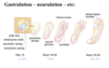
Gastrulation to Neurulation
•at 2 weeks See primitive streak and as it draws back the part at head finishes gastrulation and
- at 3 weeks forms neural groove and forms around and gets neural tube w/opening at one end w/brain and then other end closes up and somites in middle
- Spina biffada is where other end fails to close
- If no brain forms and unfinished neural tube then get anencephaly - no brain
- at 3.5 weeks the pharyngeal arches are seen
•Needs to zip closed at both ends
Neural Tube Defects
- around 3 weeks can see anencephaly or spina bifida
- Fewer neural tube defects if take folic acid a vitamin B supplement which seems to help w/and replication and some blood cell formation
- Failure to close at head end of neural tube then have anencephaly
-
Fail to close at other end it depends how far down it has closed for severity of spinabiffada disorder
- Could get bubble w/cerebral spinal fluid and problems in lower body; but sometimes fails to close at end and little opening and dr. can close that up by folding up into the tube

Week 4
- Limb bud starting to appear and heart coming along and will start beating
- Connection to umbilical cord
- Remnant of yolk sac is compressed into umbilical cord and is absorbed and closed up
- Digestive tract
- And allantois is 4th embryonic membrane – stores nitrogenous waste
- Placenta has thin walls between baby’s and mothers circulation – waste can diffuse across most of waste

Weeks 5-6
- yolk sac is shunk and absorbed and thin tube and now yolk space is becoming digestive tract
- limbs more formed but hands and feet are flippers
- retina forming

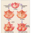
External Genitalia- at 7 weeks uncomitted
- At 7 weeks form of genitalia structures are uncommitted
- If Y chromosome is present then penis will develop around 10 weeks more formed through birth
- w/out Y then same structures but clitoris forms and remains open not closed
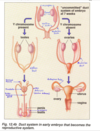
Internal Reproductive Structures
- 7 week embryo there are tubes that developed that are uncommitted – wolfian duct and mullerian duct and pre-gonad structures (testes and ovaries develop in same spot in abd) – gonad become testes and wolfian duct becomes vas deferins and mullerian ducts fade if Y chromosome and then area at bottom becomes prostate and urethra
- If no Y chromosome – get wolfian ducts that go away and mullerian ducts become the fallopian tubes and area below becomes uterus and vagina and gonad becomes ovaries
Embryonic Membranes and Placenta
•Embryo w/inner cell mass beginning to drop to make amniotic cavity
- Sintrophoblasts begin
- Chorion beginning to form and make chorionic villi and maternal blood is hooked up to spaces that connect between spaces of villi and called blood islands so maternal blood can be right against villi to help < distance for diffusion; mother’s circulation breaks down some surrounding layers around the blood vessel (usually endoderm and connective tissue) but here blood just spills into blood islands – call maternal layer of placenta the decidua (to drop a couple layers to get its blood right against chorionic villi)
- Embryo is connected by body stalk that becomes the umbilical chord and still little yolk sac that becomes incorporated into umbilical chord but not major source of nutrition that is the placenta’s job
- Placenta works as waste diffusal system
- Allantois is inside the embryo and was storage for nitrogenous waste but is shrinking now
- Yolk sac still present
- Embryo layers of endoderm, mesoderm, ectoderm on top and little nervous system is forming
- Other stuff at bottom (extra embryonic mesoderm – don’t worry about it)

Placental Circulation
- Umbilical chord
- Chorionic villi w/fetal circulation going through; surrounding that are spaces w/maternal blood called maternal blood islands
- Never are they continuous w/each other
- Placenta ends up on one side of the embryo
- Embryo was embedded completely and then bulges out into uterin space and surrounded by amnion (constituted from the urine of the child)
- fused amnion and chorionic membranes