Rheumatology Flashcards

Common and chronic conditions (plus some potential rogue ones) to know

1
Q

List common differentials for an acutely swollen joint

A
  • Most important to rule out –> septic arthritis
  • trauma & haemarthrosis
  • bursitis
  • tendinopathy
  • reactive arthritis
  • gout
  • pseudogout
  • enteropathic arthritis
2
Q

Define gout

What is the pathophysiology?

A

Gout = crystal arthropathy resulting from excess uric acid leading to precipitation in joints and tissues.

Pathophysiology –> disorder of purine metabolism, uric acid is the breakdown product of purines and is predominantly excreted via the kidneys. Imbalance between production and excretion of uric acid leading to deposits in joints and soft tissues.

3 main mechanisms - increased production with increased cell turnover e.g. psoriasis, chemoT

Increased purine intake - seafood red meat alcohol

Decreased uric acid secretion e.g. furosemide, thiazides, AKI, CKD

3
Q

Risk factors for gout?

A

Fuck My HOT And Chronic gout

Family history

Male

High purine diet/ hyperlipidaemia

Obesity

Thiazides

CKD/ CVD/ Chemotherapy / DBM

4
Q

Presentation of gout

A

Rapid onset of pain - typically over 1 hour

Monoarticular - often 1st MTP joint (may affect wrists/ carpometacarpal joint/ knee/ ankle)

or oligoarticular (< 4 joints)

joint stiffness, swelling, erythema & warmth

effusion

Gout tophi - often on extensor surfaces - elbow, knee, achilles, helix of ear

FHx of gout

5
Q

Examination features of gout

A
  • warm red swollen
  • considerable tenderness
  • limited ROM
  • Hard subcutaenous Tophi on extensors - elbow/ knees / achilles/ helix of ears
6
Q

Investigations for Gout

A
  • Gout is a clinical diagnosis but joint aspiration can be used to confirm it

Bedside:

  • BM - Diabetes = RF
  • Urine dip - for CKD (protein blood etc)

Bloods:

  • FBC
  • U& E –> check renal function
  • LFTs –> premedication baseline
  • Blood cultures –> exclude septicaemia
  • Serum Uric acid –> taken 4-6 weeks after acute attack, normal serum uric acid does not exclude gout during an acute attack as plasma urate often falls.
  • Serum Calcium

Imaging:

  • USS –> more sensitive in Xray in detecting erosions, tophi and gout specific double contour sign
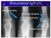
  • Xray –> typically normal in acute episodes of gout. In Chronic gout:
      • lytic lesions
      • punched out erosions
      • erosion with sclerotic borders
      • joint space maintained

Special test:

  • Joint arthocentesis –> gold standard investigation, exclude septic arthritis
  • MCS of joint aspirate –> crystal microscopy will show needle shaped monosodium urate crystals, negatively birefringent of polarised light. No bacterial growth
7
Q

Management of gout

A

Conservative:

  • Rest, ice, elevation
  • Weight loss
  • Low purine diet
  • Reduced alcohol consumption

Medical:

For acute episode:

  • NSAID 1st line, Colchicine 2nd line, Steroid 3rd line
  • NSAID CI in significant HD or renal impairment, then use oral colchicine
  • notable SE colchicine is diarrhoea which is dose dependent.
  • If NSAID and colchicine is CI then short course oral steroid or intraarticular steroid

For chronic management:

  • urate lowering therapy usually started once initial attack has resolved to enable the patient to make the decision to start urate lowering therapy without any pain
  • First line Allopurinol = xanthine oxidase inhibitor that prevents conversion of metabolites to uric acid
  • colchicine cover considered for the first 6 months
  • Caution with allopurinol in renal impairment
  • Second line = Febuxostat (also xanthine oxidase inhibitor) considered if allopurinol is CI.
8
Q

Define pseudogout

A

Pseudo gout = Microcrystal synovitis caused by the deposition of calcium pyrophosphate crystals in the synovium

9
Q

What is the pathophysiology of pseudogout

What are the risk factors

A
  • Pathophysiology:
    • excess CPP production leads to supersaturation and crystal formation / deposition in the synovium. There is an inflammatory response to calcium in the synovium
  • Risk factors: usually pseudogout is associated with increasing age therefore patients developing it at a younger age have a secondary condition:
    • haemochromatosis
    • hyperparathyroidism
    • hypomagnesaemia
    • familial CPPD disease (calcium pyrophosphate disease)
    • acromegaly
    • wilsons
    • gout
10
Q

Presentation of pseudogout

A

Asymmetrical arthropathy of larger peripheral joints

key risk factors of increasing age, family history, previous inury or surgery to the joint, metabolic disorders

often affects knee 50%, can affect the shoulders, elbows, wrists, hips, ankles and feet

sudden worsening of osteoarthritis and involvement of joints not commonly involved in OA e.g wrists and shoulders may suggest CPP arthritis

joint swelling

erythema

restricted ROM

with or without fever

11
Q

Investigations for pseudogout

A
  • Bedside: BM ( diabetes), Urine dipstick (kidney function)
  • Bloods:
    • FBC
    • U&Es (kidney function)
    • LFTs baseline premedication
    • Blood cultures –> rule out septicaemia
    • Serum calcium / bone profile (check for hyperparathyroidism)
    • Serum urate
  • Imaging:
    • Xray –> chondrocalcinosis is the classical XR finding (pathognomonic of gout)
    • Other XR changes similar to OA (LOSS) (Loss of joint space, osteophytes, subchondral sclerosis, subchondral cysts)
  • Special tests:
    • Synovial fluid analysis –> rhomboid shaped crystals positively birefringent in polarised light with no bacterial growth
12
Q

Management of pseudogout

A
  • Flares usually self resolve within a few weeks
  • Conservative:
    • ice packs, immobilisation, rest for first 48 hours
  • Medical management:
    • NSAID - naproxen first line
    • colchicine - if NSAID CI
    • Steroids PO or joint injection if no response to above
    • Recurrent episodes may need prolonged colchicine
  • Surgical:
    • Joint washout in severe cases
13
Q

Define reactive arthritis

A

Reactive arthritis - one of the group of seronegative spondyloarthropathies, arthritis occurring after an infection with “Sterile inflammation”.

Occurs several weeks following an infection with organisms that infect the urogenital or GI tract

14
Q

Common organisms causing reactive arthritis?

A
  • Urogenital infections –> chlaymidia, gonorrhoea, Ureaplasma urealyticum
  • GI –> campylobacter jejuni, shigella, salmonella, yersinia, C diff
  • Rare – >TB
15
Q

Presentation of Reactive arthritis

A

Key: cant see, pee, or climb a tree

  • Conjunctivitis
  • urethritis
  • acute monoarthritis
  • Patients with clear history of preceding infection either diarrhoea or urethritis
  • often 1- 4 weeks before onset of joint pain
  • Peripheral asymmetrical oligoarthritis most common
  • can cause monoarticular or polyarticular arthritis
  • acutely hot swollen red painful joint
  • worse in the morning
  • axial arthritis –> inflammatory back pain, buttock pain, stiffness at rest, relieved by exercise
  • enthesitis - often achilles
  • dactylitis

Extraarticular manifestations:

  • fever
  • fatigue
  • ocular –> bilateral conjunctivitis
  • oral ulcers
  • Skin –> circinate balanitis (inflammation of head of penis) and keratoderma blenorrhagica, erythema nodosum
  • GI –> diarrhoea
  • Urogenital –> urethritis & dysuria
16
Q

Investigations : reactive arthritis

A
  • Bedside:
    • urine dipstick & urinalysis for source of infection
    • Stool culture (often negative at onset of arthritis)
    • urethral / cervical swab for STI screen
  • Bloods:
    • FBC
    • U&E
    • LFT
    • CRP/ESR
    • blood cultures - rule out septic arthritis
    • STI screen - HIV/ hep/ syphilis
    • ANA (should be -ve)
    • RF (should be -ve)
  • Imaging/ special test:
    • Xray of pelvis –> asymmetric sacroilities or enthesitis of achilles tendon
    • Joint aspiration to exclude septic arthritis, gout and pseudogout
17
Q

Management : reactive arthritis

A
  • Conservative: RICE
  • Medical –> 1) indentify and eradicate underying infection, 2) NSAID& paracetamol, 3) Steroid PO or joint injection
  • Medical if chronic ReA –> use DMARDs methotrexate or sulfasalazine

Often self limiting but up to 20% develop chronic arthritis

18
Q

Key risk factors reactive arthritis

DDX reactive arthritis

A

Key risk factors: Male, HLA B27 +VE, preceding chlaymidia or GI infection

DDX:

Septic arthritis (key)

disseminated gonococcal infection

Gout

pseudogout

RA

SLE

Traumatic arthritis / haemarthrosis

19
Q

Differential diagnosis for chronic joint pain

A
  • Monoarthritis:
    • OA
    • Chronic tophaceous gout
  • Polyarthritis:
    • RA
    • Ankylosing spondylitis
    • psoriatic arthritis
    • Systemic sclerosis
    • SLE
    • Sjogrens
    • PMR
    • Fibromyalgia
20
Q

Define osteoarthritis

A
  • Osteoarthritis is a chronic degenerative joint disorder caused by degeneration of the cartilage within a joint, leading to exposure of the underlying bone and bone - bone interaction. Leads to pain, stiffness and reduced function of the joint involved.
21
Q

Risk factors for OA

A
  • Age
  • Female
  • Fhx
  • Physically demanding occupation or sport
  • anything leading to altered joint loading –> hypermobility, osteoporosis, trauma within the joint e.g. fracture, meniscal or ligament tears
22
Q

Presentation of OA (symptoms)

A
  • Pain and stiffness within joints
  • worsened by activity and weight bearing
  • background ache at rest
  • lasts less than 30 mins in the morning
  • pain better with rest
  • pain not present at night unless advanced
  • often affects larger weight bearing joints : Hips, knees, SI joints, cervical spine
  • Hands –> DIPs and 1st CMC joint (base of the thumb) & wrist
  • often > 50 years
  • slow onset over months - years
  • functional difficulty
23
Q

Signs of OA on examination?

A
  • Check weight, height, calculate BMI
  • Watch for gait abnormality and malalignment of the joints
  • Look:
    • joint swelling
    • erythema
    • bony swellings –> osteophytes
    • effusion
    • fixed flexion deformity at the knee or hip (+ve thomas test)
  • Feel:
    • crepitus
    • limited ROM
    • Effusion
    • Joint line tenderness
  • Move:
    • assess ROM, both active and passive (will be reduced)
    • crepitus felt
    • assess for ligament laxity (may be a cause)
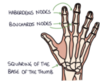
  • Hand signs:
    • Heberdens DIP
    • Bouchards PIP
    • Squaring of 1st CMC joint
    • Weakened grip
    • reduced ROM
24
Q

Diagnosis of OA?

A

Diagnosis of OA is often a clinical diagnosis based of age, symptoms and examination findings. Clinical diagnosis is considered in a patient with a typical OA presentation:

Activity related joint pain

morning stiffness no longer than 30 mins

> 45 yrs

25
Q

Investigations for OA

A

Bedside: BMI

Bloods:

  • To exclude RA:
    • CRP
    • ESR
    • RF
    • AntiCCP
  • XR of affected joint:
    • Loss of joint space
    • Osteophytes
    • Subchondral sclerosis
    • Subchondral cysts
  • MRI if soft tissue injury is suggested in the history e.g with locking or instability of the joint
  • MRI of the spine if there is any neurological deficit and spinal OA
26
Q

Management of OA

A

Conservative:

  • Weight loss to reduce any load on the joint
  • physiotherapy and occupational therapy
  • orthotics to support function
  • low impact exercise such as swimming or cycling

Medical:

Stepwise analgesia:

  • paracetamol + topical NSAID/ topical capsaicin
  • oral NSAID + PPI omeprazole
  • Consider stronger opiates e.g. codeine/ morphine –> caution with dependence tolerance and withdrawal
  • Intraarticular steroid injections

Surgical:

Joint replacement of hip or knee in severe OA that is not responding to conservative measures & is severely impacting the patients QOL

27
Q

Define rheumatoid arthritis

Quick pathophysiology

A

RA = chronic symmetrical polyarthritis with systemic inflammation

Pathophysiology: autoimmune disease with development of autoantibodies - RF and Anti CCP. Leads to inflammation in the synvoium of joints, activation of osteoclasts leading to bone and cartilage breakdown, formation of a pannus of proliferating synoviocytes & immune cells. Antigen immune complexes in the joint leads to chronic inflammation and joint destruction. Chronic inflammation leads to increased vascular permeability and joint effusion.

28
Q

Key risk factors for RA

A
  • 20-50 yrs
  • white
  • female
  • HLA DR4 HLA DR1
  • family history
  • other AI condition
  • Environmental trigger e.g. smoking or infections
29
Q

Presentation of RA - symptoms

A
  • Swollen painful stiff joints in hands and feet
  • DIPS rarely affected
  • PIP, MCP and wrist common, MTP common
  • joints are hot, red, swollen, with stiffness worse in the morning, lasting > 1hr
  • (due to drop in cortisol overnight allowing more inflammation)
  • gradually worsens with larger joints involved - knee hip elbow, shoulders, ankle, TMJ, atlantoaxial joint, cervical spine
  • presentation over a few months
  • Systemic symptoms:
    • fatigue
    • low grade fever
    • weight loss
    • myalgia
    • low mood
30
Q

Signs of RA on examination

A
  • Look: Hands
    • Z shaped deformity of thumb
    • swan neck deformity
    • boutonniere deformity
    • ulnar deviation of fingers at knuckles
    • muscle atrophy –> guttering between the extensor tendons in the hands due to wasting of interossei muscles
  • Feel:
    • boggy joint swelling
    • pain on palpation
    • +ve squeeze test of metacarpal and metatarsal joints
  • Move:
    • reduced ROM
    • Difficulty with fine motor tasks
31
Q

Extraarticular manifestations of RA?

A
  • Neurological - atlanto axial subluxation with impingement of cervical spine, CTS
  • Eyes - dry eyes, (sicca syndrome), episcleritis, scleritis
  • Oral - dry mouth, oral ulcers
  • Pulmonary - interstital lung disease, inflammation of the pleura, costochondritis
  • cardiac - pericarditis, myocarditis, endocarditis
  • Increased risk IHD - due to inflammation of arteries
  • Renal - glomerulonephritis
  • Haem - anaemia, neutropenia and splenomegaly (feltys syndrome), thrombocytopenia or cytosis
  • Derm - rheumatoid nodules on extensor surfaces
  • MSK - increased risk osteopenia, muscle weakness, tenosynovitis and trigger finger
32
Q

Diagnosis of RA?

A

All patients with suspected TA need to be referred to a rheumatologist for further assessment, refer any patient with persistent synovitis. Urgent referral if there is a delay between presentation and symptom onset > 3 months or if affecting small joints of hands / feet.

33
Q

Investigations for RA

A

Bedside

Bloods:

  • FBC - anaemia
  • ESR (raised)
  • CRP (Raised)
  • U&E - glomerulonephritis
  • Ca2+ and bone profile
  • LFTs - feltys syndrome
  • Rheumatoid factor 70% positive but non specific
  • Anti CCP - very specific but low sensitivity, if positive it predicts worse disease and can be detected up to 10 yrs before symptoms. If negative for RF test Anti ccp.

Imaging:

  • LESS
  • Loss of joint - destruction and deformity
  • erosions of bone
  • soft tissue swelling
  • soft bones - periarticular osteopenia
  • Other –> USS for effusion, tendonitis or MRI for cervical spine

Special tests –> joint aspiration if effusion is present

34
Q

Management of RA

A

Conservative management:

  • MDT - specialist nurses, physiotherapy, OT, psychological support, podiatry
  • Screening for complications e.g HTN, IHD, osteoporosis and depression

Medical management:

  • Acute flare: NSAID, PPIs, steroid e.g. prednisilone
  • Initial therapy –> DMARD, usually methotrexate 1st line, with bridging steroid therapy 2-3 m
  • If this fails to achieve remission then escalate DMARDs:
    • 1st line methotrexate
    • 2nd line methotrexate plus sulfasalzine/ leflunomide/ hydroxycholorquine
    • 3rd line above plus anti TNF biologic if 2x DMARDs have failed (infliximab, etanercept, adalimumab)
    • 4th line Methotrexate plus rituximab Anti CD20

Surgical management:

Joint replacement if there is progressive deformity with worsening function

DAS 28 scoring in clinic to monitor response to treatment

35
Q

Counselling station

Methotrexate

A

Methotrexate is what we call a “Disease modifying” drug. This means it reduces inflammation and suppresses your immune system.

It is taken once weekly, with folic acid 5mg taken at another time usually 24 hours after. The dose is built up slowly.

It is usually taken as a tablet but injections are also available.

The length of treatment tends to be long term with RA. It can take up to 4-6 months to have a proper effect on your symptoms.

Before you take methotrexate we need to take some blood tests to monitor the drug as it can affect certain organs in your body including your liver and kidneys. Therefore we need to take FBC, U&E, LFTs prior to you beginning methotrexate. We then need to take blood tests every 2 weeks until the levels have stabilised, then every 2-3 months.

Important side effects to warn you about : alopecia, headache and GI disturbance

Most important is suppression of your immune system, it can put you at higher risk of serious infection therefore if you are feeling unwell with a fever please go to A& E. It may also cause unexpected bleeding or bruising, and fatigue as it can cause anaemia.

Other important side effects to warn you about, but that we will be monitoring is liver toxicity and pulmonary toxicity, therefore please tell us if you ever feel SOB.

Pregnancy - important not to get pregnant whilst on methotrexate as it will damage the fetus. You need 6 months off methotrexate before trying to conceive (both men and women, men must use condoms for 6 months). You cannot breastfeed on methotrexate.

Additionally avoid NSAIDs - ibuprofen and aspirin as they can increase the chances of toxic effects.

Get your annual flu jab

If you would like further information arthritisreasearch.org.uk or NHS website has some good information.

36
Q

History taking in rheumatology

Key questions

A

HPC: Joint pain - socrates

  • site
  • onset - when did it first start, is it sudden or gradual, how long have they been experiencing pain?
  • character of the pain
  • Radiation
  • associated features - joint stiffness? Fever? Rashes?
  • Time course:
  • How has the pain changed overtime? Worse at a particular part of the day? (inflammatory arthritis worse in the morning, associated with stiffness >1hr, osteoarthritis worse later in the day with activity)/
  • Exacerbating or relieving factors - what makes it worse/ better?
  • Severity 0-10?

Rheum screen:

  • Systemic features —> fever, fatigue and weight loss?
  • Muscle pain or other join pain?
  • Rashes - malar rash, photosensitivity, discoid
  • Skin changes - tight skin/ dry skin/ rheumatoid nodules
  • changes to their nails (psoriatic arthritis)
  • dry eyes or dry mouth?
  • mouth ulcers?
  • raynauds - changes to the colour of your fingers in cold? (CREST)
  • ever find it difficult to swallow fluid or food? (CREST)
  • any change to urine (any blood) (Glomerulonephritis SLE/ scleroderma)
  • GI - any nausea or abdo pain
  • cardiac - any chest pain or SOB (pericarditis or myocarditis in RA)
  • fatigued more easily or bruising easily? (Anaemia)

Screen for malignancy as rheumatological disease can present similar to malignancy —> unintentional weight loss? night sweats? change in appetite?

ICE here - often patients know of a family member affected

PMH, current conditions, any hospitalisations, prev surgeries, current medications, OTC, allergies, what kind of reaction, how well controlled are those conditions?

Family history

Social history:

  • How are they finding their symptoms are impacting them day to day?
  • Occupation
  • Homelife
  • Family
  • Smoking
  • Alc
  • Recreational drugs
37
Q

Define ankylosing spondylitis

Common features of all seronegative spondyloarthropathies?

A

Ankylosing spondylitis is one of the seronegative spondyloarthropathies, characterised by chronic inflammation affecting the axial skeleton - both the spine and SI joints. Characterised by chronic lower back pain.

Common features to all seronegative spondyloarthropathies:

1) RF -ve
2) Associated with HLA B27
3) axial arthritis - affects spine and SI joints
4) Asymmetrical, large joint oligoarthritis (> 5 joints) or monoarthritis
5) enthesitis - inflammation of the site of insertion or ligament or tendon onto bone
6) dactylitis - inflammation of entire digit
7) Extra articular manifestations - uveitis, psoriasis, bowel symptoms, cardio pulmonary disease

38
Q

Name all 4 seronegative spondyloarthropathies

A
  • reactive arthritis
  • enteropathic arthritis
  • psoriatic arthritis
  • ankylosing spondylitis
39
Q

Presentation of ankylosing spondylitis?

(on history)

A
  • Risk factors present - e.g. +ve family history / HLA B27 +Ve , usually males affected earlier e.g. late teens to early 20s , women later
  • inflammatory back pain -
    • early morning stiffness (30 mins)
    • improvement with exercise
    • insidious onset
    • age at onset < 40 years
    • back pain > 3 months
  • lower back pain and buttock pain with SI joint involvement
  • progressive loss of spinal movement, decreased thoracic expansion
  • pain worse at night and early morning, can disrupt sleep
  • symptoms can fluctuate with flares
  • systemic features –> weight loss and fatigue, sleep disturbance
  • Extraarticular manifestations:
    • Iritis / uveitis
    • enthesitis - inflammation often achilles tendonitis
    • chest pain with costovertebral and costosternal joint involement
    • dactylitis
    • fatigue, pallor, sob with anaemia
    • lungs - sob with pulmonary fibrosis
    • IBD - change in bowel habit, blood, mucus, abdo pain
40
Q

Examination features Ankylosing spondylitis?

A
  • Look:
    • loss of lumbar lordosis
    • exagerrated kyphosis in advanced disease
    • SOB from the kyphosis or costochondral involvement)
    • psoriatic rash
    • uveitis
    • dactylitis
  • Feel:
    • SI joint tenderness
    • reduced lumbar flexion meausred with Schobers test –> find L5/ SI joint, measure 10cm above and 5cm below, when patient bends forwards the distance should become at least 20cm. If less, reduced movement - AS
  • move:
    • reduced lateral flexion, forward flexion, reduced chest expansion
41
Q

Common Extra articular features of Ankylosing spondylitis:

6 A’s

A

Apical fibrosis

Anterior uveitis

Aortic regurgitation

achilles tendonitis

AV node block

Amyloidosis - of kidney - frothy urine

42
Q

Investigations in ankylosing spondylitis

A
  • Bedside:
    • ECG - for AV node involvement
    • Urine - if worried about kidney involvement
  • Bloods:
    • FBC -anaemia
    • ESR and CRP - often raised, markers of disease activity
    • RF and Anti CCP- negative to rule out RA
    • LFTs - raised ALP
    • U&E - kidney involvement and baseline pre tx
    • HLA B27 genetic testing but of limited use as 90% +Ve with AS, 10% positive with no AS.
  • Imaging: X ray of the spine & pelvis
    • Sacroilitis with subchondral erosions and sclerosis
    • Squaring of the vertebral bodies
    • vertebral syndesmophytes - ossification of the annulus fibrosis
    • ossification of ligaments, discs and joints - dagger sign ossification of interspinous ligament
    • fusion of syndesmophytes with vertebral body above (ankylosis) + calcification leading to bamboo spine
    • CXR: apical fibrosis
    • If xray is negative for AS but high index of suspicion use MRI to detect active inflammation

Special test - spirometry for restrictive lung disease in apical fibrosis or due to kyphosis

43
Q

Management of ankylosing spondylitis

A
  • Conservative:
    • MDT approach to care - rheumatologist, specialist nurses, GP, physiotherapy, OT
    • physiotherapy and exercise for backache to maintain posture and mobility
    • Analgesia - NSAIDS
    • OT
    • avoid smoking
    • hydrotherapy - swimming very beneficial for AS
  • Medical:
    • 1st line NSAID +/- paracetamol and PPI
    • 2nd line - oral prednisilone or intraarticular hydrocortisone
    • 3rd line biologics - after failure of 2 NSAIDs use TNF alpha inhibitors - Infliximab, etanercept, adalimumab
    • 4th line secukinumab (anti IL17)
  • Bisphosphonates for osteoporosis

Surgical:

  • spinal osteotomy or joint replacement if affecting the hip e.g. THR
44
Q

Define psoriatic arthritis

Part of what?

What presentations?

A

Psoriatic arthritis - is an inflammatory arthritis associated with psoriasis. Occurs in 10-20% of people with psoriasis, often before skin changes or within 10 years.

One of the seronegative spondyloarthropathies. Patterns include:

1) Axial SpA - SI joint and spine
2) distal arthritis - DIPs
3) Symmetrical polyarthritis - similar to RA
4) asymmetrical polyarthritis < 5 joints
5) arthritis mutilans - deforming and destructive arthritis

45
Q

Presentation of psoriatic arthritis?

Screening tool?

A

Patterns of psoriatic arthritis:

  • Axial SpA → involvement of SI joints and axial skeleton (more common in men
  • Distal arthritis - DIP joint involvement (10%)
  • Symmetrical polyarthritis - similar to RA (most common, 40%)
  • Asymmetrical polyarthritis - typically < 5 joints (30%)
  • Arthritis mutilans - deforming & destructive arthritis
  • Other signs:
    • psoriatic skin lesions
    • enthesitis - achilles tendonitis, plantar fascitis, tenosynovitis of the hands
    • dactylitis
    • nail pitting and onycholyisis
    • eye disease - conjunctivitis anterior uveitis
    • aortitis
    • amyloidosis
  • Screening tool - PEST - psoriasis epidemiological screening tool - done for those with psoriasis, asked about joint pain/ swelling/ nail changes and if high score refer to rheumatology
46
Q

Investigations for psoriatic arthritis

A
  • Bloods: FBC for anaemia, CRP and ESR often raised can be normal, RF negative, genetic testing HLA B27 +ve
  • Imaging: XR:
    • periositis - inflammation of the periosteum
    • ankylosis (fusion of bones)
    • osteolysis (Destruction of bone)
    • dactylitis (soft tissue swelling entire digit)
    • pencil in cup appearance
    • arthritis mutilans (Severe psoriatic arthritis with destruction of the ends of digits that shrink and shorten, skin folds in on itself with telescopic finger)
47
Q

Management of psoriatic arthritis

A

Conservative:

  • Physiotherapy and hand splints/ exercises
  • occupational therapy and aids around the house

Medical:

  • NSAIDs + PPI for analgesia
  • Steroid injections
  • DMARDs - methotrexate, sulfasalazine, leflunomide
  • biologics if two DMARDs fail - antiTNF (infliximab, etanercept, adalimumab) or 4th line monoclonal antibodies e.g. ustekinumab and secukinumab
48
Q

Define SLE

What are the risk factors?

A

SLE is an autoimmune inflammatory connective tissue disease that affects multiple organs and systems. It has a relapsing remitting course with flares and periods where symptoms improve.

Risk factors:

Female: Male ratio 9:1

Afro caribbean and asian

onset 20-40 yrs

Genetic predisposition ( HLA B8/ DR2/DR3 )

smoking and sunlight are triggers

49
Q

What are the main presenting symptoms of SLE?

A

SLE affects multiple systems:

Systemic symptoms –> Fatigue, fever, weight loss, lymphadenopathy, splenomegaly.

Skin & eyes –> malar rash, discoid rash, photosensitivity, raynauds, alopecia, livedo reticularis, dry eyes, mouth ulcers

MSK–> arthralgia, myalgia, non erositve arthritis - symmetrical small joint polyarticular

CV/ Haem –> anaemia of chronic disease (SOB), Vasculitis (antiphospholipid), reduced WBC/ platelets, pericarditis, myocarditis, heart block

Resp –> SOB, pleuritic chest pain and pulmonary fibrosis

Renal –> glomerulonephritis, proteinuria and haematuria

Neuro & psychiatric –> anxiety and depression, fluctating cognition, ataxia, psychosis, seizures

50
Q

What autoantibodies are related to SLE?

A

ANA - 85% positive for ANA (initial screening test but it is not specific)

Anti dsDNA - specific to SLE, 70% positive & levels vary with disease activity therefore useful for monitoring of treatment

Anti smith antibodies - highly specific to SLE but not sensitive

Antiphospholipid antibodies can occur secondary to SLE

51
Q

Investigations for SLE?

A

Bedside:

  • ECG - SLE can cause heartblock
  • Urinalysis - protein in glomerulonephritis

Bloods:

  • FBC - anaemia of chronic disease
  • ESR/ CRO - raised
  • Complement often reduced
  • U&E - lupus nephritis
  • LFT
  • Clotting screen - hypercoaguable with antiphospholipid syndrome
  • Creatinine kinase if myalgia
  • TFT - exclude hypothyroidism
  • Immune screen: ANA, Anti dsDNA, Anti Smith, Antiphospholipid antibodies

Imaging:

  • CXR - for all patients with cardioresp symptoms
  • XR of affected jpints
  • Renal uss if abnormal renal function
  • Echo if ? pericarditis/ pericardial effusion
  • CT thorax for lung fibrosis

Special tests:

  • Lung function test - restrictive pattern
  • skin biopsy - immune deposits
  • renal biopsy - immune deposits and abnormal kidney function
52
Q

Management of SLE

A
  • Conservative:
    • healthy lifestyle measures - exercise, balanced diet
    • avoid smoking (increased risk of CVD)
    • Monitor for complications
      • CV –> hypertension, coronary artery disease
      • infections due to immunosuppression
      • anaemia
      • pulmonary fibrosis and pleuritis
      • lupus nephritis
      • neuropsychiatric
      • recurrent miscarriage and VTE secondary to antiphospholipid syndrome
    • avoid excessive sun exposure and SPF
  • Medical:
    • induction therapy with steroid prednisilone and DMARD hydroxychloroquine
    • may need additional immunosuppressants depending on the severity (add methotrexate, azathioprine, mycophenolate, ciclosporin, if refractory monoclonal antibodies e.g. anti CD20 rituximab)
    • once symptoms are in remission then maitenance therapy usually with hydroxchloroquine continuing
53
Q

Define systemic sclerosis

A

Systemic sclerosis / scleroderma is an autoimmune disease characterised by structural abnormalities of blood vessels and fibrosis of the skin & internal organs.

Scleroderma refers to thickened and hardened skin and most patients with scleroderma have systemic sclerosis.

54
Q

Who is commonly affected by systemic sclerosis?

What forms are there of the disease?

A
  • Women affected more than men BUT tend to have the limited form of the disease
  • men present with more diffuse & severe disease
  • Majority between 20-60 yrs
  • Family history
  • other autoimmune disease/ ANA +ve
  • two main patterns:
    • Limited cutaneous systemic sclerosis CREST syndrome
    • Diffuse cutaenous systemic sclerosis
55
Q

What are the core features of CREST syndrome?

A

Calcinosis

Raynauds

Eosophageal dysmotility

Sclerodactyly

Telangieectasia

56
Q

What antibodies are related to systemic sclerosis?

CREST / limited form?

Diffuse form?

A
  • Anti centromere - CREST / limited
  • Anti Scl 70 - diffuse SSC
57
Q

Presentation of systemic sclerosis?

A
  • Fatigue
  • weight loss
  • Hands:
    • Hand stiffness & swelling - worse in the morning improves in the day
    • Raynauds
    • Skin thickening and tightness
    • loss of tight grasp, reduced ROM
    • loss of fat pads and ulceration
    • Calcinosis
    • telangiectasia
  • GI
    • heartburn and reflux
    • dysphagia
    • distention & constipation - small bowel disease
  • MSK:
    • arthralgia
    • myalgia
    • muscle weakness
  • resp:
    • sob
    • dry cough
  • CV:
    • severe HTN
  • renal failure
58
Q

Examination features of systemic sclerosis

A

Skin changes:

  • perioral tightening and reduced opening
  • telangiectasia
  • calcinosis
  • puffy oedematous appearance
  • salt and pepper appearance - hypo and hyperpigmentation
  • loss of hair
  • dryness
  • ulceration
  • change to capillary bed
  • Sclerodactyly
  • subcutaneous calcinosis

Other:

  • dry crackles at lung bases - ILD
  • inflammatory arthritis
  • HTN
59
Q

Investigations: systemic sclerosis

A
  • Bedside:
    • urine –> kidney involvement
    • ECG
    • nailfold capillaroscopy –> magnifies nailbed, normal capillaries have healthy loops, with systemic sclerosis there will be calcinosis and loss of loops/ microhaemorrhages
  • Bloods:
    • FBC - anaemia of chronic disease
    • ESR & CRP
    • U&E - kidney involvement
    • LFTs - prior to immunosuppression
    • TFT - rule out hypothyroidism
    • Autoantibodies - ANA, anticentromere, antiScl 70
    • screen other autoantibodies:
      • Anti dsDNA for SLE
      • Anti CCP for RA
  • Imaging:
    • Hand XR
    • barium swallow for dysmotility
    • CXR - ILD
    • Renal uss/ biopsy for renal involvement
    • Lung function tests
60
Q

Management systemic sclerosis

A

Referral to rheumatology for specialist MDT management

Conservative measures

  • avoid smoking
  • gentle skin stretching to maintain ROM
  • regular emollients
  • avoid cold trigger
  • physiotherapy
  • OT for ADL adaptation

Medical measures:

  • For limited SSC manage vascular complications
  • for diffuse SSC manage vascular complications and consider systemic immunosuppression (methotrexate, steroid etc)
  • Raynauds –> treat with CCB e.g. nifedipine, 2nd line PDE5 inhibitor (sildenafil)
  • PPI and promotility agents e.g. domperidone or metoclopramide for dysphagia
  • analgesia for joint pain
  • ACEi for HTN/ renal crisis
61
Q

Define sjogrens

A

Sjogrens - systemic autoimmune disorder characterised by the prescence of dry eyes and dry mouth as a consequence of lymphocytic infiltration into the lacrimal and salivary glands

62
Q

Sjogrens:

Autoantibodies involved

Risk factors

A

Autoantibodies:

RF 60%

Anti Ro / Anti La 60-80% with sjogrens syndrome

Risk factors:

  • Female
  • 20-30 or post menopausal
  • other AI disease - SLE/ RA/ Systemic sclerosis
  • HLA class II markers - DR3/DQ2/DR2
  • Anti Ro or Anti La
63
Q

Presentation of sjogrens syndrome : history

A
  • Eyes - dry eyes, gritty foreign body sensation
    • blurry vision, eye itching, photophobia, corneal erosion or ulceration, reduced tear production (schirmer test)
  • Mouth - dry mouth, increased dental caries , oral infections e.g. candidiasis, diffculty eating dry food or speaking for long periods, salivary gland enlargement

Lymphoma - B symptoms - fever, night sweats, weight loss. Persistent gland swelling, hard nodular glands, new enlarged lymph nodes

Less common:

  • Skin - dry skin, purpura, raynauds
  • MSK - arthralgia, myopathy
  • Resp - ILD (SOB & cough)
  • CV - pericarditis/ myocarditis / heart block (chest pain and palpitations)
  • Renal - interstitial nephritis
  • GI - dysphagia, coeliac disease, primary biliary cholangitis, autoimmune hepatitis
  • Peripheral neuropathy
  • Gynae - vulvovaginal dryness, dyspareunia
64
Q

examination of sjogrens syndrome

A

Schirmer test - folded piece of filter paper under lower eyelid left for 5 mins tears should travel 15 mm in health adult, less than 5 mm is significant

eyes - corneal ulceration

dry oral mucosa

dental decay

oral candidiasis

salivary gland swelling

65
Q

Investigations in sjogrens?

A

Bedside:

  • Lacrimal gland function - Schirmer test
  • Salivary gland function - sialometry → measure of saliva production, assess basal saliva production from submandibular and sublingual glands
  • ECG - if ? heart involvement
  • Urinalysis - if ? kidney involvement

Bloods:

  • FBC - normocytic normochromic anaemia
  • U&Es
  • ESR/CRP
  • LFT
  • TFT - rule out hypothyroid / concomitant AI thyroid disease
  • Coeliac screen
  • Autoantibodies:
    • ANA
    • RhF / anti CCP - RA
  • ENA:
    • Anti Ro Anti La (can cross placental tissues and lead to fetal loss)
  • Complement
  • Immunoglobulins

Imaging:

  • Salivary gland imaging and biopsy (biopsy will show focal T cell/ B cell infiltration).
66
Q

Management of sjogrens

A

Conservative

  • Humidifed environment
  • punctal plugs & glasses to protect eyes
  • non pharma stimulation of saliva - sugar free sweats and gum
  • tear and saliva substitutes
  • good oral hygiene and regular check ups
  • Monitoring for any complication - the most severe of which is lymphoma (40-60 times increased risk of non hodgkins lymphoma)

Medical

  • Artificial saliva
  • topical eye drops with steroid (short term only), topical cyclosporin
  • if there is severe disease and associated arthralgia then immunosuppresion
  • 1st line - Steroid
  • 2nd line DMARD- hydroxychloroquine
  • 3rd - anti CD20 rituximab
67
Q

Define polymyalgia rheumatica

What condition is associated?

what do these conditions respond to?

A

PMR - inflammatory condition causing pain and stiffness in the shoulders pelvic girdle and neck

Strong association with Giant cell arteritis and the two conditions often occur together. Both respond well to steroids

68
Q

Who does PMR typically affect?

A
  • older adults > 50 yrs
  • peak 70-80 yrs
  • women 2-3 x more likely
  • more common caucasian
69
Q

Core features PMR?

Systemic features PMR?

A
  • Should be present for at least 2 weeks:
    • bilateral shoulder pain
    • bilateral pelvic girdle pain
    • worse with movement and worse in the morning
    • interferes with sleep
    • stiffness about 45 mins in the morning
    • affects shoulders, hips, neck and torso
  • systemic features:
    • WL
    • Fatigue
    • fever
    • low mood
    • upper arm tenderness
    • arthritis in knees and wrists
    • CTS
70
Q

examination features PMR

A
  • Reduced ROM in shoulders, cervical spine and hips
  • inability to abduct shoulder past 90 degrees
  • synovitis & swelling
  • muscles often normal but movement is limited to pain
  • 10% develop GCA:
    • Unilateral headache
    • temporal artery tenderness
    • scalp pain
    • jaw claudication
71
Q

Diagnosis in PMR?

A

Based off clinical presentation and response to steroids

Other conditions need to be excluded to make diagnosis of PMR

ESR and CRP usually raised but normal does not exclude PMR.

Key factors:

  • Age > 50 yrs
  • Typical symptoms - bilateral symmetrical shoulder and or hip girdle pain with stiffness
  • Duration > 2 weeks and lasting > 45 mins
  • Raised ESR/CRP
  • Rapid resolution of symptoms with corticosteroids
72
Q

DDX for polymyalgia rheumatica?

A

MSK - frozen shoulder, rotator cuff injury, RA, OA, inflammatory myositis

Thyroid/ parathyroid disease

statin induced myalgia

fibromyalgia

Active infection e.g. TB

haematological malignancy

73
Q

Investigations PMR

A

Bedside

  • Urinalysis

Bloods:

  • FBC
  • ESR / CRP (raised)
  • U&E
  • LFT
  • TFT - rule out hypothyroidism
  • CK - rule out myositis
  • RF/AntiCCP - rule out RA
  • Calcium & bone profile - rule out hyperparathyroidism and osteomalacia
  • serum protein electrophoresis - rule out myeloma
  • Autoantibody screen:
    • ANA - SLE
    • Anti CCP - RA

Imaging:

  • CXR - lung & mediastinal abnormalities
  • Shoulder XR ? OA of shoulder
  • USS of painful joints to assess for synovitis/ rotator cuff tear/ bursitis
74
Q

Management PMR

A
  • Medical management:
    • oral prednisilone 15 mg daily
    • review in 1 week for response
    • if there is no response within a week unlikely to be PMR then refer to rheum and stop steroids
    • assess again 3-4 weeks after starting steroids, should be 70% improvement in symptoms and inflammatory markers return to normal
    • if improvement at 3-4 weeks then start reducing regime starting at 15 mg until full control, then reduce very slowly over weeks.
    • if unable to wean off steroids after 2 years referral to rheum
    • long term steroid therefore need:
      • PPI for gastric protection
      • Bisphosphonates and vitamin D for bone protection
      • monitor BM
      • steroid emergency card: inform patient not to stop abruptly
    • Dont S T O P

Dont stop abruptly after 3 weeks of treatment as will be steroid dependent (risk of adrenal crisis)

Sick day rules - increase dose if unwell

Treatment card 0 provide steroid treatment card

Osteoporosis prevention - bisphosphonates, calcium and vitamin D

Proton pump inhibitor - gastric protection with omeprazole