S9) The Wrist Flashcards
(33 cards)
What is the wrist joint?
The wrist joint (aka radiocarpal joint) is a synovial joint in the upper limb, marking the area of transition between the forearm and the hand

What are the structures forming the wrist joint?
- Distally – proximal row of the carpal bones (except the pisiform)
- Proximally – distal end of the radius and the articular disk

Does the ulna form part of the radiocarpal joint?
No, it articulates with the radius at the distal radioulnar joint but is prevented from articulating with the carpal bones by the articular disk

Describe the blood supply to the wrist joint
Arterial supply via branches of the dorsal and palmar carpal arches, which are derived from the ulnar and radial arteries

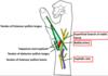
Describe the innervation of the wrist joint
- Median nerve – anterior interosseous branch
- Radial nerve – posterior interosseous branch
- Ulnar nerve – deep and dorsal branches

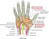
Identify the 4 ligaments which ensure the stability of the wrist joint

Describe the location and function of the palmar radiocarpal ligament
- Location: anterior side of the hand, passes from the radius to both rows of carpal bones
- Function: increases stability, ensures the hand follows the forearm during supination
Describe the location and function of the dorsal radiocarpal ligament
- Location: posterior side of the hand, passes from the radius to both rows of carpal bones
- Function: increases stability of the wrist, ensures that the hand follows the forearm during pronation

Describe the location and function of the ulnar collateral ligament
- Location: runs from the ulnar styloid process to the triquetrum and pisiform
- Function: prevents excessive medial joint displacement (in union with other collateral ligament)

Describe the location and function of the radial collateral ligament
- Location: runs from the radial styloid process to the scaphoid and trapezium
- Function: prevents excessive lateral joint displacement (in union with other collateral ligament)

What type of joint is the wrist joint?
The wrist is an ellipsoid type synovial joint, allowing for movement along two axes
Identify the four different types of movement possible at the radiocarpal joint as well as the individual muscles involved
- Flexion – flexor carpi ulnaris and flexor carpi radialis (assistance from the flexor digitorum superficialis)
- Extension – extensor carpi radialis longus and brevis and extensor carpi ulnaris (assistance from the extensor digitorum)
- Adduction – extensor carpi ulnaris and flexor carpi ulnaris
- Abduction – abductor pollicis longus, flexor carpi radialis, extensor carpi radialis longus and brevis
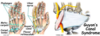
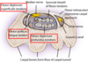
What is the carpal tunnel?
The carpal tunnel is a narrow passageway on the anterior portion of the wrist, serving as the entrance to the palm for several tendons and the median nerve

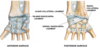
Describe the borders of the carpal tunnel
- The deep carpal arch (formed by carpal bones) forms a concave surface acting as the floor
- The overlying superficial flexor retinaculum acts as the roof

Identify the carpal bones forming the carpal arch laterally and medially
- Lateral: scaphoid and trapezium tubercles
- Medial: hook of hamate and pisiform

State the structure and function of the flexor retinaculum
- Structure: thick connective tissue
- Function: turns the carpal arch into the carpal tunnel
State the origin and attachment of the flexor retinaculum
- Origin: lateral side of carpal arch
- Attachment: medial side of the carpal arch
Briefly describe the contents of the carpal tunnel
The carpal tunnel contains a total of 9 tendons, surrounded by synovial sheaths, and the median nerve

Identify the different tendons found in the carpal tunnel
- The tendon of flexor pollicis longus
- Four tendons of flexor digitorum profundus
- Four tendons of flexor digitorum superficialis
The synovial sheaths in the carpal tunnel allow free movement of the tendons.
How many are there?
- Tendons of the flexor digitorum profundus and flexor digitorum superficialis are surrounded by a single synovial sheath
- The tendon of flexor pollicis longus is surrounded by its own synovial sheath

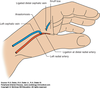
Describe the course of the median nerve once it passes through the carpal tunnel
Median nerve divides into 2 branches: recurrent branch and palmar digital branch

Which structures do the palmar digital and recurrent branches of the median nerve innervate respectively?
- Palmar digital nerves give sensory innervation to the palmar skin and dorsal nail beds of the lateral 3½ digits and motor innervation to the lateral two lumbricals
- Recurrent branch supplies the thenar muscle group
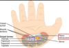
What is the anatomical snuffbox?
The anatomical snuffbox is a triangular depression found on the lateral aspect of the dorsum of the hand, at the level of the carpal bones

Identify the different borders of the anatomical snuffbox as well as the structures forming them
- Medial border: tendon of extensor pollicis longus
- Lateral border: tendons of abductor pollicis longus and extensor pollicis brevis
- Proximal border: styloid process of the radius
- Floor: carpal bones; scaphoid and trapezium
- Roof: skin