Salivary Gland Lesions 1 Flashcards
(23 cards)
Reactive Lesions
Mucocele
Ranula
Sialolithiasis
Necrotizing sialometaplasia
Infectious Lesions
Bacterial sialadenitis
Viral sialadenitis (mumps)
Autoimmune Lesions
Sarcoidosis
Sjorgen’s syndrome
Other
Xerostomia
Sialorrhea

Mucocele
Children/Young adults 80% seen in lower lip
Dome-shaped swelling, bluish-translucent hue
Older lesions may appear fibrosed and firm
Area of spilled mucin surrounded by granulation tissue
Some lesions may rupture and heal. Surgical excision

Ranula (derived from rana = frog)
Term used for mucocele occuring in the floor of the mouth
Dome-shaped swelling in the floor of mouth, usually lateral to midline
Plunging ranula: spilled mucin dissects through the mylohyoid muscle
Area of spilled mucin surrounded by granulation tissue
Treatment: Removal of feeding gland, marsupialization, removal of lesion’s roof, small, superficial ranulas

Sialolith
Calcified structure within salivary duct system
Deposition of calcium salts around a niduc of debris
Round, oval or cylindrical, yellow hard mass
Appear as radiopaque masses on radiographs
Treatment: small stones can be “milked out”. Larger stones may need surgery

Necrotizing Sialometaplasia
Locally destructive inflammatory process probably due to ischemia and infraction
75% of cases occur in posterior hard palate mimics a malignant process!
Squamous metaplasia of salivary ducts and acinar necrosis
Treatment: must always be biopsied. Once diagnosis is estabhlished, no treatment. Heals in 5-6 weeks.
Blockage of salivary gland ducts
Sialolith, Congenital stricture, adjacent tumor
Decreased salivary flow
dehydration, debilitation, medications
Retrograde spread of bacteria, especially S. aureus
Acute and Chronic
Bacterial Sialadenitis

Acute sialadenitis
unilateral parotid swelling. Swollen gland, skin warm/red
Fever and purulence often are present
Treatment: antibiotics + rehydration
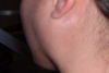
Acute sialadenitis

Chronic sialadenitis
often due to sialoliths swelling and pain (mealtime)
Treatment: conservative - surgical

Viral Sialadenitis (Mumps)
Caused by a paramyxovirus, genus rubulavirus
Low grade fever, headache, malaise, anorexia, and myalgia
Pain, discomfort and swelling from ear to mandibular area
Salivary gland enlargement is usually bilateral (75% of cases)
Diagnosis: based on clinical findings (epidemic). Serological studies (IgG or IgM) helpful in isolated cases
Treatment: Palliative treatment - analgesic, antipyretic and rest
Multisystem disorder of unknown cause
Formation of non-caseating granulomas
10-17x more common in blacks
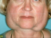
Lupus pernio: violaceous, indurated lesions. Frequent in nose, ears, lips and face
Eyes involved seen in 25% of cases. Xerophthalmia
Salivary gland involvement can cause Xerostomia
Oral manifestations include massess, papules or ulcerations. Color may be brown-red, violaceous or hyerkeratotic
Diagnosis: clinical radiographic and microscopic examination. Elevated angiotensin-converting enzyme levels
Treatment: 50% of cases remission in 3 years. Corticosteroids first line of therapy
Sarcoidosis

Sarcoidosis
lupus pernio

Sarcoidosis
eyes involved in 25% of cases - xerophthalmia

Sarcoidosis
Salivary gland involvment can cause xerostomia

Sarcoidosis
oral manifestations include massess, papules or ulcerations
color may be brown-red, violaceous or hyperkeratotic
Sjogren Syndrome
Autoimmune disorder affecting salivary and lacrimal glands
primary sjogren = sicca syndrome: dry eyes + dry mouth
secondary sjorgen: sicca symdrome + auto-immune disease
diffuse, firm, bilateral enlargement of major salivary glands in 30-50% of pts
Main symptom is dry mouth. Fissured tongue and atrophy papillae
Schirmer test: used to confirm decreased tear secretion
Supportive treatment (artificial tears/saliva). Increased caries and candida risk. 40x risk of lymphoma

Xerostomia - Dry mouth
1 in 4 adults report xerostomia
500 drugs reported to cause xerostomia
Treatment: difficult and often unsatisfactory. Modification of medication. Artificial saliva, pilocarpine

Xerostomia

Sialorrhea
Patients with certain neurological disorders may drool, but have normal saliva quantity
Peri-oral skin may become ulcerated and secondarily infected
Treatment: if transient - no treatment needed. Medication, speech therapy, surgery
In conclusion…
A variety of diseases can affect salivary glands
Primary or secondary to other diseases
detailed clinical history and exam required
Histopathological exam frequently needed
