Salt reabsorption 2 Flashcards
(25 cards)
What is the airway surface liquid layer (ASL)
Layer of liquid that lies over the epithelial cells of the lower and upper respiratory tract. In the upper tract there is a mucous layer which lies on top of the liquid layer, the liquid layer is called the pericellular layer and it contains salts. Cilia project from the ASL and beat, moving the liquid layer to the top of the tract and we swallow it along with any pathogens as a first line of defence.
What is the importance of the height of the ASL
Too high and the mucous movement is slowed down, too low and the cilia become bent and are not capable of moving the liquid up the respiratory tract.
What is the problem in the ASL height due to movement up the airways
The lower respiratory tract has a much larger surface layer (2m squared) compared to the upper airway (50cm squared). However the ASL optimum height remains the same between the two. Therefore, if nothing was in place to maintain this height, the ASL would become taller in the upper airways and reduce the movement of mucosa. There must be a mechanism to absorb excess fluid in the respiratory tract to maintain the height of the ASL.
What is the passive mechanism of ASL control q
The mucous acts as a resevoir and takes up and excess liquid to maintain the ASL height. This acts in both ways but probably only plays a small role in comparison to the active mechanism
What is the active mechanism of ASL height control
Theres an active ion transport by the epithelial cells. They control how much salt is present in the layer. They control the balance between chloride secretion and sodium reabsorption.
What experiment was performed on cultured epithelial airway cells to show the height of the ASL
A salt solution was placed on top of the cells which was too high. They started with a height of around 30 microns and left the cells, monitoring the height of the layers which came down to around 7 microns. This allowed the cilia to function in vitro, in vivo studies the height is around 14 microns.
What experiment was done to show the role of chloride and sodium channels in maintaining the ASL
They investigated two compounds; amiloride and bumetanide (a Cl channel blocker) They observed the % inhibtion of the Vte by the two compounds at 0 and 48 hours. (at 48 hours the optimum height has been reached, at 0 it is too high)
At time 0, bumetanide gives around 18% inhibition, i.e. not much Cl secretion (or water secretion), helping to bring the height of the layer down. At 48Hrs the amount of secretion has greatly increased (around 55% inhibition)
Opposite seen for amiloride - high inhibition to start (prevent water reabsorption) and low at the end.
What is the balance between sodium and chloride secretion atthe correct ASL height
chloride secretion is slightly higher than sodium reabsorption. If this balance is lost then the height of the liquid layer changes and the efficiency of the liquid layers movement up the tract is effected.
What is the importance of ENaC at birth
Important for lung fluid clearence when there is excess fluid in the lungs. ENaC is massively upregulated just before birth
Describe the cell model of the epithelia of the upper respiratory tract
Basolateral - NaKATPase and NKCC1 (recycle Na) and a K channel (allows for recycling of K)
Apical - ENaC and CFTR
What is RSV?
Respiratory syncytial virus
Disrupts fluid balance in the respiratory tract (why we get a runny nose)
Symptoms include nasal congestion, broncholitis in children and pneumonia in adults.
What experiment was done in mouse trachea to show the effect of RSV
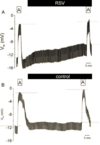
Measured the Vte and the amiloride sensitive SCC in mouse trachea that was either infected with RSV or not. In both cells, amilorde is added, then washed away, after an hour amilorde is added again, this shows the function of ENaC in these cells. In the RSV infected cell (RSV is applied after first amiloride application), over time the Vte shifts in a positive direction and there is a smaller shift in response to second amiloride exposure. This suggests that RSV inhibits ENaC function and at around 50% inhibition.
What effect does PKC have on ENaC and how does it respond to RSV
PKC inhibits the function of ENaC by preventing its expression (ERK pathway), and preventing its insertion by intefering with trafficking. BIM is a PKC inhibitor, which when added alongside RSV, relieves the inhibition of ENaC i.e. RSV is activating PKC in order to inactivate ENaC
What relationship does RSV have to cell surface glycoproteins in ENaC inhibition
Neuroaminidase (NA) inhibits the binding of RSV to glycoproteins on the cell surface. When co-applied with RSV, ENaC inhibition is still seen, so RSV does not interact with cell surface glycoproteins to infect the cell.
How does RSV interact with glycolipids in order to inhibit ENaC function
PPMP is an inhibitor of virus binding to glycolipids. See a smiliar response to RSV compared to BIM (the PKC inhibitor) - Suggests RSV binds to glycolipids to infect the host cells where it then interacts wtih PKC to inhibit ENaC function.
Give three examples of glycoproteins that bind cell surface proteins and inhibit ENaC in influenza
- M1 protein
- Haemagglutin - binds to sialic acid residues which activates PKC
- M2 protein is inserted into the apical membrane and forms an acid activated proton channel. This causes a pH change which inhibits the function of ENaC
What experiment was performed to investigate long term infection of M2
Overexpression of M2 which is tagged with GFP inhibits ENaC function. Seen by single channel patch clamp. There are much fewer deflections showing the channels have a reduced Po. The number of channels is also reduced which is shown by western blot analysis - Density of the ENaC band is much less after M2 expression.
What mechanism does M2 have in ENaC inhibition
M2 promotes the endocytosis of ENaC. In Liddle’s patients, endocytosis of ENaC is not possible. There is little ENaC inhibition by M2 in Liddle’s syndrome cells. Can conclude that influenza causes the endocytosis of ENaC. Do Liddle’s patients not suffer from influenza infection?
What is the PKC signalling pathway in ENaC inhibition
Cells were made to express RFP, or RFP-M2. RFP-M2 induces lots of ROSs. If you inhibit ROS by adding GSH (an antioxidant) then the inhibition is removed, this is also seen if you block PKC with BIM.
Influenza inserts the M2 protein into the apical membrane of respiratory tract cells which generate ROS and activates PKC. This blocks ENaC, reduces the Po and pulls ENaC out of the membrane. No water reabsorption = tall ASL = runny nose.
What are characteristics of PHA1
Salt wasting
hypotension
Hyperkalaemia
Metabolic acidosis - can’t secrete H ions
High renin and aldosterone - try to compensate for ENaC
What are the characteristics of the autosomal dominant form
Renal, localised to the kidney
Mineralocorticoid receptor gene mutations (aldosterone receptor)
What are the characteristics of the PHA1 recessive form
Systemic, multiple organs effected
ENaG gene mutants in all subunits, patients have frequent lower respiratory tract illnesses
How does the wet weight of systemic PHA1 patients differ to renal.
Patients have red and inflamed skin under their nose due to a constant runny nose. The wet weight in the systemic form is much higher than the renal form. There is also a higher Na concentration in the nasal fluid of systemic patients.
How does the Vte of the nasal epithelia compare in normal and systemic PHA1 patients.
Normal = around -24mV
systemic = around -6mV as Na reabsorption is not happening.
Amiloride inhibition is much less in systemic patients too (only 6% inhibition compared to 58% in normal/renal)
ENaC not functioning, chloride secretion still occuring so the ASL becomes too high - retention of pathogens means these patients often suffer from infection.
