Secret Papers Flashcards
(194 cards)
- According to Okada, et al. (JAVMA, 2010), what is a characteristic feature of myelomalacia on T2w images?
- Also, what changes were noted in CSF?
- A different study by Gilmour, et al. (VRU, 2015), found what factor(s) to be predictive of myelomalacia using T2w images and SSTSE sequences?
-
Okada: T2w-hyperintensity > 6x the length of L2
- Xanthochromia
-
Gilmour:
- CSF:L2SSTSE < 7.4 – unlikely to develop myelomalacia
- CSF:L2SSTSE > 12.5 – suggested cut-off to minimize number of false positives (100% specific)
Okada, M., Kitagawa, M. & Ito, D. (2010) Magnetic resonance imaging features and clinical signs associated with presumptive and confirmed progressive myelomalacia in dogs: 12 cases (1997–2008). Journal of the American Veterinary Medical Association 237, 1160–1165
Gilmour, L.J., Jeffery, N.D., Miles, K., et al. (2017) SINGLE-SHOT TURBO SPIN ECHO PULSE SEQUENCE FINDINGS IN DOGS WITH AND WITHOUT PROGRESSIVE MYELOMALACIA. Veterinary Radiology & Ultrasound 58, 197–205
Name the labeled structures and determine the anomaly.

- CVC
- Ao
- PV
Intrahepatic PSS
Bruehschwein, A., Foltin, I., Flatz, K., et al. (2010) Contrast-enhanced magnetic resonance angiography for diagnosis of portosystemic shunts in 10 dogs. Veterinary Radiology and Ultrasound 51, 116–121
- What is the most frequent location for a gastric leiomyoma/sarcoma?
- Although there is a great deal of overlap, and there are no specific differentiating characteristics, what are some features of intestinal neoplasia can help to differentiate inflammatory disease from neoplasia?
- (T/F) Intestinal carcinoma is most often solitary but is also commonly multifocal.
- What CT features can help distinguish gastric lymphoma from other types of neoplasia?
- What is the most frequent location for a gastric leiomyoma/sarcoma? Pylorus
- Although there is a great deal of overlap, and there are no specific differentiating characteristics, what are some features of intestinal neoplasia can help to differentiate inflammatory disease from neoplasia?
- Neoplastic infiltration has a median wall thickness statistically greater than inflammatory disease (15mm vs. 6mm)
- Loss of wall layering
- Intestinal carcinoma is most often solitary.
- What CT features can help distinguish gastric lymphoma from other types of neoplasia?
- Lower mean attenuation on both early and delayed post-contrast phases
- More widespread lymphadenopathy with larger lymph nodes
Simeoni, F., Signore, F. Del, Terragni, R., et al. (2020) Diagnostic Imaging of Gastrointestinal Tumours in Dogs and Cats : A Review. American Journal of Animal and Veterinary Sciences 15, 89–101
More GB sludge:
- Serial US over the course of a year showed what changes to GB sludge?
- No significant difference in median amount of sludge
- Most dogs remained asymptomatic
- Some dogs had more sludge and some had sludge that became non-dependent
DeMonaco, S.M., Grant, D.C., Larson, M.M., et al. (2016) Spontaneous Course of Biliary Sludge Over 12 Months in Dogs with Ultrasonographically Identified Biliary Sludge. Journal of Veterinary Internal Medicine 30, 771–778
Regarding radiographic changes associated with mycobacterium infection in cats (Bennett, 2011 JFMS):
- What were the most common changes in the thorax?
- Abdomen?
- Appendicular skeleton?
- What are some of the less common findings described in this report?
- Thorax:
- Commonly mixed bronchial, alveolar, and nodular interstitial patterns
- Perihilar and sternal lymphadenopathy
- Abdomen:
- Uncommon, but include hepatosplenomegaly
- Skeletal:
- Permeative osteolysis, periosteal reaction, soft tissue swelling
- Less common findings:
- Mineralization of the great vessels
- Dystrophic soft tissue mineralization
- Submandibular soft tissue swelling
Bennett, A.D., Lalor, S., Schwarz, T., et al. (2011) Radiographic Findings in Cats with Mycobacterial Infections. Journal of Feline Medicine & Surgery 13, 718–724

Likely diagnosis?

Gastroesophageal intussusception
Pollard, R.E. (2012) Imaging Evaluation of Dogs and Cats with Dysphagia. ISRN Veterinary Science 2012, 1–15
Regarding the sonographic features of thymomas and mediastinal lymphoma:
- What are the typical sonographic characteristics of each tumor?
- Which features were significant differentiators?
- Lymphoma:
- About 50/50 hypoechoic vs. heterogeneously echogenic
- Mostly solid and commonly lobulated
- Thymoma:
- Almost all were heterogeneously echogenic
- 60% cystic
- Mostly lobulated shape with irregular/indistinct margins
- Which features were significant differentiators?
- Heterogeneity and cysts (NS) more suggestive of thymoma
Patterson, M.M.E. & Marolf, A.J. (2014) Sonographic characteristics of thymoma compared with mediastinal lymphoma. Journal of the American Animal Hospital Association 50, 409–413
Describe the normal progression (phases) of a excretory urogram
- Arterial phase: arterial renal blood flow, is extremely brief, and usually has already passed when the first image is made
- Nephrogram phase: contrast accumulation in the renal tubules
- Pyelogram phase: contrast accumulation in the collecting system (diverticuli, pelvis, ureters)
- Cystogram: contrast accumulation in the bladder
Pugh, C.R., Rhodes, W.H. & Biery, D.N. (1993) Contrast Studies of the Urogenital System. Veterinary Clinics of NA: Small Animal Practice 23, 281–306
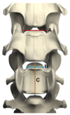
Regarding MRI of vertebral endplate changes in dogs (Gendron, VRU 2012):
- What were the imaging characteristics of the following categories of endplate changes?
- Reactive changes
- Fatty infiltration
- Sclerosis
- Osteochondrosis
- Schmorl’s node
- What other category of endplate changes were evaluated?
- Reactive changes: T2w/STIR hyperintense, T1w-hypointense, +/- CE
- Fatty infiltration: T1 & T2w-hyperintensities nulled on STIR
- Sclerosis: T2/T1w-hypointense
- Osteochondrosis: defect on dorsal edge of endplate +/- fragment, material filling the gap is isointense to disc
- Schmorl’s node: centrally-located, well-marginated, focal endplate defect contiguous with disc and filled with disc-isointense material
- Also looked at disko
GENDRON, K., DOHERR, M.G., Gavin, P., et al. (2012) MAGNETIC RESONANCE IMAGING CHARACTERIZATION OF VERTEBRAL ENDPLATE CHANGES IN THE DOG. Veterinary Radiology & Ultrasound 53, 50–56
Regarding MRI of dogs with head trauma:
- What features were linked to prognosis?
- How did the patterns of injury in dogs compare to those in humans?
- Prognosis:
- Injuries affecting the caudal fossa or causing a severe midline shft were associated with increased mortality
- Herniation through an open fontanelle was somehow associated with a good outcome
- Different patterns of intracranial trauma in dogs than humans
Yanai, H., Tapia-Nieto, R. & Cherubini, G.B. (2015) Results of magnetic resonance imaging performed within 48 hours after head trauma in dogs and association with outcome: 18 cases (2007–2012). Journal of the American Veterinary Medical Association 246, 1222–1229

Some normal measurements of the horse LUT:
- Bladder wall
- Ureteral wall
- Urethral wall
- Bladder wall: 3mm
- Ureteral wall: 1.8 mm
- Urethral wall: 5mm
- What is the most common distribution of thyoid disease in cats (i.e., bilateral/unilateral)?
- How common is ectopic thyroidal tissue in cats?
- What is the most common distribution of thyoid disease in cats (i.e., bilateral/unilateral)? IN ORDER
- Bilateral asymmetric
- Unilateral
- Bilateral symmetric
- Multifocal
- How common is ectopic thyroidal tissue in cats?
- Uncommon – about 4% of cats
Peterson, M.E. & Broome, M.R. (2015) Thyroid scintigraphy findings in 2096 cats with hyperthyroidism. Veterinary Radiology and Ultrasound 56, 84–95
Positive contrast MRI bursography for assessement of the structures of the foot:
- What dose of saline/contrast solution achieved distension of the navicular bursa such all structures of interest were separated from one another?
- (T/F): Structures of the distal aspect of the navicular bursa distend with lower volumes of solution and structures of the proximal aspect require larger volumes.
- What abnormalities can be identified using this procedure?
- What dose of saline/contrast solution achieved distension of the navicular bursa such all structures of interest were separated from one another? 6 ml
- (F): Structures of the distal aspect of the navicular bursa distend with lower volumes of solution and structures of the proximal aspect require larger volumes.
- Opposite – proximal filled first
- What abnormalities can be identified using this procedure?
- Can rule out adhesions if structures separate with distension
- Can increase index of suspicion for adhesions if they fail to separate
- Flexor erosions previously obscured due to close proximity of DDFT
MAHER, M.C., Werpy, N.M., Goodrich, L.R., et al. (2011) POSITIVE CONTRAST MAGNETIC RESONANCE BURSOGRAPHY FOR ASSESSMENT OF THE NAVICULAR BURSA AND SURROUNDING SOFT TISSUES. Veterinary Radiology & Ultrasound 52, 385–393
What is the normal appearance of horse urine on US?
Swirling, echogenic. Likely due to mucus and calcium carbonate.
doi: 10.1111/j.1740-8261.2007.00297.x
- What are differentials for this image of a 12yr Thoroughbred gelding?
- Given the ultimate diagnosis, what was the prognosis?
- What treatment was initiated?

- DDX:
- Fungal pneumonia
- Interstitial pneumonia
- Equine multinodular pulmonary fibrosis; associated with EHV-5
- Pulmonary neoplasia
- Given the ultimate diagnosis, what was the prognosis?
- Fair to poor
- 3/5 euthanized due to failure to improve/deterioration
- 2/5 responded favorably to treatment
- Fair to poor
- What treatment was initiated?
- Corticosteroids
- Anti-virals
- Sildenafil and lasix in a horse with PHT
Wong, D.M., Belgrave, R.L., Williams, K.J., et al. (2008) Multinodular pulmonary fibrosis in five horses. Journal of the American Veterinary Medical Association 232, 898–905
Regarding the appearance of muscular metastasis on CT:
- What was the typical appearance of muscular metastatic lesions?
- Where were they most commonly located?
- What was different about metastatic lesions in the myocardium?
- What was the typical appearance of muscular metastatic lesions?
- Well-defined, oval to round, isodense pre- and contrast-enhancing post C+ (homogeneous/heterogeneous/ring)
- Where were they most commonly located?
- Epaxial/paraspinal muscles > thoracic wall > scapula/shoulder = hind limb > abdominal wall
- What was different about metastatic lesions in the myocardium?
- Isodense pre-C, hypodense post-C
Vignoli, M., Terragni, R., Rossi, F., et al. (2013) WHOLE BODY COMPUTED TOMOGRAPHIC CHARACTERISTICS OF SKELETAL AND CARDIAC MUSCULAR METASTATIC NEOPLASIA IN DOGS AND CATS. Veterinary Radiology & Ultrasound 54, 223–230
Regarding the CT features of lipomatous masses in dogs:
- What features differentiate lipomas from infiltrative lipomas?
- What features differentiate liposarcomas?
- Lipoma: round or ovoid, well-marginated, fat attenuating, non-CE
- Infiltrative lipoma: homogeneous, fat-attenuating, non-CE but with irregular shape and linear hyperattenuating components
- Liposarcomas: heterogeneous, primarily soft-tissue attenuating with some foci of fat, CE, nodular masses +/- mineralization or regional lymphadenopathy
Tumor definition and shape were most useful for differentiating lipoma vs. infiltrative lipoma
Soft tissue, heterogeneous mass with mineralization and regional lymphadenopathy was useful for liposarcoma
Spoldi, E., Schwarz, T., Sabattini, S., et al. (2017) Comparisons Among Computed Tomographic Features of Adipose Masses in Dogs and Cats. Veterinary Radiology and Ultrasound 58, 29–37
- What are the MRI features of septic arthritis in horses?
- What are the MRI features of septic and non-septic arthritis in foals?
- What are the “rim sign” and “penumbra sign” in this context?
- Horses and foals with septic arthritis:
- Diffuse hyperintensity in bone and extracapsular tissues on FS images – foals also had hypointense halo
- Joint effusion – fluid is less T2 hyperintense than normal synovial fluid
- Synovial proliferation – low T2w-signal, (+) CE
- Capsular thickening
- Foals have subchondral hemorrhage
- Non-infectious foals:
- Joint effusion
- No bone lesions, no synovial thickening
- Osteomyelitis has characteristic rim sign (hypointense halo surrounding active disease) and penumbra sign (zone of transition of increasing signal intensity and the surrounding sclerotic bone on T1 images)
EASLEY, J.T., BROKKEN, M.T., Zubrod, C.J., et al. (2011) MAGNETIC RESONANCE IMAGING FINDINGS IN HORSES WITH SEPTIC ARTHRITIS. Veterinary Radiology & Ultrasound 52, 402–408
GASCHEN, L., LEROUX, A., TRICHEL, J., et al. (2011) MAGNETIC RESONANCE IMAGING IN FOALS WITH INFECTIOUS ARTHRITIS. Veterinary Radiology & Ultrasound 52, 627–633

- What are the CT findings associated with laryngeal collapse?
- What concomitant abnormalities were noted in these dogs?
- What are the CT findings associated with laryngeal paralysis?
- What are the CT findings associated with laryngeal collapse? What concomitant abnormalities were noted in these dogs?
- MUST be assessed with 3D internal volume rendering
- Everted laryngeal saccules
- Medial +/- ventral collapse of cuneiform processes
- All dogs with LC were brachycephalic
- Concomitant abnormalities:
- Bronchial collapse
- Elongated soft palate (extending past epiglottis)
- Tracheal hypoplasia +/- collapse
- Laryngeal paralysis
- Failure to abduct arytenoids
- Collapse of rima glottis
- Stenosis of laryngeal inlet
- Air in ventricles
Stadler, K.L., HARTMAN, S., MATHESON, J., et al. (2011) COMPUTED TOMOGRAPHIC IMAGING OF DOGS WITH PRIMARY LARYNGEAL OR TRACHEAL AIRWAY OBSTRUCTION. Veterinary Radiology & Ultrasound 52, 377–384
Zwingenberger, A.L., Marks, S.L., Baker, T.W., et al. (2010) Ultrasonographic Evaluation of the Muscularis Propria in Cats with Diffuse Small Intestinal Lymphoma or Inflammatory Bowel Disease. Journal of Veterinary Internal Medicine 24, 289–292
This paper had discordant results about the sonographic appearance of IBD and LSA in comparison to other studies. What was the significant difference in this patient population?
IBD was not associated with muscularis thickening.
Regarding incomplete longitudinal fractures of the proximopalmar aspect of MCIII in horses:
- What was the most common orientation and location for a stress-related injury? What forms did these injuries take?
- What was the correlation of these findings with scintigraphy?
- What was the most common orientation and location for a stress-related injury? What forms did these injuries take?
- Incomplete fracture and/or increased opacity in a proximodistal orientation, more often medial
- What was the correlation of these findings with scintigraphy?
- In 50% with IRU, there were rad abnormalities and in 50% there were no rad abnormalities.
- Majority of horses with a fracture line had moderate to marked IRU, however this was not significant.
Morgan, R. & Dyson, S. (2012) Incomplete longitudinal fractures and fatigue injury of the proximopalmar medial aspect of the third metacarpal bone in 55 horses. Equine Veterinary Journal 44, 64–70
Regarding aortic lesions in dogs with spirocercosis:
- What are the common CT features?
- What was the average length of CT lesions of the aorta?
- How well did radiography predict the lesions identified on CT?
- What CT feature was significantly associated with neoplastic transformation of esophageal nodules?
- T/F: dogs with spirocercosis tend to be hypocoagulable
- What are the common CT features?
- Aortic mineralization
- Aortic aneurysm
- Aortic thrombi
- What was the average length of CT lesions of the aorta? 4 vertebral bodies
- Radiography overdiagnosed aneurysms but underdiagnosed mineralization
- What CT feature was significantly associated with neoplastic transformation of esophageal nodules? Mineralization
- HYPERcoagulable
Kirberger, R.M., Stander, N., Cassel, N., et al. (2013) Computed tomographic and radiographic characteristics of aortic lesions in 42 dogs with spirocercosis. Veterinary Radiology and Ultrasound 54, 212–222
- What is the likely diagnosis in this patient given the abnormality indicated by the black arrow?

- Laryngeal paralysis – air-filled ventricles
- Laryngeal collapse – no gas in ventricles, increased soft tissue opacity of larynx
Stadler, K.L., HARTMAN, S., MATHESON, J., et al. (2011) COMPUTED TOMOGRAPHIC IMAGING OF DOGS WITH PRIMARY LARYNGEAL OR TRACHEAL AIRWAY OBSTRUCTION. Veterinary Radiology & Ultrasound 52, 377–384