Senses Flashcards
(535 cards)
The middle ear begins at the tympanic membrane (eardrum) and ends at a bony wall containing two small openings covered by membranes. These openings are called the oval window and the round window. Three small bones are found between the tympanic membrane and the oval window. Collectively, they are called the ossicles. Individually, they are called the malleus (hammer), the incus (anvil), and the stapes (stirrup) because their shapes resemble these objects. The malleus adheres to the Page 320tympanic membrane, and the stapes touches the oval window. An auditory tube, also called the eustachian or pharyngotympanic tube, extends from the middle ear to the nasopharynx. Its purpose is to equalize air pressure across the tympanic membrane. When changing elevation, such as in an airplane, the act of chewing gum, yawning, or swallowing opens the auditory tubes wider. As this occurs, we often feel the ears “pop.”
Whereas the outer ear and the middle ear contain air, the inner ear is filled with fluid. The inner ear has three areas: The semicircular canals and the vestibule are concerned with equilibrium; the cochlea is concerned with hearing. The cochlea resembles the shell of a snail because it spirals.
reversed prompt
Sensory receptors – dendrites specialized to detect certain types of stimuli – Exteroceptors: detect stimuli from outside the body (e.g., taste, hearing, vision) – Interoceptors: receive stimuli from inside the body (e.g., change in blood pressure) • Directly involved in homeostasis and a part of a negative feedback loop
CHECK YOUR PROGRESS 15.6
State the location and function of the structures involved in maintaining balance.
Answer
All structures are in the inner ear and involve mechanoreceptors. For rotational equilibrium—semicircular canals, ampullae, cupula, stereocilia, hair cells, vestibular nerve, supporting cells, and endolymph; for gravitational equilibrium—utricle, saccule, otoliths, otolithic membrane, hair cells, supporting cells, and vestibular nerve.
Exteroceptors are sensory receptors that detect stimuli from outside the body, such as those that result in taste, smell, vision, hearing, and equilibrium
15.5 Sense of Hearing
LEARNING OUTCOMES
Upon completion of this section, you should be able to
Identify the structures of the ear involved in hearing.
Summarize how sound waves are converted into nerve signals.
Describe the pathway of sensory information from the ear to the brain.
The ear has two sensory functions: hearing and balance (equilibrium). The sensory receptors for both of these are located in the inner ear. Each consists of hair cells with stereocilia (sing., stereocilium), which are long, stiff microvilli that are sensitive to mechanical stimulation. The stereocilia act as mechanoreceptors.
Anatomy and Physiology of the Ear
Figure 15.12 shows that the ear has three divisions: outer, middle, and inner. The outer ear consists of the pinna (external flap) and the auditory canal. The opening of the auditory canal is lined with fine hairs and sweat glands. Modified sweat glands are located in the upper wall of the canal. They secrete earwax, a substance that helps guard the ear against the entrance of foreign materials, such as air pollutants.
Figure 15.12 The three divisions of the human ear. The external ear consists of the pinna (the structure commonly referred to as the “ear”) and the auditory canal. The tympanic membrane separates the external ear from the middle ear. In the middle ear, the malleus (hammer), the incus (anvil), and the stapes (stirrup) amplify sound waves. In the inner ear, the mechanoreceptors for equilibrium are in the semicircular canals and the vestibule. The mechanoreceptors for hearing are in the cochlea.
The middle ear begins at the tympanic membrane (eardrum) and ends at a bony wall containing two small openings covered by membranes. These openings are called the oval window and the round window. Three small bones are found between the tympanic membrane and the oval window. Collectively, they are called the ossicles. Individually, they are called the malleus (hammer), the incus (anvil), and the stapes (stirrup) because their shapes resemble these objects. The malleus adheres to the Page 320tympanic membrane, and the stapes touches the oval window. An auditory tube, also called the eustachian or pharyngotympanic tube, extends from the middle ear to the nasopharynx. Its purpose is to equalize air pressure across the tympanic membrane. When changing elevation, such as in an airplane, the act of chewing gum, yawning, or swallowing opens the auditory tubes wider. As this occurs, we often feel the ears “pop.”
Whereas the outer ear and the middle ear contain air, the inner ear is filled with fluid. The inner ear has three areas: The semicircular canals and the vestibule are concerned with equilibrium; the cochlea is concerned with hearing. The cochlea resembles the shell of a snail because it spirals.
Auditory Pathway to the Brain
The auditory pathway begins with the auditory canal. Thereafter, hearing requires the other parts of the ear, the cochlear nerve, and the brain.
Through the Auditory Canal and Middle Ear
The process of hearing begins when sound waves enter the auditory canal. Just as ripples travel across the surface of a pond, sound waves travel by the successive vibrations of molecules. Ordinarily, sound waves do not carry much energy. However, when a large number of waves strike the tympanic membrane, it moves back and forth (vibrates) ever so slightly. As you know, the auditory ossicles attach to one another: malleus to incus, incus to stapes. The malleus is attached to the inner wall of the tympanic membrane. Thus, vibrations of the tympanic membrane cause vibration of the malleus and, in turn, the incus and stapes. The magnitude of the original pressure wave increases significantly as the vibrations move along the auditory ossicles. The pressure is multiplied about 20 times. Finally, the stapes strikes the membrane of the oval window, causing it to vibrate. In this way, the pressure is passed to the fluid within the cochlea.
SCIENCE IN YOUR LIFE
What are “ear tubes”?
The auditory tubes of children tend to be oriented more horizontally than those of adults. Because of this, fluid may accumulate in the tubes, allowing for an infection to occur. These infections are called otitis media, and they are often painful. Extended cases of otitis media may produce long-term hearing loss.
A procedure called a tympanostomy places small tubes in the tympanic membrane, allowing these fluids to drain more easily, thus reducing the chance of infection. In most cases, the tubes fall out of the membrane over time, but sometimes they need to be removed by a physician.
From the Cochlea to the Auditory Cortex
By examining the cochlea in cross-section (Fig. 15.13), you can see that it has three canals. The sensory organ for hearing, called the spiral organ (or the organ of Corti), is located in the cochlear canal. The spiral organ consists of little hair cells and a gelatinous material called the tectorial membrane. The hair cells sit on the basilar membrane, and their stereocilia are embedded in the tectorial membrane.
Figure 15.13 How the spiral organ (organ of Corti) translates sound waves into nerve signals. a. The spiral organ (organ of Corti) is located within the (b) cochlea. c. The spiral organ consists of hair cells resting on the basilar membrane, with the tectorial membrane above. Pressure waves moving through the canals cause the basilar membrane to vibrate. This causes the stereocilia embedded in the tectorial membrane to bend. Nerve impulses traveling in the cochlear nerve result in hearing. d. A micrograph of the stereocilia.
(photo): ©P. Motta/SPL/Science Source
Page 322When the stapes strikes the membrane of the oval window, pressure waves move from the vestibular canal to the tympanic canal across the basilar membrane. The basilar membrane moves up and down, and the stereocilia of the hair cells embedded in the tectorial membrane bend. Then, nerve signals begin in the cochlear nerve and travel to the brain. When they reach the auditory cortex in the temporal lobe, they are interpreted as a sound.
Effect of Sound Waves
Each part of the spiral organ is sensitive to different wave frequencies, or pitch. Near the tip, the spiral organ responds to low pitches, such as those of a tuba. Near the base (beginning), it responds to higher pitches, such as those of a bell or a whistle. The nerve fibers from each region along the length of the spiral organ lead to slightly different areas in the auditory cortex. The pitch sensation we experience depends upon which region of the basilar membrane vibrates and which area of the auditory cortex is stimulated.
Volume is a function of the amplitude (strength) of sound waves. Loud noises cause the fluid within the vestibular canal to exert more pressure and the basilar membrane to vibrate to a greater extent. The resulting increased stimulation is interpreted by the brain as volume. As discussed in the Health feature “Noise Pollution,” noise levels above 85 decibels (Table 15.3) may cause permanent hearing loss.
BIOLOGY TODAY Health
Noise Pollution
Though we can sometimes tune out its presence, unwanted noise is all around us. Noise pollution is noise from the environment that is annoying, distracting, and potentially harmful. It comes from airplanes, cars, lawn mowers, machinery, and our own loud music and that of our neighbors. It is present at our workplaces, in public spaces like amusement parks, and at home. Its prevalence allows loud noise to have a potentially high impact on our welfare.
Noise and Health
How does noise affect human health? Perhaps the greatest worry about noise pollution is that exposure to loud (over 85 decibels) or chronic noises can damage cells of the inner ear and cause hearing loss (Fig. 15B). When we are young, we often do not consider the damage that noise may be doing to our spiral organ. The stimulation of loud music is often sought by young people at rock concerts without regard to the possibility that their hearing may be diminished as a result. Over the years, loud noises can bring deafness and accompanying depression when we are older adults.
Figure 15B Loud noise damages the hair cells in the spiral organ. a. Normal hair cells in the spiral organ of a guinea pig. b. Damaged cells. This damage occurred after 24-hour exposure to a noise level equivalent to that at a rock concert (see Table 15.3). Hearing is permanently impaired because lost cells will not be replaced, and damaged cells may also die.
(both): ©Dr. Yeohash Raphael, Kresge Institute/University of Michigan, Ann Arbor
Noise can affect well-being by other means, too. Data from studies of environmental noise can be difficult to interpret because of the presence of other confounding factors, including physical or chemical pollution. The tolerance level for noise also varies from person to person. Nonetheless, laboratory and field studies show that noise may be detrimental in nonauditory ways. Its effects on mental health include annoyance, inability to concentrate, and increased irritability. Long-term noise exposure from air or car traffic may impair cognitive ability, language learning, and memory in children. Noise often causes loss of sleep and reduced productivity and can induce stress. Additionally, several studies have demonstrated a link between noise pollution and cardiovascular health, specifically hypertension.
Regulating Noise Pollution
Noise pollution has been a concern for several decades. In 1972, the Noise Control Act was passed as a means for coordinating federal noise control and research and to develop noise emission standards. The aim was to protect Americans from “noise that jeopardizes their health or welfare.” The Environmental Protection Agency (EPA) had federal authority to regulate noise pollution, and its Office of Noise Abatement and Control (ONAC) worked on establishing noise guidelines. However, the activities of the ONAC were transferred to state and local governments in 1981. Today, there is no national noise policy, although the EPA does maintain standards on noise pollution on its website: www.epa.gov.
Workplace noise exposure is controlled by the Occupational Safety and Health Administration (OSHA). OSHA has set guidelines for workplace noise. OSHA regulations require that protective gear be provided if sound levels exceed certain values. This may include noise-reducing earmuffs and other protective methods for people who work around big equipment. However, OSHA guidelines don’t cover things like telephone ringing and computer noise that may be present in a nonindustrial environment such as an open-plan office. Aviation noise and traffic noise reduction plans are overseen by the Department of Transportation, the Federal Aviation Administration (FAA), and the Federal Highway Administration (FHWA), respectively. Local governments often have legislation that controls noise levels in public places, such as downtown areas and public parks. However, without national standards, the laws vary by location.
Questions to Consider
Given that noise pollution induces stress, what other body systems may be affected?
At a local level, what do you think could be done to curb noise pollution in your neighborhood?
Table 15.3Noises That Affect Hearing
Table Summary: Table lists the different types of noises in column 1. Other information related to each type of noise appears in columns 2 and 3.
Type of NoiseSound Level (Decibels)Effect
“Boom car,” jet engine, shotgun, rock concertOver 125Beyond threshold of pain; potential for hearing loss high
Nightclub, thunderclapOver 120Hearing loss likely
Earbuds in external ear canal110–120Hearing loss likely
Chain saw, pneumatic drill, jackhammer, symphony orchestra, snowmobile, garbage truck, cement mixer100–200Regular exposure of more than 1 min risks permanent hearing loss
Farm tractor, newspaper press, subway, motorcycle90–100Fifteen minutes of unprotected exposure potentially harmful
Lawn mower, food blender85–90Continuous daily exposure for more than 8 hr can cause hearing damage
Diesel truck, average city traffic noise80–85Annoying; constant exposure may cause hearing damage
CHECK YOUR PROGRESS 15.5
Identify the structures of the ear involved in hearing and provide a function for each.
Answer
The outer ear directs sound into the middle ear, causing vibrations in the tympanic membrane and the ossicles that attach to the inner ear, where fluid stimulates receptors that generate impulses in nerves, sending signals to the brain.
Describe the role of mechanoreceptors in the sense of hearing.
Answer
The hair cells located in the spiral organ of the cochlea are mechanoreceptors, which are sensitive to the movements of fluid in the inner ear.
Summarize how the spiral organ translates sound waves to nerve impulses.
Answer
Pressure waves move through the canals, causing the basilar membrane to vibrate. This causes the stereocilia embedded in the tectorial membrane to bend, generating nerve impulses that travel to the brain.
CONNECTING THE CONCEPTS
For more information on the material in this section, refer to the following discussions:
Section 14.2 describes the function of the cerebral cortex area of the brain in hearing.
Figure 14.15 illustrates the structure of a nerve.
Reverse.Prompt
Figure 15.14 The mechanoreceptors of the inner ear and the sense of balance. a. Rotational equilibrium is coordinated by receptors in the ampullae of the semicircular canals. b. Gravitational equilibrium is coordinated by receptors in the utricule and saccule located near the semicircular canals.
The vestibular nerve originates in the semicircular canals, saccule, and utricle. It takes nerve signals to the brain stem and cerebellum (Fig. 15.14). Through its communication with the brain, the vestibular nerve helps us achieve equilibrium, but other structures in the body are also involved. For example, in Section 15.5, we mentioned that proprioceptors are necessary for maintaining our equilibrium. Vision, if available, usually provides extremely helpful input the brain can act upon. To explain, let’s take a look at the two sets of mechanoreceptors for equilibrium.
- Whereas the outer ear and the middle ear contain air,
- the inner ear is filled with fluid.
- The inner ear has three areas:
- concerned with equilibrium;
- The semicircular canals and the
- vestibule are the
- concerned with hearing: The cochlea
- resembles the shell of a snail because it spirals.
convert a signal from the environment, called a stimulus, into a nerve impulse.
This conversion is commonly referred to as sensory transduction. Some sensory receptors are modified neurons, and others are specialized cells closely associated with neurons.
Sensory receptors may detect stimuli originating from both the internal and external environments. Exteroceptors are sensory receptors that detect stimuli from outside the body, such as those that result in taste, smell, vision, hearing, and equilibrium (Table 15.1). Interoceptors receive stimuli from inside the body. Examples of interoceptors are the baroreceptors (also called pressoreceptors) that respond to changes in blood pressure, osmoreceptors that monitor the body’s water-salt balance, and chemoreceptors that monitor the pH of the blood.
sensory receptor
Reverse.Prompt
Sensation Visual
15.1

15.5 Sense of Hearing
LEARNING OUTCOMES
Upon completion of this section, you should be able to
Identify the structures of the ear involved in hearing.
Summarize how sound waves are converted into nerve signals.
Describe the pathway of sensory information from the ear to the brain.
The ear has two sensory functions: hearing and balance (equilibrium). The sensory receptors for both of these are located in the inner ear. Each consists of hair cells with stereocilia (sing., stereocilium), which are long, stiff microvilli that are sensitive to mechanical stimulation. The stereocilia act as mechanoreceptors.
Anatomy and Physiology of the Ear
Figure 15.12 shows that the ear has three divisions: outer, middle, and inner. The outer ear consists of the pinna (external flap) and the auditory canal. The opening of the auditory canal is lined with fine hairs and sweat glands. Modified sweat glands are located in the upper wall of the canal. They secrete earwax, a substance that helps guard the ear against the entrance of foreign materials, such as air pollutants.
Figure 15.12 The three divisions of the human ear. The external ear consists of the pinna (the structure commonly referred to as the “ear”) and the auditory canal. The tympanic membrane separates the external ear from the middle ear. In the middle ear, the malleus (hammer), the incus (anvil), and the stapes (stirrup) amplify sound waves. In the inner ear, the mechanoreceptors for equilibrium are in the semicircular canals and the vestibule. The mechanoreceptors for hearing are in the cochlea.
The middle ear begins at the tympanic membrane (eardrum) and ends at a bony wall containing two small openings covered by membranes. These openings are called the oval window and the round window. Three small bones are found between the tympanic membrane and the oval window. Collectively, they are called the ossicles. Individually, they are called the malleus (hammer), the incus (anvil), and the stapes (stirrup) because their shapes resemble these objects. The malleus adheres to the Page 320tympanic membrane, and the stapes touches the oval window. An auditory tube, also called the eustachian or pharyngotympanic tube, extends from the middle ear to the nasopharynx. Its purpose is to equalize air pressure across the tympanic membrane. When changing elevation, such as in an airplane, the act of chewing gum, yawning, or swallowing opens the auditory tubes wider. As this occurs, we often feel the ears “pop.”
Whereas the outer ear and the middle ear contain air, the inner ear is filled with fluid. The inner ear has three areas: The semicircular canals and the vestibule are concerned with equilibrium; the cochlea is concerned with hearing. The cochlea resembles the shell of a snail because it spirals.
Auditory Pathway to the Brain
The auditory pathway begins with the auditory canal. Thereafter, hearing requires the other parts of the ear, the cochlear nerve, and the brain.
Through the Auditory Canal and Middle Ear
The process of hearing begins when sound waves enter the auditory canal. Just as ripples travel across the surface of a pond, sound waves travel by the successive vibrations of molecules. Ordinarily, sound waves do not carry much energy. However, when a large number of waves strike the tympanic membrane, it moves back and forth (vibrates) ever so slightly. As you know, the auditory ossicles attach to one another: malleus to incus, incus to stapes. The malleus is attached to the inner wall of the tympanic membrane. Thus, vibrations of the tympanic membrane cause vibration of the malleus and, in turn, the incus and stapes. The magnitude of the original pressure wave increases significantly as the vibrations move along the auditory ossicles. The pressure is multiplied about 20 times. Finally, the stapes strikes the membrane of the oval window, causing it to vibrate. In this way, the pressure is passed to the fluid within the cochlea.
SCIENCE IN YOUR LIFE
What are “ear tubes”?
The auditory tubes of children tend to be oriented more horizontally than those of adults. Because of this, fluid may accumulate in the tubes, allowing for an infection to occur. These infections are called otitis media, and they are often painful. Extended cases of otitis media may produce long-term hearing loss.
A procedure called a tympanostomy places small tubes in the tympanic membrane, allowing these fluids to drain more easily, thus reducing the chance of infection. In most cases, the tubes fall out of the membrane over time, but sometimes they need to be removed by a physician.
From the Cochlea to the Auditory Cortex
By examining the cochlea in cross-section (Fig. 15.13), you can see that it has three canals. The sensory organ for hearing, called the spiral organ (or the organ of Corti), is located in the cochlear canal. The spiral organ consists of little hair cells and a gelatinous material called the tectorial membrane. The hair cells sit on the basilar membrane, and their stereocilia are embedded in the tectorial membrane.
Figure 15.13 How the spiral organ (organ of Corti) translates sound waves into nerve signals. a. The spiral organ (organ of Corti) is located within the (b) cochlea. c. The spiral organ consists of hair cells resting on the basilar membrane, with the tectorial membrane above. Pressure waves moving through the canals cause the basilar membrane to vibrate. This causes the stereocilia embedded in the tectorial membrane to bend. Nerve impulses traveling in the cochlear nerve result in hearing. d. A micrograph of the stereocilia.
(photo): ©P. Motta/SPL/Science Source
Page 322When the stapes strikes the membrane of the oval window, pressure waves move from the vestibular canal to the tympanic canal across the basilar membrane. The basilar membrane moves up and down, and the stereocilia of the hair cells embedded in the tectorial membrane bend. Then, nerve signals begin in the cochlear nerve and travel to the brain. When they reach the auditory cortex in the temporal lobe, they are interpreted as a sound.
Effect of Sound Waves
Each part of the spiral organ is sensitive to different wave frequencies, or pitch. Near the tip, the spiral organ responds to low pitches, such as those of a tuba. Near the base (beginning), it responds to higher pitches, such as those of a bell or a whistle. The nerve fibers from each region along the length of the spiral organ lead to slightly different areas in the auditory cortex. The pitch sensation we experience depends upon which region of the basilar membrane vibrates and which area of the auditory cortex is stimulated.
Volume is a function of the amplitude (strength) of sound waves. Loud noises cause the fluid within the vestibular canal to exert more pressure and the basilar membrane to vibrate to a greater extent. The resulting increased stimulation is interpreted by the brain as volume. As discussed in the Health feature “Noise Pollution,” noise levels above 85 decibels (Table 15.3) may cause permanent hearing loss.
BIOLOGY TODAY Health
Noise Pollution
Though we can sometimes tune out its presence, unwanted noise is all around us. Noise pollution is noise from the environment that is annoying, distracting, and potentially harmful. It comes from airplanes, cars, lawn mowers, machinery, and our own loud music and that of our neighbors. It is present at our workplaces, in public spaces like amusement parks, and at home. Its prevalence allows loud noise to have a potentially high impact on our welfare.
Noise and Health
How does noise affect human health? Perhaps the greatest worry about noise pollution is that exposure to loud (over 85 decibels) or chronic noises can damage cells of the inner ear and cause hearing loss (Fig. 15B). When we are young, we often do not consider the damage that noise may be doing to our spiral organ. The stimulation of loud music is often sought by young people at rock concerts without regard to the possibility that their hearing may be diminished as a result. Over the years, loud noises can bring deafness and accompanying depression when we are older adults.
Figure 15B Loud noise damages the hair cells in the spiral organ. a. Normal hair cells in the spiral organ of a guinea pig. b. Damaged cells. This damage occurred after 24-hour exposure to a noise level equivalent to that at a rock concert (see Table 15.3). Hearing is permanently impaired because lost cells will not be replaced, and damaged cells may also die.
(both): ©Dr. Yeohash Raphael, Kresge Institute/University of Michigan, Ann Arbor
Noise can affect well-being by other means, too. Data from studies of environmental noise can be difficult to interpret because of the presence of other confounding factors, including physical or chemical pollution. The tolerance level for noise also varies from person to person. Nonetheless, laboratory and field studies show that noise may be detrimental in nonauditory ways. Its effects on mental health include annoyance, inability to concentrate, and increased irritability. Long-term noise exposure from air or car traffic may impair cognitive ability, language learning, and memory in children. Noise often causes loss of sleep and reduced productivity and can induce stress. Additionally, several studies have demonstrated a link between noise pollution and cardiovascular health, specifically hypertension.
Regulating Noise Pollution
Noise pollution has been a concern for several decades. In 1972, the Noise Control Act was passed as a means for coordinating federal noise control and research and to develop noise emission standards. The aim was to protect Americans from “noise that jeopardizes their health or welfare.” The Environmental Protection Agency (EPA) had federal authority to regulate noise pollution, and its Office of Noise Abatement and Control (ONAC) worked on establishing noise guidelines. However, the activities of the ONAC were transferred to state and local governments in 1981. Today, there is no national noise policy, although the EPA does maintain standards on noise pollution on its website: www.epa.gov.
Workplace noise exposure is controlled by the Occupational Safety and Health Administration (OSHA). OSHA has set guidelines for workplace noise. OSHA regulations require that protective gear be provided if sound levels exceed certain values. This may include noise-reducing earmuffs and other protective methods for people who work around big equipment. However, OSHA guidelines don’t cover things like telephone ringing and computer noise that may be present in a nonindustrial environment such as an open-plan office. Aviation noise and traffic noise reduction plans are overseen by the Department of Transportation, the Federal Aviation Administration (FAA), and the Federal Highway Administration (FHWA), respectively. Local governments often have legislation that controls noise levels in public places, such as downtown areas and public parks. However, without national standards, the laws vary by location.
Questions to Consider
Given that noise pollution induces stress, what other body systems may be affected?
At a local level, what do you think could be done to curb noise pollution in your neighborhood?
Table 15.3Noises That Affect Hearing
Table Summary: Table lists the different types of noises in column 1. Other information related to each type of noise appears in columns 2 and 3.
Type of NoiseSound Level (Decibels)Effect
“Boom car,” jet engine, shotgun, rock concertOver 125Beyond threshold of pain; potential for hearing loss high
Nightclub, thunderclapOver 120Hearing loss likely
Earbuds in external ear canal110–120Hearing loss likely
Chain saw, pneumatic drill, jackhammer, symphony orchestra, snowmobile, garbage truck, cement mixer100–200Regular exposure of more than 1 min risks permanent hearing loss
Farm tractor, newspaper press, subway, motorcycle90–100Fifteen minutes of unprotected exposure potentially harmful
Lawn mower, food blender85–90Continuous daily exposure for more than 8 hr can cause hearing damage
Diesel truck, average city traffic noise80–85Annoying; constant exposure may cause hearing damage
CHECK YOUR PROGRESS 15.5
Identify the structures of the ear involved in hearing and provide a function for each.
Answer
The outer ear directs sound into the middle ear, causing vibrations in the tympanic membrane and the ossicles that attach to the inner ear, where fluid stimulates receptors that generate impulses in nerves, sending signals to the brain.
Describe the role of mechanoreceptors in the sense of hearing.
Answer
The hair cells located in the spiral organ of the cochlea are mechanoreceptors, which are sensitive to the movements of fluid in the inner ear.
Summarize how the spiral organ translates sound waves to nerve impulses.
Answer
Pressure waves move through the canals, causing the basilar membrane to vibrate. This causes the stereocilia embedded in the tectorial membrane to bend, generating nerve impulses that travel to the brain.
CONNECTING THE CONCEPTS
For more information on the material in this section, refer to the following discussions:
Section 14.2 describes the function of the cerebral cortex area of the brain in hearing.
Figure 14.15 illustrates the structure of a nerve.
Reverse.Prompt
- The cornea, assisted by the
- lens and the
- humors,
- focuses images on the retina.
- Process/Steps:
- Focusing starts with the cornea and
- continues as the rays pass through the lens and the humors.
- The image produced is much smaller than the object,
- because light rays are bent (refracted) when they are brought into focus.
- If the eye is too long or too short, the person may need corrective lenses to bring the image into focus.
- The image on the retina is inverted (upside down) and reversed from left to right.
- Visual accommodation occurs for close vision.
- During visual accommodation,
- the lens changes its shape to bring the image into focus on the retina.
- The shape of the lens is controlled by the ciliary muscle, within the ciliary body.
- When we view a distant object,
- the ciliary muscle is relaxed,
- causing the suspensory ligaments attached to the ciliary body to be taut.
- The ligaments put tension on the lens and cause it to remain relatively flat (Fig. 15.7a).
- When we view a near object,
- the ciliary muscle contracts,
- releasing the tension on the suspensory ligaments.
- The lens becomes round and thick due to its natural elasticity (Fig. 15.7b).
- When we view a distant object,
- Thus, contraction or relaxation of the ciliary muscle allows the image to be focused on the retina.
- During visual accommodation,
Function of the Lens
- Close work requires contraction of the ciliary muscle,
- so it often causes muscle fatigue, known as eyestrain.
- Eyestrain is more common after the age of 40,
- because the lens loses some of its elasticity and is unable to accommodate.
- It is also common among those who work with computers,
- because the intense focusing causes the person to blink less, allowing the eyes to dry out.
- Eyedrops and/or corrective lenses, either eyeglasses or contact lenses, may be necessary to reduce eyestrain.
Eyestrain
Figure 15.7 Focusing light on the retina. Light rays from each point on an object are bent by the cornea and the lens in such a way that an inverted and reversed image of the object forms on the retina. a. When focusing on a distant object, the lens is flat because the ciliary muscle is relaxed and the suspensory ligament is taut. b. When focusing on a near object, the lens accommodates—it becomes rounded because the ciliary muscle contracts, causing the suspensory ligament to relax.
Anatomy of the eye • 2 compartments 1. Anterior compartment: between the cornea and lens; filled with a clear fluid called aqueous humor – this liquid is continuously produced each day and drains through small ducts 2. Posterior compartment: most of the eye, behind the lens; contains a gelatinous material called vitreous humor that holds the retina in place and supports the lens – this liquid you are born with and remains; no more is produced
reversed prompt
What are 3 types of receptors:
* Sensory receptors
* Exteroreceptors
* Interoreceptors
Chapter 15.1 slide 1.2
There are three of us:
dendrites specialized to detect certain types of stimuli –
We detect stimuli from outside the body
* (e.g., taste, hearing, vision)
We receive stimuli from inside the body
* (e.g., change in blood pressure)
* Directly involved in homeostasis and a part of a negative feedback loop
15.2 Somatic SensesLEARNING OUTCOMES
Upon completion of this section, you should be able to
Distinguish between proprioceptors and cutaneous receptors with regard to function.
State the location and general function of each type of cutaneous receptor.
Explain the role of nociceptors and summarize the type of sensory input they detect.
Senses whose receptors are associated with the skin, muscles, joints, and viscera are termed the somatic senses. These receptors can be categorized into three types: proprioceptors, cutaneous Page 309receptors, and pain receptors. All of these send nerve impulses via the spinal cord to the primary somatosensory areas of the cerebral cortex (see Fig. 14.11).
Proprioceptors
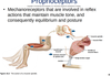
Proprioceptors are mechanoreceptors involved in reflex actions that maintain muscle tone, and thereby the body’s equilibrium and posture. For example, proprioceptors called muscle spindles are embedded in muscle fibers (Fig. 15.2). If a muscle relaxes too much, the muscle spindle stretches, generating nerve impulses that cause the muscle to contract slightly. Conversely, when muscles are stretched too much, proprioceptors called Golgi tendon organs, buried in the tendons that attach muscles to bones, generate nerve impulses that cause the muscles to relax. Both types of receptors act together to maintain a functional degree of muscle tone. The knee-jerk reflex, which involves muscle spindles, offers an opportunity for physicians to test a reflex action. The information sent by muscle spindles to the CNS is used to maintain the body’s equilibrium and posture. Proper balance and body position are maintained, despite the force of gravity always acting on the skeleton and muscles.
Figure 15.2 The action of proprioceptors. 1. When a muscle is stretched, muscle spindles send sensory nerve impulses to the spinal cord. 2. Motor nerve impulses from the spinal cord cause slight muscle contraction. 3. When tendons are stretched excessively, Golgi tendon organs cause muscle relaxation.
Cutaneous Receptors
The skin is composed of two layers: the epidermis and the dermis (see Section 4.6). The dermis contains cutaneous receptors (Fig. 15.3), which make the skin sensitive to touch, pressure, pain, and temperature (warmth and cold). The dermis is a mosaic of these tiny receptors, as you can determine by slowly passing a metal probe over your skin. At certain points, you will feel touch or pressure; at others, you will feel heat or cold (depending on the probe’s temperature).
Figure 15.3 Sensory receptors of the skin. The general function of each sensory receptor is shown here. However, receptors are not always this specialized. For example, microscopic examination of the skin of the ear shows only free nerve endings (pain receptors), yet the skin of the ear is sensitive to all sensations.
Several types of cutaneous receptors are sensitive to fine touch. These receptors give a person specific information, such as the location of the touch, as well as its shape, size, and texture. Meissner corpuscles and Krause end bulbs are concentrated in the fingertips, palms, lips, tongue, nipples, penis, and clitoris. Merkel discs are found where the epidermis meets the dermis. A free nerve ending called a root hair plexus winds around the base of a hair follicle. This receptor responds if the hair is touched.
Two types of cutaneous receptors sensitive to pressure are Pacinian corpuscles and Ruffini endings. Pacinian corpuscles are onion-shaped sensory receptors that lie deep inside the dermis. Ruffini endings are encapsulated by sheaths of connective tissue and contain lacy networks of nerve fibers.
Temperature receptors are simply free nerve endings in the epidermis. Some free nerve endings are responsive to cold; others respond to warmth. Cold receptors are far more numerous than warmth receptors, but the two types have no known structural differences.
Structure/Function
Sclera: Protects and supports the eye
Cornea: Refracts light rays
Pupil: Admits light
Choroid: Absorbs stray light
Ciliary body: Holds lens in place, accommodation
Iris: Regulates light entrance
Retina: Contains photoreceptors for sight
Rod cells: Make black-and-white vision possible
Cone cells: Make color and acute vision possible
Fovea centralis: Contains mostly cones for acute vision
Other
Lens: Refracts and focuses light rays
Humors: Transmit light rays and support the eye
Optic nerve:
Transmits impulses to the visual cortex
See Table 15.2
Table 15.2 summarizes the major structures of the eye and their functions.
Table 15.2Structures of the Eye
Table Summary: Table is divided into information for different structures of the eye grouped under sclera, choroid, retina, and other in column 1. The functions of each structure appear in the next column.
reversed prompt
Communication - from peripheral nervous system deteching stimuli
Odor
Reverse.Prompt
vestibular nerve
Figure 15.14 The mechanoreceptors of the inner ear and the sense of balance. a. Rotational equilibrium is coordinated by receptors in the ampullae of the semicircular canals. b. Gravitational equilibrium is coordinated by receptors in the utricule and saccule located near the semicircular canals.
- originates in the semicircular canals, saccule, and utricle.
- It takes nerve signals to the brain stem and cerebellum
- Through its communication with the brain, helps us achieve equilibrium, but other structures in the body are also involved.
- proprioceptors
- Vision, if available, usually provides extremely helpful input the brain can act upon.
- To explain, let’s take a look at the two sets of mechanoreceptors for equilibrium.
Sensory ReceptorStimulusCategorySenseSensory OrganTaste cellsChemicalsChemoreceptorTasteTaste budOlfactory cellsChemicalsChemoreceptorSmellOlfactory epitheliumRod cells and cone cells in retinaLight raysPhotoreceptorVisionEyeHair cells in spiral organ of the inner earSound wavesMechanoreceptorHearingEarHair cells in semicircular canals of the inner earMotionMechanoreceptorRotational equilibriumEarHair cells in vestibule of the inner earGravityMechanoreceptorGravitational equilibriumEarTable 15.1ExteroceptorsTable Summary: Table lists the names of different types of sensory receptors in column 1. Other information like their stimulus, category in which they fall into, and so on appear in the other columns.
- Layers of the eye: Retina •
- Sensory receptors from the retina form the optic nerve that takes impulses to the brain. •
- The blind spot is the optic disc, and is
- where the optic nerve attaches;
- it lacks photoreceptors, therefore consequently nothing can be visually detected at this location.
reversed prompt
Sensory receptors • Sensory receptors – dendrites specialized to detect certain types of stimuli – Exteroceptors: detect stimuli from outside the body (e.g., taste, hearing, vision) – Interoceptors: receive stimuli from inside the body (e.g., change in blood pressure) • Directly involved in homeostasis and a part of a negative feedback loop
reversed prompt
Chemoreceptors – respond to nearby chemicals –
Nociceptors (pain receptors) – chemoreceptors that respond to chemicals released by damaged tissue
Photoreceptors – respond to light energy
Mechanoreceptors – respond to mechanical forces such as pressure
Thermoreceptors – stimulated by temperature changes
We are 4 types of sensory receptors and what we do!
15.1 Overview of Sensory Receptors and Sensations, 15.1 Lecture, 1.3