Session 7 Flashcards
(42 cards)
Function of the liver
Catabolism:
RBC breakdown -> biliverdin -> billurubin -> conjugated bilirubin (WATER SOLUBLE glucoronic acid by UDP glucuronyl transferse) -> bile -> SI (stercobillin/ urobilinogen)
Anabolism
- Albumin
- Glycogen
- Numerous coagulation factors
- Haematopoiesis in fetus (can be revived in adult if bone marrow failing)
- cholesterol
Catabolism/breakdown/toxin degredation
- Drugs/poisons (cytochrome P450)
- Hormones
- Haemoglobin
- Can take over removal of aged red cells after splenectomy
- Storage -> glycogen, triglyceride, protein

Liver function tests

- ALP present is present in the ? Thus a raised ALP can be due to ?
- Using these blood test results state what type of jaundice the patient has
Blood test results:
Bilirubin 63 (3-17 mol/L)
Alanine transaminase (ALT) 3100 (3-40 iu/L) 155
Alkaline phosphotase (ALP) 155 (\<150 iu/L)
Haemoglobin (Hb) 145 (135-180 g/L)
- In the diseases in the table state if ALP, ALT or Gamma GT would be inc or dec

- liver canaliculi, bile ducts & bone
Bone disease, liver disease, particularly with cholestasis or biliary obstruction
Growth

Q. Ultrasonography is a key investigation in the investigation of liver disease. Why?
A. – Because it is very sensitive in detecting biliary obstruction
– Because it can detect:
- hepatic fibrosis
– cirrhosis
– fatty infiltration of the liver
– portal hypertension
– ascites
– gallstones in the gallbladder
– Because it can detect liver metastases
- Distinguish pre-hepatic, hepatic and post-hepatic jaundice
- Pre-hepatic jaundice: Too much bilirubin (e.g. haemolytic anemia)
Intra-hepatic: Failure of hepatocytes to conjugate and/or secrete most of the bilirubin presented to them (e.g hepatitis, cirrhosis). Stasis within the liver is called cholestasis
Post-hepatic jaundice: Failure of the biliary tree to convey the conjugated bilirubin to the doudenum (e.g. biliary tree obstruction)

- Conjugated bilirubin is water soluble and if serum levels are raised it will be excreted in the urine. What is the clinical significance?
- Can be measured with a dipstick. In which of three types of jaudice will this be seen?

- Turn the urine dark yellow
- Post-hepatic jaundice, Excess urobilinogen will not noticeably colour the urine but can be measured with a dipstick.


- What are the effects chronic alcohol consumption? (4) Explain how they occur.
- How do you treat alcohol dependence? Explain how it works.
- Pathology
- Causes of hepatitis (4)
- The underlying pathology is?
- Hepatitis B – the acute mortality from liver failure is 1%. However?
- Disulfiram, inhibitor of aldehyde dehydrogenase, acetaldehyde will accumulate causing symptoms of a hangover
- Inflamed &/or necrotic hepatocytes
- – Viral (Hepatitis A, B, C etc..) – Acute alcohol intake – Fatty liver disease – Drugs/toxins
- Inflamed &/or necrotic hepatocytes that cannot function normally
- 30% of individuals may go on to develop cirrhosis or hepatocellular cancer
(Risk of dying low but long term condition, where vaccination can prevent cancer)

- Liver failure:
- Symptoms of hepatitis ( viral, alcoholic, drug and fatty)
- Typical blood test findings in acute hepatitis
Describe the consequences of cirrhosis of the liver LO
- Cirrhosis is due to
- Difference between cirrhosis and fibrosis
- Increased susceptibility to infections - 80% are bacterial but also fungal. High mortality!
Increased susceptibility to toxins and drugs
Increased blood ammonia due to failure to clear ammonia via urea cycle. Ammonia is
produced by colonic bacteria and deamination of amino acids – AMMONIA CAUSES HEPATIC ENCEPHALOPATHY - toxins accumulate in brain - • Feels generally unwell, particularly if viraemic
- Anorexia ( not metabolising absorbs contents)
- Fever (IL6 by macrophages inc CRP and fribrinogen)
- Right upper quadrant pain (impinge on visceral peritoneumj
- Dark urine
- Jaundice
- • Normal albumin and INR
• High serum bilirubin
• Conjugated bilirubin present in the urine
• Very high serum ALT
• Normal or only very slightly raised ALP
• Normal or only very slightly raised Gamma GT - liver fibrosis producing a shrunken hard nodular liver
- Fibrosis is the first stage of liver scarring.
Scar tissue builds up and takes over most of the liver - cirrhosis.
- Portal hypertension is defined as portal venous pressure > 20mmHg. It can be caused by?
- Alcohol, viral hep, fatty liver disease -> Fibrosis leads to (-> cirrhosis) how does this lead to portal hypertension?
- Sites of portosystemic anastomoses
- Associated pathology hypertension may lead to?
- What is the problem with having oesophageal varices ?
- What is this image showing

- o Obstruction of the portal vein
Congenital, thrombosis or extrinsic compression
o Obstruction of flow within the liver
Cirrhosis, hepatoportal sclerosis, Schistosomiasis, sarcoidosis) - Pressure & occlusion of the hepatic sinusoids -> portal hypertension -> portosystemic shunting, including oesophageal varices. Portosystemic shunting also diverts nutrient-carrying blood away from the liver.
- Image
- Haemorrhoids,
Oesophageal varices
Caput Medusa - Can get Bleeding oesophageal varices -> INR is prolonged (same with haemorrhoids)
- Caput Medusa (blood flowing in different directions in ligamentum teres

- Blood goes from Portal -> Systemic state the veins the blood backs up through to form
oesophageal varices, rectal varices, caput medusae - Symptoms of cirrhosis (8)
- Typical blood test findings in cirrhosis
- Fibrosis leads to:
- Left Gastric -> Azygous/Oesophageal = Oesophageal Varices
Superior rectal -> Inferior rectal = Rectal Varices
Paraumbilical -> Small epigastric of abdominal wall = Caput Medusae
Colic/Splenic/Portal -> Retroperitoneal veins of posterior abdominal wall or diaphragm
Portal veins here are on the posterior aspects (bare areas) of secondarily retroperitoneal viscera or the liver.
- •Fatigue/weakness
•Bleeding and bruising easily
•Swollen abdomen (ascites) (Dec albumin, inc venous pressure)
•Swollen legs (hypoproteinaemia)
•Weight loss ( to absorb nutrient)
•Jaundice
•Haematemesis and or melaena (dark black stools)
•Confusion, drowsiness and slurred speech (hepatic encephalopathy) (so much ammonia in blood) - • normal!!
• low albumin and or prolonged INR
• raised bilirubin
• rise in ALT
• Alk Phos usually normal or very mildly raised if a degree of cholestasis
• Gamma GT may be raised if the underlying problem is alcohol & gamma GT has been induced by alcohol - • Pressure on bile canaliculi: reduced ability to excrete toxins, bilirubin
- Replacement of hepatocytes by fibrous tissue : reduced albumin & clotting factors
- Ascites - The high pressure in the portal venous system means blood is backed up into the abdomen. The increase in hydrostatic pressure in the abdomen means less fluid is reabsorbed into blood vessels at the end of capillary beds. If the liver is damaged, reduced oncotic pressure inside the vessels, due to lack of plasma proteins, may also contribute.
- Splenomegaly – Due to subsequent increased B.P. in the spleen
Hep, fibrosis, cirrhosis -> causes, pathology, symptoms, typical blood test finding,

Describe the causes and consequences of gallstones LO
- What are the causes of gallstones (think of composition)
- Biliary duct obstruction. The two main causes are:
- What are the typical laboratory findings in gallstones which obstruct the biliary tract?
- 4/5 = excess cholesterol
1/5 = excess levels of bilirubin - – Gallstones migrating from the gallbladder into the common bile duct
– Carcinoma of the head of pancreas - • Tests for hepatocyte inflammation/necrosis (serum ALT) normal or very slightly raised
• Serum bilirubin very high
• Conjugated bilirubin present in the urine
• Tests for bile duct cell dysfunction raised (Alk phos & Gamma GT)
- What is the consequence of the gallstone obstructing the common bile duct?
- Why is bilary colic worse after eating?
- What is Acute cholecystitis?
-
ascending cholangitis
- bilary colic
- Acute cholecystitis
-
ascending cholangitis
- Move into the neck of the gall bladder/ biliary tree, inflammation (Cholecystitis) and infection of the Gall Bladder. Pain from Gallstones can be worse after eating, as the secretion of cholecystokinin (CCK) will cause the gall bladder to contract.
- • If a gallstone obstructs the cystic duct then there is stasis of the gallbladder contents – infection risk!!
• The infecting organism is usually E. Coli
Patient presents with severe gall bladder pain but in addition:
– Is systemically unwell
- pyrexic
- The liver is a common site for metastases. Why? And from which organs?
- Laboratory findings in liver metastases
Describe the causes & consequences of acute pancreatitis LO
- Comment on prevalence, cause, problems, pathology of acute/chronic pancreatitis

- Dual blood supply, breast, colon, lung
- • Raised serum bilirubin
• Conjugated bilirubin present in the urine
• Raised Alk Phos
• ALT and Gamma GT may be slightly raised
• Serum albumin and INR usually normal - Erythema ab igne

- Image?
- Explain how alcohol and gallstones cause acute pancreatitis

- Pancreatic pseudocyst: collection of fluid around the pancreas, cyst contains pancreatic enzymes, blood, and necrotic tissue, typically located in the lesser sac of the abdomen.
(usually complications of acute/chronic pancreatitis)
- • Alcohol - alters the balance between proteolytic enzymes & protease inhibitors, thus triggering enzyme activation, autodigestion and cell destruction
• Gallstones - obstruction in the ampulla of vater/pancreatic duct & a toxic effect of bile salts contribute to activation of pancreatic proteases

- Symptoms of acute pancreatitis
- Diagnosis of acute pancreatitis
- • Raised serum amylase or serum lipase
• CT scan may be used in moderate/severe cases to look for pancreatic necrosis/ pseudocyst - Treatment of acute pancreatitis
Describe the presentation of carcinoma of the pancreas LO
- Carcinoma of the pancreas
• Nearly all are ?
• ? % are in the head of the pancreas
Why is there a 1 year survival 20% and 5 year survival 3%?
- • Epigastric pain that goes through to the back
• Vomiting
(Severe: Grey-Turner’s sign (hemorrhagic discoloration of the flanks), Cullen’s sign (hemorrhagic discoloration of the umbilicus))
- • Analgesia, supportive treatment, in particular fluid resuscitation because these patients can sequester many litres of fluid in their retroperitoneum
- treat underlying cause
- nutrition - ductal adenocarcinomas, 70, Metastasises early, presents late

Q. Clinical presentation of pancreas cancer LO
A. • Anorexia, malaise , fatigue
• Significant weight loss
• Epigastric and/or back pain
• Dark urine (conjugated billirubin)
• Pale stools (bile does not go into gut)
• Pruritis (bile salts)
PRESENTS LIKE EXTRA HEPATIC JAUNDICE
(last 3 can be due to common bile duct obstruction and/or liver metastases)
Over age of 40
- The liver is located where?
- What surfaces does the liver have?
- Why is the posteroinferior surface irregular & flat?
Identify and describe the position of: the falciform ligament and ligamentum teres, the coronary, right and left triangular ligaments & the bare area of the liver LO
- What are the Ligaments of the Liver
- Function of the falciform ligament?
- What is the function of the Coronary ligaments (anterior and posterior folds) & Triangular ligaments (left and right)
- Function of the Lesser omentum
- right hypochondrium, epigastric areas, extending into the left hypochondrium
- the diaphragmatic (anterosuperior) & the visceral (posteroinferior)
- Contact: oesophagus, right kidney, right adrenal gland, right colic flexure, duodenum, gallbladder and the stomach.
- Falciform ligament, Coronary ligaments (anterior and posterior folds), Triangular ligaments (left & right), Lesser omentum, hepatogastric ligament
- attaches the anterior surface of the liver to the anterior abdominal wall (the free edge of this ligament contains the ligamentum teres, a remnant of the umbilical vein)
- attach the superior surface of the liver to the diaphragm
- hepatoduodenal ligament & hepatogastric ligament

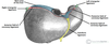
- What is the bare area?
- The visceral surface of the liver is covered with peritoneum, except at the?
- The Diaphragmatic surface of the liver is covered with visceral peritoneum, except posteriorly in the Bare Area of the liver, where it lies in direct contact with the diaphragm. There is a deep groove in the bare area, where the inferior vena cava travels.
- fossa for the gallbladder
- porta hepatis a transverse fissure where vessels (hepatic portal vein, hepatic artery and lymphatics) that supply and drain the liver enter & leave it.
- fossa for the gallbladder
Identify & describe the position of the left, right, caudate and quadrate lobes of the liver LO
- The liver is split into what lobes?
- The attachment of the ? divides the Right Lobe from the much smaller Left Lobe.
- On the visceral surface, the right and left sagittal fissures split the right lobe with the porta hepatis into the
- Describe in more detail the location of the caudate lobe
- Describe in more detail the location of the quadrate lobe
- What are the three types of hepatic recesses?

- Two anatomical ( R&L) & two accessory lobes (caudate and quadrate)
- Falciform ligament
- Quadrate (anterior and inferiorly)
Caudate Lobe (posteriorly and superiorly)
- Between the IVC & a fossa produced by the ligamentum venosum (a remnant of the fetal ductus venosus)
- between the gallbladder and a fossa produced by the ligamentum teres (a remnant of the fetal umbilical vein)
- Subphrenic spaces (left and right), Subhepatic space, Morisons pouch

- Where is the subphrenic recess located?
- Where is the subhepatic space recess located?
- Where is Morisons pouch located?
- Clinical significance
- between the diaphragm and liver, either side of the falciform ligament
- between the inferior surface of the liver & the transverse colon
- Posterosuperior aspect of the right subhepatic space,
Between the visceral surface of the liver & the right kidney
- Deepest part of the peritoneal cavity when supine (lying flat), and this is where fluid is likely to collect in a bedridden patienty
Identify and describe the structures (such as the hepatic portal vein, hepatic artery & bile duct) in the porta hepatis LO
- Where is the location of the porta hepatis?
- The liver has a unique dual blood supply:

- Hepatic artery proper – supplies the liver with arterial blood. It is derived from the coeliac trunk.
Hepatic portal vein – deoxygenated blood, nutrients (detoxification, metabolism)
Q. Which main veins drain into the hepatic portal vein?

- Which main veins drain into the hepatic portal vein?
- The Porta hepatis transmits the following (anterior to posterior order)
- common hepatic duct (leaving) - anterior, right
- hepatic artery proper (entering) - left
- hepatic portal vein (entering) - behind, between duct and artery
nerves and lymphatics
Sympathetic nerves - these provide afferent pain impulses from the liver and gall bladder to the brain. Pain may be referred to the lower pole of the right scapula (T7).
Hepatic branch of the vagus nerve (CN X).


