Skull Flashcards

(89 cards)

1
Q

Bregma of skull

A

at the junction of sagittal and frontal suture

2
Q

Glabella of Skull

A

medium elevation between eye brows (supraciliary arches)

3
Q

nasion of skull

A

at the junction of internasal and frontonasal sutures

4
Q

pterion

A

a circular area formed by the junction of

  1. frontal bone
  2. sphenoid bone
  3. parietal and squamous part of temporal bone
5
Q

What bones form the pterion?

A
  1. frontal bone
  2. sphenoid bone
  3. parietal and squamous part of temporal bone
6
Q

What does the pterion mark?

A

marks the anterior branch of the middle meningeal artery

7
Q

lambda

A

at the junction of the lambdoid and sagittal sutures

8
Q

What are the eight paired bones of the skull?

A

a. parietal - top of the skull
b. temporal - below parietal on the side of the skull
c. zygomatic - cheek bone
d. nasal - forming bridge of the nose
e. lacrimal - on the medial wall of the orbit
f. inferior concha - lower part of nasal cavity
g. palatine - posterior part of hard palate
h. maxilla - forming upper jaw

9
Q

What are the six unpaired bones of the skull?

A

a. frontal - forehead
b. ethmoid - forming medial wall of the orbit and lateral wall of the nasal cavity
c. occipital - posterior aspect of skull - surrounds foramen magnum
d. sphenoid - a bat shaped bone, in most part located deep in the skull
e. vomer - forming inferior posterior part of nasal septum
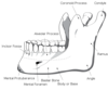
f. mandible - lower jaw

10
Q

What paired bones enclose the cranial cavity?

A

a. parietal - top of head
b. temporal - side of head

11
Q

What unpaired bones enclose the cranial cavity?

A

a. frontal - forehead region
b. occipital - back of head
c. sphenoid - base of cranial cavity
d. ethmoid - related to nose

12
Q

What are the paired bones of the face?

A

a. zygomatic - cheek bones
b. maxilla - upper jaw
c. nasal - nose
d. lacrimal - in orbit
e. palatine - roof of mouth
f. inferior concha - in nose

13
Q

What are the unpaired bones of the face?

A

a. vomer - nasal septum
b. mandible - lower jaw

14
Q

What is endochondral bone formation?

A

has a cartilaginous model before bone is
formed

15
Q

what is intramembraneous bone formation?

A

NO cartilaginous model
a. bone is formed directly from mesenchyme

16
Q

What type of joints join the bones of the skull?

A

fibrous type of
joints - synchondroses

17
Q

What are the three large important sutures of the skull?

A

a. sagittal suture - between parietal bones
b. coronal suture - between frontal and parietal bones
c. lambdoid suture - separates occipital bone from parietal and temporal
bones

18
Q

What important formina can be seen from the superior aspect of the skull, and what passes through here?

A

parietal - for emissary veins

19
Q

What bones can be see in the posterior region of the skull?

A
  1. squamous/flat part of occipital bone
  2. parietal
  3. mastoid process of the temporal bone
20
Q

What sutures can be seen from the posterior aspect of the skull?

A
  1. lambdoid
  2. parietomastoid
  3. occipitomastoid
21
Q

What is the median occipital crest?

A

it descends from external occipital
protuberance to the foramen magnum
a. provides attachment to ligamentum nuchae

22
Q

What bones can be seen from the lateral aspect of the skull?

A
  1. frontal
  2. parietal
  3. temporal
  4. temporalis process of zygomatic bone
  5. sphenoid bone
  6. occipital bone
  7. mandible
  8. maxilla
23
Q

What parts of the temporal bone can be seen when viewing the skull laterally?

A

a. squamous part - zygomatic process
b. tympanic part - external auditory meatus
c. mastoid process

24
Q

What part of the spenoid bone can be seen when viewing laterally?

A

a. greater wing (probably lateral plate as well)

25
What parts of the mandible can be seen from the lateral view of the skull?
a. condyle b. coronoid process c. ramus d. angle of mandible
26
What are the major structures of the lateral part of the skull?
1. zygomatic arch 2. temporal line 3. external auditory meatus
27
What is the temporal line of the skull?
begins at zygomatic process of frontal bone, arches backward across frontal and parietal bone a. provides attachment to temporalis fascia
28
What are the borders of the infratemporal fossa?
a. anterior - posterior surface of maxilla b. posterior - tympanic plate, mastoid and styloid process of the temporal bone c. superior/roof - infratemporal surface of the greater wing of the sphenoid d. inferior - the level where the medial pterygoid muscle attaches to the mandible near the angle e. medial - lateral pterygoid plate of the sphenoid bone f. lateral - ramus and coronoid process of mandible
29
Through what structures does the infratemporal fossa communicate with other regions of the skull?
a. superolaterally - temporal fossa through gap bounded by the zygomatic arch b. anteriorly - orbit through inferior orbital fissure c. medially - pterygopalatine fossa through pterygomaxillary fissure d. superiorly - middle cranial fossa through foramen ovale and foramen spinosum e. inferiorly - neck
30
Where is the pterygopalatine fossa located?
located below the apex of the orbit, between the pterygoid plates of the sphenoid and palatine bones. This fossa is difficult to see..
31
What are the boundaries of the pterygopalatine fossa?
a. superior - body of sphenoid, but mostly inferior orbital fissure b. anterior - posterior surface of maxilla (posterior wall of maxillary sinus) c. posterior - pterygoid process and the root of the greater wing of the sphenoid d. medial - perpendicular plate of palatine bone e. inferior - anterior and posterior walls come together and continue as pterygopalatine canal f. lateral - opens into pterygomaxillary fissure
32
Through what structures does the pterygopalatine fossa communicate with other regions of the skull?
a. superiorly - inferior orbital fissure to orbit b. inferiorly - greater and lesser palatine canals to palate c. posteriorly (1) foramen rotundum to middle cranial fossa (2) pterygoid canal to foramen lacerum HA-18-4 (3) pharyngeal canal to posterior nasal aperture d. medially - sphenopalatine foramen to nasal cavity e. laterally - pterygomaxillary fissure to infratemporal fossa f. anteriorly - none
33
What are the supracilliary arches?
extend laterally from glabella
34
What is the supraorbital notch?
in upper margin of orbit
35
What are the parts of the anterior maxilla?
body, zygomatic process, frontal process, palatine process, alveolar process, intermaxillary suture
36
What are the parts of the body of the maxilla?
(1) nasal surface - contributes to lateral wall of nasal cavity (2) orbital surface - forms the floor of the orbit (3) infratemporal surface - anterior wall of the infratemporal fossa (4) anterior surface - contains infraorbital foramen
37
What are the two parts of the zygomatic bone?
a. frontal process articulates with frontal bone b. temporal process - forms part of the zygomatic arch
38
What are the parts of the mandible?
body, ramus, condylar process, angle of mandible
39
What are the parts of the body of the mandible?
(1) symphysis menti - the median region (2) mental protuberance - triangular lower end of symphysis menti (3) mental foramen - below 2nd pre-molar (4) oblique line - run upward and backward from mental protuberance to anterior border of ramus (5) alveolar process - contains lower teeth (6) digastric fossa - near the symphysis
40
What are the two parts of the ramus of the mandible?
(1) mandibular notch - upper border of ramus (2) coronoid process - anterior to the notch
41
What are the two parts to the condylar process?
(1) head/condyle - part of the temporomandibular joint (2) neck of mandible - below the head
42
What are the strunctures on the interior of the mandible?
body and ramus
43
What are the important structures on the interior body of the mandible?
(1) genial tubercle/mental spine - behind symphysis (2) mylohyoid line - an oblique ridge that runs backward and upward HA-18-5 (3). submandibular fossa - below mylohyoid line
44
What are the important structures on the internal aspect of of ramus of the mandible?
1) mandibular foramen - in the middle of the ramus - leads to mandibular canal (2) lingula - medial projection on mandibular foramen (3) mylohyoid groove - from lingula to submandibular fossa
45
Describe the structure of each orbit.
a. each orbit is like a cone b. the medial walls of two orbits are parallel, whereas lateral walls are at right angles to each other
46
What forms the supraorbital margin?
formed by frontal bone
47
Where is the supraorbital notch (foramen)?
located at medial 1/3 of the margin
48
What forms the lateral margin of the orbit?
formed by zygomatic process of frontal and frontal process of zygomatic
49
What forms the infra-orbital margin of the orbit?
formed by the zygomatic bone and maxilla
50
What forms the medial margins of the orbits?
formed by maxilla, lacrimal, and frontal bones
51
Where is the anterior lacrimal crest?
on the frontal process of the maxilla
52
Where is the posterior lacrimal crest?
on the lacrimal bone
53
Where is the fossa for the lacrimal sac?
between anterior and posterior lacrimal crests
54
What is the nasolacrimal canal?
downward continuation of the fossa
55
What froms the superior wall of the orbit?
formed by orbital plate of frontal and lesser wing of sphenoid
56
Where is the fossa for the lacrimal gland?
at anterolateral angle of orbit
57
Where is the trochlear fossa of the orbit?
at anteromedial angle
58
Where is the optic canal?
between the two roots of lesser wings of sphenoid, situated at the extreme back of the roof (near the apex of the cone)
59
What forms the lateral wall of the orbit?
formed by the frontal process of the zygomatic bone and greater wing of sphenoid
60
Where is the location of the superiororbital fissure?
between lesser and greater wings of sphenoid
61
Where is the inferior orbital fissure supposed to be located?
between greater wing of sphenoid above and maxilla and palatine bones below
62
What forms the inferior wall of the orbit?
formed by zygomatic and maxilla bones and orbital process of the palatine bone
63
Where is the infra-orbital groove and canal?
continues from inferior orbital fissure
64
Where can you find the nasolacrimal canal?
continues from its opening in the medial margin
65
What froms the medial wall of the orbit?
formed by the orbital plate of ethmoid bone, lacrimal bone and frontal process of the maxilla and a small portion of the body of sphenoid bone
66
Which wall of the orbit is the thinnest?
medial wall
67
Where are the anterior and posterior ethmoidal foramina?
small foramina at the junction of the medial and the superior wall
68
What else is found in the medial wall of the orbit?
ethmoidal air cells
69
What bones form the inferior aspect of the skull?
occipital bone, temporal bone, sphenoid bone, posterior nasal aperture (choanae), vomer, palatine bone, maxilla,
70
What parts of the occipital bone are important on the inferior aspect of the skull?
a. foramen magnum, b. squamous (flat) part, c. lateral (condylar or jugular) part, d. basilar part - (body of occipital bone)
71
What structures are on the lateral (jugular or condylar part) of the occipital bone on the inferior aspect of the skull?
(1) occipital condyles - two large protuberances at the sides of foramen magnum (2) condylar fossa - behind each condyle (3) condylar canal - in the fossa - not seen in all skulls (4) hypoglossal canal - above the front of each condyle (5) jugular foramen - between the occipital and petrous part of temporal bones (6) jugular notch - at posterior edge of jugular foramen
72
What structures are seen on the basilar part of the occipital bone on the inferior aspect of the skull?
(1) pharyngeal tubercle - approximately in the center of the basilar part (2) foramen lacerum - on each side of the basilar part
73
What parts of the temporal bone can be seen on the inferior aspect of the skull?
squamous part, tympanic part, styloid part , mastoid part, petrous part/petrous portion of temporal bone, foramen lacerum
74
What structures are important on the squamous part of the temporal bone on the inferior aspect of the skull?
(1) zygomatic arch (2) mandibular fossa - for temporomandibular joint (3) articular tubercle - anterior to the fossa
75
What structures are important on the tympanic part of the temporal bone on the inferior aspect of the skull?
forms floor and anterior wall of the external auditory meatus (1) squamotympanic fissure - in the mandibular fossa separates squamous part from tympanic part (2) the fissure divides into the petrosquamous and petrotympanic fissures
76
What structures are important on the mastoid process of the temporal bone on the inferior portion of the skull?
(1) mastoid notch - situated medially (2) groove for occipital artery (3) stylomastoid foramen - located between styloid and mastoid processes
77
What are the important structures on the petrous part of the temporal bone on the inferior aspect of the skull?
(1) jugular foramen - anterior and lateral to foramen magnum (2) carotid canal - medial to styloid process and anterior to jugular foramen (3) foramen lacerum - opening between petrous part of the temporal, the greater wing of the sphenoid and the basilar part of the occipital bone
78
What does the sphenoid bone consist of?
consists of a body, lesser wings, greater wings and pterygoid processes
79
What are the important structures of the spenoid bone on the inferior aspect of the skull?
greater wings, pterygoid processes
80
What are the important structures seen on the greater wings of the spenoid bones on the inferior aspect of the skull?
(1) located lateral to lateral pterygoid plate and forms the roof of the infratemporal fossa (2) foramina (a) foramen ovale - posterior to lateral pterygoid plate (b) foramen spinosum - small hole located posterior to foramen ovale (3) spine of sphenoid - posterior to foramen spinosum
81
What are the important structures of the pterygoid process on the sphenoid bones on the inferior aspect of the skull?
from base of the bone extends inferiorly or downward on each side of greater wings (1) these lie behind the maxilla and lateral to posterior aperture of the nose (2) at the lower end, the processes are separated from each other by the pyramidal part of the palatine bone (3) divides into two plates (a) lateral pterygoid plate - articulates with tuber of maxilla (b) medial pterygoid plate - prolongs downward as the pterygoid hamulus
82
Where is the pterygoid fossa on the inferior aspect of the skull?
between lateral and medial pterygoid plates
83
Where is the scaphoid fossa of the skull?
a depression located at the base of the superior/upper end of the posterior border of the medial pterygoid plate
84
What is the choanae?
posterior opening of the nasal cavity
85
What is the vomer?
forms the posterior inferior part of the bony nasal septum
86
What are the important parts of the palatine bone?
a. perpendicular plate - located at medial aspect of posterior maxilla b. horizontal plate - two plates join to form the posterior part of the bony palate
87
What are the parts of the horizontal plate of the palatine bone?
(1) pyramidal process - at the junction of two pterygoid plates (2) cruciform suture - between palatine processes of maxilla and horizontal processes of palatine bone (3) greater and lesser palatine foramina - medial to last molar tooth
88
What are the important parts of the maxilla on the inferior aspect of the skull?
a. palatine process - extends horizontally to meet the opposite side process to form the anterior greater part of the hard palate b. incisive foramen - in midline behind incisive teeth (1) separates premaxilla (primary maxilla) from maxilla
89