SM MSK Anatomy - Upper Limb: Axilla, Shoulder, Arm, Forearm, Hand Flashcards
SM 222a, Lab 2, Lab 3, Lab 4, Lab 5,
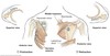
Which nerve might be injured by a fracture of the hook of the hamate?
Which functions would be compromised?
Ulnar nerve
- Finger abduction, adduction would be lost
- Interossei
- Flexion of 3rd-5th MCP, extension of 3rd-5th PIP and DIP would be weakened
- Lumbricals
- Thumb adduction
- Adductor pollicis
- Sensation over the skin of the medial 1.5 fingers, palm, and dorsum
- Cutaneous branches

Damage to which nerve would result in pain or sensory loss on the medial side of the forearm?
Medial cutaneous nerve
Note - if a cutaneous nerve is damaged, there will be no motor loss because it is a cutaneous nerve
(superficial branch of the ulnar nerve)
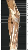
What structure is labeled by #8?

Long head of triceps brachii

This image shows the deepest layer of the anterior compartment of the arm
Which structure is labeled by #40?

Flexor digitorum profundus
(The only muscle that can flex the fingers at the DIP joints!)

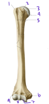
Identify #1:
What muscle attaches to #1?

1: Greater tubercle of the humerus
Supraspinatus, infraspinatus, and teres minor attach here

What is the function of the lumbricals of the hand?
What is their innervation?
Radialy deviate the fingers, flex MCP, extend PIP and DIP
Median nerve for 1-2
Ulnar nerve for 3-4
Following a dog bite to the arm, a patient reports a loss of sensation over the 5th digit (pinkie). Cutaneous branches of which nerve may have been injured?
Cutaneous branches of the ulnar nerve
When the arm is abducted 180 degrees, _____ degrees occurs by rotation of the scapula and ______ degrees occurs by rotation of the humerus at the shoulder joint
When the arm is abducted 180 degrees, 60** degrees occurs by rotation of the scapula and **120 degrees occurs by rotation of the humerus at the shoulder joint
The scapula and the humerus move in a 1:2 ratio
What innervates adductor pollicis?
Ulnar nerve
If the median nerve (or recurrent branch of the median nerve) is damaged, the abductor pollicis brevis will no longer work; the thumb will be pressed against the hand by adductor policis
Which muscles protract the scapula?
Serratus anterior
Pectoralis minor
Describe the structure labeled by #3
- Muscle:
- Function:
- Attachments:
- Innervation:

- Muscle: Brachioradialis
- Function: Flex the elbow
- Attachments: Humerus, distal radius
- Innervation: Radial nerve

Which vein is labeled by #2?

Cephalic vein
(Empties into the axillary vein)

A man suffering from entrapment of the ulnar nerve at the medial epicondyle gets a medial condylar osteotomy. During the procedure the muscular attachments to the medial epicondyle are accidentally cut. Which of the following muscles may have been damaged?
A) Supinator
B) Flexor carpi radialis
C) Brachioradialis
D) Extensor carpi ulnaris
E) Flexor pollicis longus
B) Flexor carpi radialis
Muscles of the superficial layer of the anterior forearm:
- Pronator teres (Creates the medial border of the cubital fossa)
- Flexor carpi radialis
- Palmaris longus
- Flexor carpi ulnaris

Which structure is labeled by #11?

Ulnar nerve
Travels closely with the ulnar artery (17)

Which nerve supplies the structures in purple (labeled #4)?

Medial cutaneous nerve of the forearm
(A branch from the brachial plexus)

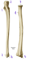
Which structure is labeled by #5?

Head of the ulna
Fig. 11.4 Adapted from Gilroy et al. Atlas of Anatomy, second edition, Figs. 22.1A, 22.1B.

The vessels labeled by #4 most recently originated from which vessel?

Subscapular artery
(Posterior = circumflex scapular artery, anterior = thoracodorsal artery)
Fig. 16.2 Adapted from Gilroy et al. Atlas of Anatomy, second edition, Fig. 24.1B.

Describe the muscle labeled by #6
- Muscle:
- Function:
- Attachments:
- Innervation:

- Muscle: Supinator
- Function: Supinate the forearm
- Attachments: Lateral epicondyle of humerus, radius
- Innervation: Radial nerve

Which structure is labeled by #1?

Surgical neck of the humerus

In the brachial plexus, the medial cord branches into the…
Ulnar and median nerves

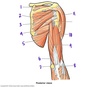
Describe the deltoid muscle
- Function:
- Innervation:
- Attachments
Deltoid
- Function: Arm abduction, flexion, internal rotation
- Innervation: Axiliary nerve (C5-C6)
- Attachments: Clavicle, acromion, scapular spine, humerus
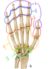
Together, the structures labeled #4 make up the…

Metacarpals
Fig. 11.5 Adapted from Gilroy et al. Atlas of Anatomy, second edition, Fig. 23.2.

Which structure is labeled by #4?

Coracoacromial ligament

Which structure is labeled by #7?

Pronator quadratus

Which vein is labeled by #2?
How does it enter the deep venous system?

Basilic vein
Empties into the brachial vein just above the cubital fossa

Describe thumb flexion/extension
Describe thrumb abduction/adduction
Which muscles accomplish these movements?
Starting with the hand in anatomical position:
- Flexion is moving the thumb across the palm
- Flexor pollicis brevis (thenar compartment)
- Flexor pollicis longus (anterior forearm)
- Extension is the opposite of flexion
- Extensor pollicis brevis (posterior forearm)
- Extensor pollicis longus (posterior forearm)
- Abduction is moving the thumb away from the palm
- Abductor pollicis brevis (thenar compartment)
- Abductor pollicis longus (posterior forearm)
- Adduction is the opposite of abduction
- Adductor pollicis (in the palm)

In the brachial plexus, the lateral cord branches into the…
Musculocutaneous and median nerves

Which bone is labeled by #7?

Radius (radial tuberosity)

Which structure is labeled by #6?

Medial head of triceps brachii

Which structure is labeled by #8?

Tendons of flexor digitorum superficialis

Which structure of the nervous system is labeled by #2?

Axillary nerve

Which actions are controlled by the musculocutaneous nerve?
- Flexion
- Shoulder
- Elbow
- Supination
- Biceps brachii
Which structure is labeled by #9?

Long head of triceps brachii

Which muscle is labeled by #3?
Which nerve supplies it?

Biceps brachii, short head
Musculocutaneous nerve

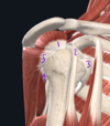
Which joint in the body has the largest range of motion?
Glenohumeral joint
Ball and socket joint between the head of the humerus and the glenoid of the scapula
List the visible muscles of the thenar compartment

- Abductor pollicis brevis (11)
- Flexor pollicis brevis (13)

Which muscles attach to the coracoid process?
Short head of biceps brachii
Coracobrachialis
Which muscles function to extend the wrist?
Which nerve innervates these muscles?
- Wrist extension only
- Extensor carpi radialis
- Wrist and digit extension
- Extensor digitorum
- Extensor pollicis
Radial nerve (posterior comartment of the forearm)

Which structure is labeled by #7?
What does it innervate?

Ulnar nerve
- Anterior forearm
- Flexor carpi ulnaris, flexor digitorum profundus
- Hand
- Intrinsic muscles of the hand

Why do most shoulder dislocations occur anteriorly?
The ligaments stabilizing the anterior part of the glenohumeral joint (SGHL, MGHL, IGHL) are very thin and lax - they don’t provide much stability
More stability is provided superiorly and posteriorly by the tendons of the rotator cuff muscles

Which structure is labeled by #4?
What is its innervation?

Triceps brachii (medial head)
Radial nerve

Describe teres major
- Function:
- Attachments:
- Innervation:
Teres major
- Function: Internally rotate, adduct, extend the arm
- Attachments: Scapula, humerus (lesser tuberosity)
- Innervation: Subscapular nerve

Which structure is labeled by #1?

Ulnar nerve
(Ulnar artery is next to it, but the pointer doesn’t quite reach)

If a patient cannot abduct their shoulder, which nerve is most likely injured?
Axillary nerve
Together, the structures labeled #3 make up the…

Proximal phalanges
Fig. 11.5 Adapted from Gilroy et al. Atlas of Anatomy, second edition, Fig. 23.2.
Which structure is labeled by #3?

Subscapularis
Part of the rotator cuff of the shoulder

Which structures are labeled by #2?
- Artery:
- Nerve:

- Artery: posterior humeral circumflex artery
- Nerve: Axillary nerve

Describe the structure of synovial tendon sheaths.
What is their function?
- Structure
- Visceral layer and parietal layer with synovial fluid in between
- Mesotendons provide routes for blood vessles to get to tendons
- Function
- Reduce the friction between the tendon and the surrounding are as it moves
Which muscles are in the deep layer of the anterior compartment of the forearm?
Flexor digitorum profundus
Flexor pollicis longus
Which cord from the brachial plexus forms the axillary nerve?
Posterior cord

What structure is labeled by #9?

Latissimus dorsi

Which vessel is labeled by #2?
Axillary artery
- Right subclavian -> Thoracoacromial (1) -> Axillary (2) -> Brachial (6)*
- Fig. 16.2 Adapted from Gilroy et al. Atlas of Anatomy, second edition, Fig. 24.1B.*

Which structure is labeled by #5?

Coracoacromial ligament

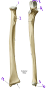
Which bone is labeled by #1?

Humerus

Which nerve supplies the structures in pink (labeled #3)?

Radial nerve (superficial branch)
Note - the superficial branch of the radial nerve only gives cutanous supply to the hand; loss of this nerve in the hand will result in loss of sensation on the dorsal surface and over the thenar compartment, but no functional deficits

Palmaris longus and flexor carpi ulnaris have been removed.
Which structure is labeled by #30?
What is its function?
Attachments?
Innervation?

Flexor digitorum superficialis
Flex the fingers at MCP and PIP
Medial epicondyle of the humerus, middle phalanges 1-4
Median nerve

Describe teres minor
- Function:
- Attachments:
- Innervations:
Teres minor
- Function: External rotation, weak adduction of the ar,
- Attachments: Scapula, greater tuberosity of the humerus
- Innervations: Axillary nerve
Part of the rotator cuff

Which structure is labeled by #10?

Radial artery

Which structure is labeled by #2?
What is its innervation?

Trapezius
Innervated by cranial nerve XI

Describe the posterior compartment of the forearm
- Muscles:
- Actions:
- Nerve:
- Atttachments:
Posterior component of the forearm
- Muscles:
- Wrist extensors - Extensor carpi radialis
- Wrist and digital extensors - extensor digitorum, extensor pollicis
- Forearm supinator - Supinator
- Thumb abductor - Abductor pollicis longus
- Actions: Extension of the wrist and/or digits, supination of the forearm, abduction of the thumb
- Nerve: Radial nerve
- Atttachments: Lateral epicondyle of the humerus

Which nerve supplies the structures in yellow (labeled #1)?

Median nerve

Which vein is labeled by #6?

Basilic vein

Which structure is labeled by #2?

Medial epicondyle of the humerus

Which ligament prevents the upward dislocation of the humerus?
Coracoacromial ligament

Which articulation connects the upper limb to the axial skeleton?
Sternoclavicular joint
The scapulothoracic articulation is free-floating; there are no attachments (not a true synovial joint)

Which structure is labeled by #1?

Supraspinatus
Part of the rotator cuff

Describe the structure labeled by #17
- Muscle:
- Function:
- Attachments:
- Innervation:

- Muscle: Extensor pollicis longus
- Function: Extend the thumb
- Attachments: Ulna, distal phalanx of the thumb
- Innervation: Radial nerve

What structure is labeled by #14?

Long head of biceps brachii?
(Short head = #13)

Which nerve innervates the compartment in blue?
Which muscles are in this compartment?

Ulnar nerve
Hypothenar muscles, medial two lumbricals, adductor pollicis

Which vessel is labeled by #6?

Brachial artery
- Right subclavian -> Thoracoacromial (1) -> Axillary (2) -> Brachial (6)*
- Fig. 16.2 Adapted from Gilroy et al. Atlas of Anatomy, second edition, Fig. 24.1B.*

What structure is labeled by #10?

Teres major

A man falls and suffers a dislocation of the greater tubercle of the humerus. Which actions would be most affected?
A) Abduction and lateral rotation
B) Protraction of the scapula
C) Flexion of the shoulder
D) medial rotation of the shoulder
E) Retraction of the scapula
Abduction and lateral rotation
The greater tubercle is the attachment site of:
- Supraspinatus (abductor)
- Infraspinatus (lateral rotator)
- Teres minor (lateral rotator)

What is the function of the palmar interossei muscles?
What is their innervation?
Adduct the fingers toward the middle finger
Ulnar nerve (deep branch)
What structure is labeled by #7?

Medial head of triceps brachii

Describe pectoralis major
- Function:
- Attachments:
- Innervation:
Pectoralis major
- Function: Shoulder adduction and flexion
- Attachments: Clavicle, sternum, greater tuberosity of humerus
- Innervation: Medial and lateral pectoral nerve

Which muscle supinates the forearm?
Which nerve innervates this muscle?
Supinator
Innervated by the radial nerve (posterior comartment of the forearm)

Which nerve innervates section 1 (red)?

Radial nerve (posterior cutaneous branch)

Which actions are controlled by the median nerve?
- Flexion
- Wrist
- Digits
- Thumb
- Pronation
- Thumb opposition, abduction
Together, structures #7 adn #8 are called….

7 = Trochlea (Articulates with the coronoid process of the ulna)
Condyle of the humerus

Which structure of the nervous system is labeled by #1?

Radial nerve

Which structure is labeled by #8?
What is its innervation?

Pectoralis minor
Medial pectoral nerve

Which muscle is labeled by #3?

Brachioradialis

Which muscles are forearm pronators?
Which nerve innervates these muscles?
- Pronator teres
- Pronator quadratus
Innervated by the median nerve (anterior compartment of the forearm)

Which structure is labeled by #11?
What is its innervation?

Deltoid
Axillary nerve

Which nerve innervates the deltoid and teres minor?
Axillary nerve

Which muscles are shown in this picture?
What is their function?
Attachments?

Lumbricals
- Originate from the tendons of flexor digitorum profundus
- Radially deviate the fingers
- Innervation
- Median nerve for the lateral two lumbricals
- Ulnar nerve for the medial two lumbricals
Describe the structure labeled by #13
- Muscle:
- Function:
- Attachments:
- Innervation:

- Muscle: Flexor pollicis brevis
- Function: Flex the thumb
- Attachments: Trapezium, flexor retinaculum, proximal phalanx of thumb
- Innervation: Median nerve (recurrent branch)

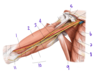
Which nerves innervate the fingertips?
Median nerve (thumb -> ring finger)
Ulnar nerve (ring finger -> pinkie)

Which structure is labeled by #1?

Head of the radius
Articulates with the capitulum of the humerus
Fig. 11.4 Adapted from Gilroy et al. Atlas of Anatomy, second edition, Figs. 22.1A, 22.1B.

Which muscle is labeled by #7?

Brachioradialis

Which nerves innervate the back of the arm?
Radial nerve - posterior cutaneous branch (red)
Musculocutaneous nerve (purple)

Which spinal nerves supply the brachial plexus?
C5-T1
Which structures are labeled by #1?

Anterior glenohumeral ligaments
(Superior, middle, and inferior)

Which nerve innervates the compartment in yellow?
Which muscles are in this compartment?

Median nerve
- Thenar muscles
- Abductor pollicis brevis, flexor pollicis brevis
- Lateral two lumbricals

Which structure is labeled by #7?

Anular ligament of the radius

Which structure is labeled by #5?

Surgical neck of the humerus

What functional deficit(s) would you expect to see in this patient?

Deficits in…
- Finger and wrist extension
- Sensory loss on the dorsal aspect of the forearm and dorsolateral hand
These deficits would result from damage to the radial nerve, which sits in the radial groove (in the mid-shaft of the humerus)
Function of the triceps brachii is usually spared, because is innervation is proximal to the fracture
What separates the compartments within any section of the upper and lower limb?
Deep fascia
Which actions are controlled by the axillary nerve?
- Shoulder abduction
- Lateral rotation
Together, the structures labeled #1 make up the…

Distal phalanges
Fig. 11.5 Adapted from Gilroy et al. Atlas of Anatomy, second edition, Fig. 23.2.

What structure is labeled by #1?

Biceps brachii

Which number labels teh radial artery?

8

Which number labels the radial nerve?

21

Which elbow flexor is in the posterior compartment of the forearm?
Brachioradialis
Which vessel is labeled by #5?

Profunda brachii artery
(Deep brachial artery)
Fig. 16.2 Adapted from Gilroy et al. Atlas of Anatomy, second edition, Fig. 24.1B.

Which numbers label the basilic vein?
Where does it enter the deep venous system?

Basilic vein = 6, 5, 2
Empties into the brachial vein just above the cubital fossa

Which structure is labeled by #8?
Capitellum of the humerus
Articulates with the head of the radius

Which structure is labeled by #6?

Greater tuberosity of the left humerus

Which structure is labeled by #2?

Olecranon process of the ulna
(The other side contains the trochlear notch, which articulates with the trochlea of the humerus)
Fig. 11.4 Adapted from Gilroy et al. Atlas of Anatomy, second edition, Figs. 22.1A, 22.1B.

What structure is labeled by #6?

Brachialis

If the median nerve is damaged, which digits can still be flexed?
4th and 5th digits
Ulnar nerve supplies medial tendons of flexor digitorum profundus
What structure is labeled by #3?

Acromion of the scapula

Which vein is labeled by #5?

Basilic vein

Which nerves innervate latissimus dorsi, teres major, and subscapularis?
Subscapular nerves

Which structure of the nervous system is labeled by #4?

Musculocutaneous nerve

Describe the structure of fibrous tendon sheaths
What is their function?
Connective tissue that wratp the synovial tendon sheaths against the bone
Act as pullies that hold the tendons against the bone

Which muscle is labeled by #1?

Latissimus dorsi
(The only spinal muscle that attaches to the humerus)

Which muscles are shown in this picture?
What is their function?
What is their innervation?

Dorsal interossei
Abduct the fingers away from the middle finger
Ulnar nerve (deep branch)
Which bone is labeled by #3?

Clavicle
Fig. 11.6 Adapted from Gilroy et al. Atlas of Anatomy, second edition, Fig. 20.2A.

What is the purpose of the acromioclavicular joint?
What type of joint is it?
The achromioclavicular joint (AC) anchors the clavicle to the scapula
Synovial joint - allows only small slidign movements between the acromion of the scapula and the distal end of the clavicle

Which muscles act to supinate the forearm
What are their innervations?
Supinator (posterior forearm, radial nerve)
Biceps brachii long head (anterior arm, musculocutaneous nerve)
Damage to which nerve results in “Ape hand”: adducted thumb with supinated, extended wrist and extended 1st and 2nd digits?
Damage to the median nerve
Describe the path of the radial nerve after it leaves the brachial plexus
Radial nerve (C5-6)
- Innervates the posterior arm
- Descends within the spiral radial groove in the humerus
- Passes anteriorly
- -> Superficial branch: cutaneous innervtion to the lateral dorsum of the hand
- -> Deep branch: Motor innervation to the posterior forearm
Which structure is labeled by #6?

Medial epicondyle of the humerus

Which vessel is labeled by #15?

Superficial palmar arch
(Gets most of its supply from the ulnar artery)
Fig. 16.2 Adapted from Gilroy et al. Atlas of Anatomy, second edition, Fig. 24.1B.

Which structure is labeled by #3?

Subscapular artery

Which structure is labeled by #4?
Which muscles attach here?

Medial epicondyle
Some wrist and digial flexors

Which nerve supplies the structures in green (labeled #5)?

Musculocutanous nerve
Via the lateral cutaneous nerve of the forearm; supplies the lateral half of the anterior suface of the forearm

Which nerves arise from the posterior cord of the brachial plexus?
What do they innervate?
Axillary nerve innervates the deltoid and teres minor
Radial nerve innervates the posterior compartments of the arm and forearm
Which muscles work to pronate the forearm?
Where are they located?
- Pronator teres
- Pronator quadratus
Both are in the flexor (anterior) compartment

Which vessel is labeled by #13?

Ulnar artery
Fig. 16.2 Adapted from Gilroy et al. Atlas of Anatomy, second edition, Fig. 24.1B.

Which nerve supplies the structures in blue (labeled #2)?

Ulnar nerve

Which muscle is labele by #6?

Biceps brachii
Fig. 11.6 Adapted from Gilroy et al. Atlas of Anatomy, second edition, Fig. 20.2A.

What is the flexor retinaculum?
Where does it attach?
The flexor retinaculum forms the anterior wall (“roof”) of the carpal tunnel
Hook of the hamate, pisiform, tubercle of the scaphoid, tubercle of the trapezoid

Which structure is labeled by #18?

Pronator teres
Most superficial muscle of the flexor goup, on the medial side of the forearm

Which arteries encircle the surgical neck of the humerus?
Anterior and posterior circumflex arteries
Branches from the brachial artery

Which vein is labeled by #5?
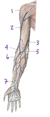
Median cubital vein
Connects the cephalic (lateral) and basilic (medial) veins

Which structure is labeled by #3?
What is its innervation?

Infraspinatus
Suprascapular nerve

Which vein is commonly used for blood draws and IV line placement?

Median cubital vein - #3

Which boney structure is labeled by #5?

Olecranon of the ulna
Fig. 11.6 Adapted from Gilroy et al. Atlas of Anatomy, second edition, Fig. 20.2A.

List the muscles of the hypothenar compartment
What innervates them?

- Abductor digiti minimi (35)
- Flexor digiti minimi brevis (36)
Ulnar nerve (deep branch)

If a patient cannot pronate their hand, which nerve is most likey injured?
Median nerve
Which muscle is labeled by #5?
Which nerve supplies it?

Brachialis
Musculocutaneous nerve

Which vessel is labeled by #7?

Lateral thoracic artery
Fig. 16.2 Adapted from Gilroy et al. Atlas of Anatomy, second edition, Fig. 24.1B.

If a patient cannot flex their ring or pinkie finger, which nerve is most likely damaged?
Ulnar nerve
Whihc structure is labeled by #10?

Radial nerve

Describe triceps brachii
- Function:
- Attachments:
- Innervation:
Triceps Brachii
- Function: Extend shoulder and forearm
- Attachments:
- Long head: Scapula, olecranon process of the ulna
- Lateral head: Humerus, olecranon process of the ulna
- Medial head: Humerus, olecranon process of the ulna
- Innervation:
- Radial nerve (C6 to C8)
Which articulation is labeled by #5?

Scapulothoracic “joint” (free floating, no attachment)

Which boney structure is labeled by #4?

Medial epicondyle of the humerus
Fig. 11.6 Adapted from Gilroy et al. Atlas of Anatomy, second edition, Fig. 20.2A.

Which structure is labeled by #4?

Coronoid process of the ulna
Fig. 11.4 Adapted from Gilroy et al. Atlas of Anatomy, second edition, Figs. 22.1A, 22.1B.

Which structure is labeled by #6?

Styloid process of the ulna
Fig. 11.4 Adapted from Gilroy et al. Atlas of Anatomy, second edition, Figs. 22.1A, 22.1B.

Which muscles functions to abduct the thumb?
Which nerve innervates these mucles?
- Abductor policis longus
- Radial nerve (posterior comartment of the forearm)
- Abductor pollicis brevis
- Recurrent branch of the median nerve (thenar compartment)

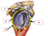
Which structure is labeled by #8?

Glenohumeral joint
(Glenoid fossa of the scapula + head of humerus)

Which structure is labeled by #2?

Infraspinatus

Which muscle is labeled by #4?
Which nerve supplies it?

Biceps brachii, long head
Musculocutaneous nerve

Which structure is labeled by #10?
What innervates it?

Triceps brachii
Radial nerve

Which structure is labeled by #10?

Flexor retinaculum

Describe the subscapularis
- Function:
- Attachments:
- Innervation:
Subscapularis (anterior part of the rotator cuff)
- Function: Internally rotate the arm at the shoulder joint
- Attachments: Scapula, humerus (lesser tuberosity)
- Innervation: Subscapular nerve

Which structure is labeled by #13?

Brachial artery
Splits into the radial (10) and ulnar (17) arteries

Which structure is labeled by #9?

Axillary artery and vein

Which structure is labeled by #6?

Radial artery

Which number labels the median nerve?
Normally, which muscles is it deep to? Superficial to?

7
Deep to flexor digitorum superficialis
Superficial to flexor digitorum profundus

Which structure is labeled by #17?

Ulnar artery

Which structure is labeled by #11?
What does it innervate?

Ulnar nerve
- Anterior forearm
- Flexor carpi ulnaris, flexor digitorum profundus
- Hand
- Intrinsic muscles of the hand

Which structure is labeled by #6?
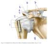
Acromioclavicular joint
(covered by the acromioclavicular ligament)

Describe pectoralis minor
- Function:
- Attachments:
- Innervation:
Pectoralis minor
- Function: Stabilize scapula against the body wall
- Attachments: Coracoid process, ribs 3-5
- Innervation: Medial pectoral nerve

Loss of extension in the digits and wrist is most likely the result of damage to which nerve?
Radial nerve
Which nerve follows the radial artery?
Superficial branch of the radial nerve (23)

In the upper arm…
- The coracobrachialis works on the ______ joint
- The brachialis works on the ____ joint
- The biceps brachii works on the _____ joint
In the upper arm…
- The coracobrachialis works on the shoulder joint
- The brachialis works on the elbow joint
- The biceps brachii works on the shoulder and elbow joint
List the muscles in the deep posterior forearm

- Extensor indicis (19)
- Extensor pollicis longus (17)
- Extensor pollicis brevis (18)
- Abductor pollicis longus (16)
- Supinator (6)

Which structure is labeled by #3?

Flexor pollicis longus

List the muscles of the rotator cuff and their actions
- Supraspinous - abduction
- Subcapsularis - medial rotation
- Infraspinatus - lateral rotation
- Teres minor - lateral rotation
What is a “sprain”?
Damage to a ligament
Graded based on the degree of the tear and resulting joint instability
Which structure is labeled by #8?
(Not the ligements, but what they are attaching to)

Glenoid labrum

List the medial rotators of the shoulder joint
What innervates them?
- Teres major - Subscapular nerve
- Subscapularis - Subscapular nerve
- Part of the rotator cuff
Damage to which nerve results in loss of thumb function and thenar atrophy?
Median nerve (recurrent branch)
Which vessel is labeled by #11?

Radial artery
Fig. 16.2 Adapted from Gilroy et al. Atlas of Anatomy, second edition, Fig. 24.1B.

What muscle is labeled by #3?

Teres minor
(part of rotator cuff)

Which structure is labeled by #7?

Coracoclavicular joint
(Made up of the coracoclavicular ligaments)

Identify the scaphoid

Fig. 11.5 Adapted from Gilroy et al. Atlas of Anatomy, second edition, Fig. 23.2.

The structures highlighted in green are the attachment points for which other structure?
List the names of the attachment points

Attachments for the flexor retinaculum (the roof of the carpal tunnel)
- Hook of the hamate
- Pisiform
- Tubercle of the trapezium
- Tubercle of the scaphoid

Describe infraspinatus
- Function:
- Attachments:
- Innervations:
Infraspinatus
- Function: Externally rotate the arm
- Attachments: Scapula, greater tuberosity
- Innervations: Suprascapular nerve
Part of the rotator cuff

Which structure of the nervous system is labeled by #3?

Median nerve
(Lateral and Medial cord join together)

Which vein is labeled by #4?

Cephalic vein

Which structure is labeled by #7?
What is its innervation?

Pectoralis major
Medial and lateral pectoral nerves

Which structure is labeled by #1?

Glenoid cavity of the scapula

Which joint in the body is most commonly dislocated?
Glenohumeral
Ball and socket joint of the shoulder, between the head of the humerus and the glenoid of the scapula
Which structure is labeled by #18?

Palmaris longus

Ligaments attach _______ to ________
Ligaments attach bone** to **bone
Which structure is labeled by #1?
Which muscles attach here?

Greater tubercle of the humerus
Supraspinatus, infraspinatus, teres minor (all rotator cuff muscles except subscapularis, which inserts on the lesser tubercle)

Which structure is labeled by #5?

Subclavian artery and vein

Which structure is labeled by #3?

Sternoclavicular joint

What muscle tendon is the examiner testing?

A) Triceps brachii
B) Coracobrachialis
C) Deltoid
D) Biceps brachii
E) Teres major
D) Biceps brachii
Supplied by the musculocutaneous nerve, C5-C7
Which structure is labeled by #6?
Which bone articulates with this structure?

Trochlea
Trochlear notch of the ulna

Which cord from the brachial plexus forms the radial nerve?
Posterior cord

Which numbers label the cephalic vein?
Where does it enter the deep venous system?

Cephalic vein = 7, 4, 1
Empties into the axillary vein at the deltopectoral triangle

A neoplasm of the acromion process completely compresses the axillary artery. Yet the patient has a normal radial pulse. What anastomosis explains this finding?
- Anterior and posterior humeral circumflex arteries
- Subscapular and brachial arteries
- Subscapular and posterior humeral circumflex arteries
- Suprascapular and subscapular arteries
- Thoracoacromial and subclavian arteries
d. Suprascapular and subscapular arteries

Which muscle is labeled by #18?
Perspective: imagine the elbow is coming out of the screen and the shoulder is going into the screen

Triceps brachii

Which nerve innervates section 5 (blue)?

Ulnar nerve

Together, the structures labeled #2 make up the…

Middle phalanges
Fig. 11.5 Adapted from Gilroy et al. Atlas of Anatomy, second edition, Fig. 23.2.

Which structure is labeled by #1?

Greater tubercle of the humerus

Which structure is labeled by #2?

Coracohumeral ligament

What structure is labeled by #12?

Subscapularis
What is the examiner doing here?

A. Testing the brachial pulse
B. Testing the femoral pulse
C. Testing the radial pulse
D. Testing the ulnar pulse
E. Testing the axillary pulse
C. Testing the radial pulse
Which vessel is labeled by #8?

Superior and inferior ulnar collateral arteries
Fig. 16.2 Adapted from Gilroy et al. Atlas of Anatomy, second edition, Fig. 24.1B.

Which articulations anchor the scapula to the clavicle?
Acromioclavicular (AC) - synovial joint
Coracoclavicular (CC) - fibrous joint

Which nerves are involved in pronation of the elbow?
Which compartment is this?
Median nerve
Anterior compartment of the forearm
- Forearm pronators – pronator teres, pronator quadratus

Which structure is labeled by #16?
Which muscles originate here?
Where do they attach?

Medial epicondyle of the humerus
- Flexor carpi radialis (4) -> 2nd and 3rd Metacarpals
- Flexor digitorum superficialis (6?) -> Middle phalanges 1-4
- Underneath palmaris longus and flexor carpi ulnaris
- Flexor carpi ulnaris (19) -> pisiform, hook of hamate
- Pronator teres (17) -> Radius
- Palmaris longus (18) -> Palmar aponeurosis
- not everyone has this!

Which number labels the infraspinatus?

3

Which number labels the long head of triceps brachii?

10

Which structure is labeled by #7?
What is its innervation?

Teres major
Lower subscapular nerve

Which muscles retract the scapula?
Middle trapezius
Rhomboids

Which muscle is labeled by #2?
Which nerve supplies it?

Latissimus dorsi
Ventral rami of a spinal nerve
(Latissimus dorsi is an extrinsic back muscle)

Which structure is labeled by 9?
Which structure is labeled by 23?
What do they innervate?

9 - superficial branch of the radial nerve; cutaneous innervation to the lateral dorsum of the hand
23 - deep branch of the radial nerve; supplies the muscles of the posterior forearm (extensors)

Which structure is labeled by #2?

Radial tuberosity
Fig. 11.4 Adapted from Gilroy et al. Atlas of Anatomy, second edition, Figs. 22.1A, 22.1B.

Which structure is labeled by #20?

Ulnar artery

Why doesn’t the middle finger have a palmar interosseus muscle?
Movement in either direction is accomplished by the dorsal interosseus muscles of the middle finger

Which vein empties into the axillary vein?
Cephalic vein (#7, 4, 1)

Which structure is labeled by #17?

Pronator teres

Which structure is labeled by #7?

Olecranon fossa of the humerus
The olecranon process of the ulna sits in here

Which structure is labeled by #7?

Radial groove of the humerus
Radial nerve sits in here
This is why humeral shaft fractures can cause damage to the radial nerve

Which nerve innervates the intrinsic muscles of the hand?
What are the consequences if this nerve is damaged?
Ulnar nerve (deep branch)
Loss of interossei (dorsal and palmar) results in loss of extension at PIP and DIP, loss of flexion at MCP
The result flexion at PIP and DIP, extension at MCP
(Extensor digitorum is till intact)

Which structure is labeled by #7?

Lesser tuberosity of the left humerus

Which cords of the brachial plexus form the median nerve?
Lateral and medial cords

Which structure of the nervous system is labeled by #5?

Posterior cord

Which articulation is labeled by #1?

Acromioclavicular joint

Which structure is labeled by #2?

Acromion of the scapula

Which structure is labeled by #5?

Coracoid process of the left scapula

Which structure is labeled by #3?

Head of the humerus

Which muscle is outlined in blue?
What is its function?
Innervation?
Attachments?

Flexor digiorum profundus
Flex fingers at MCP, PIP, and DIP
Median nerve (lateral 1/2), Ulnar nerve (medial 1/2)
Ulna, distal phalanges 1-4

Which structure is labeled by #1?

Coracoclavicular joint

Conoid ligament (left) + Trapezoid ligament (right)
Which structure is labeled by #7?

Median nerve
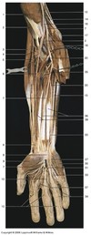
Which compartment is shown in this picture?
What are its primary actions?
What is its innervation?

Anterior compartment of the forearm
Wrist flexion, finger flextion, forearm pronation
Median nerve

Which vessel is labeled by #5?

Median cubital vein
Fig. 11.6 Adapted from Gilroy et al. Atlas of Anatomy, second edition, Fig. 20.2A.

Which structure is labeled by #1?

Supraspinatus

Which boney structure is labeled by #2?

Coracoid process of the scapula
Fig. 11.6 Adapted from Gilroy et al. Atlas of Anatomy, second edition, Fig. 20.2A.

The medial cutaneous nerve of the forearm is a terminal branch off which cord of the brachial plexus
Medial cord

Which structure is labeled by #7?

Trochlea of the humerus
Articulates with the coronoid process of the ulna

Which nerve is compressed in carpal tunnel syndrome?
What are the consequences?
The median nerve is compressed
Usually compression is due to repetitive use of the fingers; the tendons of flexor digitorum superficialis and flexor digitorum profundus run through the carpal tunnel. Overuse results in increased serous fluid, which leads to compression of the median nerve
- Loss/reduction of thumb function
- The thenar compartment is innervated by the recurrent branch of the medial nerve, which branches right after the nerve emerges from the carpal tunnel
- Pain in the cutaneous distribution of the median nerve
- Lateral palm of the hand

Which structure is labeled by #10?

Lateral head of triceps brachii

Which structure is labeled by #2?

Acromioclavicular joint
(Covered by the acromioclavicular ligament)
Note: This is the left scapula, anterior view

Which structure is labeled by #4

Greater tubercle of the humerus

Which nerve innervates section 4 (orange)?

Median nerve

Which strucutre is labeled #6?

Radius
Fig. 11.5 Adapted from Gilroy et al. Atlas of Anatomy, second edition, Fig. 23.2.

Which structure is labeled by #1?

Brachial artery

Which structure is labeled by #1?
What is its innervation?

Supraspinatus
Suprascapular nerve

Which structure is labeled by #14?

Median nerve

Describe biceps brachii
- Function:
- Attachments:
- Innervation:
Biceps brachii
- Function: Shoulder flexion, elbow fexion, supination
- Attachments:
- Short head: Coracoid process, radial tuberosity
- Long head: Supraglenoid tubercle, radial tuberosity
- Innervation: Musculocutaneous nerve
- Same as all muscles of the anterior compartment of the arm

Which structure is labeled by #4?

Radial tuberosity
Fig. 11.4 Adapted from Gilroy et al. Atlas of Anatomy, second edition, Figs. 22.1A, 22.1B.

Which structure of the nervous system is labeled by #6?

Medial cord

This image shows the deepest layer of the anterior compartment of the arm
Which structure is labeled by #14?

Tendon of flexor pollicis longus
(Muscle is 37)

Which structure is labeled by #5?

Abductor pollicis longus

Which vessel is labeled by #14?

Deep palmar arch
Gets most of its supply from the radial artery
Fig. 16.2 Adapted from Gilroy et al. Atlas of Anatomy, second edition, Fig. 24.1B.

Which nerve is labeled by #7?

Median nerve
Will go on to innervate most of the anterior compartment of the forearm

Which nerves innervate the body wall?
Spinal nerves
- Ventral ramus
- Extrinsic back muscles
- Intercostal muscles
- Limbs (extensions of the body wall) via plexi
- Anterior and lateral cutaneous nerves
- Skin and superficial fascia
- Dorsal ramus
- Intrinsic back muscles
- Posterior cutaneous nerves
- Skin and superficial fascia

Loss of senasation in the pinkie finger indicates injury to which nerve?
Ulnar

List the pathway of the vein on the radial side of the upper limb, from its most proximal point to its entry into the deep venous system
- Doral Arch
- Cerphalic Vein
- Axillary vein (part of deep venous system)

Which structures are labeled by #4?
- Artery:
- Nerve:

- Artery: Profunda brachii (Deep brachial artery)
- Nerve: Radial

Which muscles function to extend the wrist and digits?
Which nerve innervates these muscles?
- Extensor digitorum
- Extensor pollicis
Innervated by the radial nerve (posterior comartment of the forearm)

Which structure is labeled by #3?

Brachioradialis

What part of the shoulder is injured in a separated shoulder?
The ligaments of the acromioclavicular (AC) and/or coracoclavicular (CC) joints
Severity of the injury depends on whether they are stretched or torn

Which muscle is labeled by #3?

Lateral head of triceps brachii
Fig. 11.6 Adapted from Gilroy et al. Atlas of Anatomy, second edition, Fig. 20.2A.

Which cord from the brachial plexus forms the ulnar nerve?
Medial cord

Which structure is labeled by #20?
Ulnar artery

Which cord from the brachial plexus forms the musculocutaneous nerve?
Lateral cord

Which number labels the axillary nerve?

9

Which muscles are shown in this picture?
What is their function?
What is their innervation?

Palmar interossei (deep to the lumbricals)
Adduct the fingers toward the middle finger
Ulnar nerve (deep branch)
Which vessel is labeled by #10?

Posterior interosseous artery
Fig. 16.2 Adapted from Gilroy et al. Atlas of Anatomy, second edition, Fig. 24.1B.

Which nerve is labeled by #4?
It is a continuation of which nerve?

Lateral cutaneous nerve of the arm
Continuation of the musculocutaneous nerve

A girl falls on her outstretched hand. She experiences wrist pain and points to area between the extensor pollicis longus and abductor pollicis longus tendons.
Which bone is most likely broken?
What is a possible complication of this injury?
Scaphoid
Blood supply to the scapoid is through the distal side; a break in the scaphoid could cut off blood supply to the proximal scaphoid, resulting in avascular necrosis

Which vein is labeled by #3?

Basilic vein
(Empties into the brachial vein)

Which boney structure is labeled by #6?

Styloid process of the radius
Fig. 11.6 Adapted from Gilroy et al. Atlas of Anatomy, second edition, Fig. 20.2A.

List the pathway of the vein on the ulnar side of the upper limb, from its most proximal point to its entry into the deep venous system
- Dorsal arch
- Basilic vein
- Brachial vein (part of the deep venous system)

Describe the serratus anterior
- Function:
- Attachments:
- Innervation:
Serratus anterior
- Function: Protract the scapula, lower a raised arm, rotate the scapula laterally
- Attachments: Scapula, ribs
- Innervation: Long thoracic nerve

Describe the structure labeled by #36
- Muscle:
- Function:
- Attachments:
- Innervation

- Muscle: Flexor digiti minimi brevis
- Function: Flex the 5th digit
- Attachments: Hook of hamate, proximal phalanx
- Innervation: Ulnar nerve (deep branch)

Which articulation is labeled by #3?

Glenohumeral joint

Which nerve innervates section 2 (purple)?

Musculocutaneous nerve

Which number labels flexor digitorum superficialis?

30

Which structure is labeled by #6?
What is it innervated by?

Subscapularis
Subscapular nerve

Which structure is labeled by #3?

Extensor carpi radialis

Which structure is labeled by #21?

Common interosseous artery
It branches into the anterior (23) and posterior interosseous arteries

Which vein is labeled by #3?

Median cubital vein
Connects the cephalic and basilic veins
Median cubital vein is often used for blood draws and IV line placement

Which structure is labeled by #4?

Teres major

Which structure is labeled by #2?

Coracoacromial ligament

Which structure is labeled by #8?

Flexor retinaculum
(Roof of the carpal tunnel)

Which structure is labeled by #5?
Which bone articulates with this structure?

Capitellum
Head of the radius

Which number labels the ulnar nerve?
Which muscles of the anterior compartment does it supply?

13, 27
Flexor digitorum profundus (medial 1/2)
Flexor carpi ulnaris
Note: does not pass under the flexor retinaculum => it is vulnerable to cuts of the wrist

Which vessels are labeled by #3?

Anterior and posterior humeral circumflex arteries
Fig. 16.2 Adapted from Gilroy et al. Atlas of Anatomy, second edition, Fig. 24.1B.

Which structure is labeled by #4?

Brachial artery

Which structure is labeled by #3?

Coracoid process of the right scapula

In the brachial plexus, the posterior cord branches into the…
Axillary and radial nerves

What structure is labeled by #15?

Supraspinatus?

Which muscle is labeled by #7?
Which nerve supplies it?

Subscapularis
Subscapular nerve

Which vein is labeled by #7?

Cephalic vein

Which vein is labeled by #1?
How does it enter the deep venous system?

Cephalic vein
Empties into the axillary vein at the deltopectoral triangle

What is the purpose of the coracoclavicular joint?
What type of joint is it?
The coracoclavicular joint (CC) anchors the clavicle to the scapula
Fibrous joint

Which structure is labeled by #2?

Median nerve

Which structure is labeled by #2?

Lesser tubercle of the humerus

Which structures are supplied by the suprascapular nerve?
Supraspinatus
Infraspinatus
(Muscles of the rotator cuff)

Which structure is labeled by #6?

Radial collateral ligament

Which number labels the deep brachial artery?

20

Which structure is labeled by #9?

Radial artery

Which vessel is labeled by #24?
Perspective: imagine the elbow is coming out of the screen and the shoulder is going into the screen

Brachial artery

Which structure is labeled by #4?

Teres minor
Part of the rotator cuff of the shoulder

Which structure is labeled by #5?

Lateral epicondyle of the humerus

Describe the anterior compartment of the forearm
- Muscles:
- Actions:
- Nerve:
- Atttachments:
Anterior component of the forearm
- Muscles:
- Wrist flexors - Flexor carpi radialis
- Wrist and digital flexors - Flexor digitorum superficialis, flexor digitorum profundus, flexor pollicis longus
- Forearm pronators - Pronator teres, pronator quadratus
- Actions: Flexion of the wrist and/or digits, pronate the forearm
- Nerve: Median nerve
- Ulnar nerve for flexor digitorum profundus and flexor carpi ularis only
- Atttachments: medial epicondyle of the humerus

Which structure is labeled by #2?

Anatomical neck of the humerus

What structure is labeled by #4?

Brachial artery

Which structure is labeled by #5?
Infraspinatus
Part of the rotator cuff of the shoulder

Describe the structure labeled by #14
- Muscle:
- Function:
- Attachments:
- Innervation:

- Muscle: Extensor carpi radialis longus (Brevis is 15)
- Function: Extend the wrist
- Attachments: Lateral condyle of the humerus, 2nd metacarpal
- Innervation: Radial nerve

Describe coracobrachialis
- Function:
- Attachments:
- Innervation:
Coracobrachialis
- Function: Shoulder flexion, adduction, internal rotation
- Attachments: Coracoid process, humerus
- Innervation: Musculocutaneous nerve
- Same as all muscles of the anterior compartment of the arm

Which muscle is labeled by #1?

Deltoid
Fig. 11.6 Adapted from Gilroy et al. Atlas of Anatomy, second edition, Fig. 20.2A.

Describe supraspinatus
- Function:
- Attachments:
- Innervations:
Supraspinatus
- Function: Abduct the arm
- Attachments: Scapula, greater tuberosity
- Innervations: Suprascapular nerve
Part of the rotator cuff

Which nerve innervates section 3 (pink)?

Radial nerve (superficial branch)

What structure is labeled by #5?

Ulnar nerve

Which structure is labeled by #4?

Acromion of the right scapula

Which nerve is labeled by #7?
What does it innervate?

Superficial branch of the radial nerve
Sensory nerve that innervates the lateral half of the back of the hand (not including the fingertips)

Which number labels the common interosseous artery?

21
Comes from the ulnar artery (17)
Branches into the anterior (23) and posterior interosseous arteries
Note: #14 is the median nerve

Which structure is labeled by #3?

Teres minor

Which nerves are involved in flexion of the wrist and fingers?
Which compartment is this?
Median and ulnar nerves
Anterior compartment of the forearm
- Wrist flexors – Flexor carpi radialis, flexor carpi ulnaris
- Wrist and digital flexors – Flexor digitorum superficialis, flexor digitorum profundus (ulnar), flexor pollicis longus

Describe brachialis
- Function:
- Attachments:
- Innervation:
Brachialis
- Function: Elbow flexion
- Attachments: Humerus, ulnar tuberosity
- Innervation: Musculocutaneous nerve
- Same as all muscles of the anterior compartment of the arm

Which structure is labeled by #5?
What does it innervate?

Musculocutaneous nerve
Innervates the anterior compartment of the arm (biceps brachii, coracobrachialis, brachialis)
Which structure is labeled by #3?

Head of the radius
Fig. 11.4 Adapted from Gilroy et al. Atlas of Anatomy, second edition, Figs. 22.1A, 22.1B.

Which structure is labeled by #11?

Deltoid

Which structure is labeled by #5?
What is its innervation?

Triceps brachii (Lateral head)
Radial nerve

Which nerve is responsible for extension of the shoulder, elbow, wrist, and fingers?
Which compartments are involved?
Radial nerve
Posterior arm, posterior forearm

An infection in which synovial digital sheath could result in the most serious complication?
Synovial digital sheath of the 5th digit
This sheath is continuous with the common flexor synovial sheath, which contains the tendons of the wrist; the infection could spread far

Which vessel is labeled by #12?

Anterior interosseous artery
Fig. 16.2 Adapted from Gilroy et al. Atlas of Anatomy, second edition, Fig. 24.1B.

Which structure is labeled by #6?

Ulnar artery and nerve

Which structure is labeled by #7?

Medial epicondyle of the humerus

What structure is labeled by #4?

Glenoid cavity of the scapula

The head of the humerus articulates with the
_____ of the _____
The head of the humerus articulates with the
glenoid** of the **scapula

What is cubital tunnel syndrome?
Irritation of the ulnar nerve as it crosses the medial epicondyle, passing through the two heads of flexor carpi ulnaris (the cubital tunnel)
This leads to sensation similar to hitting your funny bone

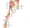
Label the indicated components of the medial plexus
- 7.

- Lateral cord
- Musculocutaneous nerve
- Lateral cord’s contribution to the median nerve
- Median nerve
- Ulnar nerve
- Medial cord’s contribution to the median nerve
- Medial cord
Not pictured: Posterior cord, which gives rise to the axillary nerve and radial nerve

Which structure is labeled by #3?

Coracoid process of the scapula

Describe the structure labeled by #16
- Muscle:
- Function:
- Attachments:
- Innervation:

- Muscle: Abductor pollicis longus
- Function: Abduct the thumb
- Attachments: Ulna, radius, interosseus membrane, 1st metacarpal
- Innervation: Radial nerve

Which structure is labeled by #8?

Medial epicondyle of the humerus

Which structure is labeled by #10?

Cephalic vein
(Joins the deep venous system via the axillary vein)

Which vein is labeled by #19?
Perspective: imagine the elbow is coming out of the screen and the shoulder is going into the screen

Cephalic vein

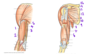
Which injury is showin in both Picture A and Picture B?
Which structures are compromised in each picture?

Both: Separated shoulder
Separation of the clavicle from the scapula
- A: Dislocation of acromioclavicular joint
- B: Dislocation of acromioclavicular joint with rupture of the coracoclavicular ligament

Which structure is labeled by #1?

Axillary artery

Which structure is labeled by #4?

Flexor digitorum profundus

Loss of sensation in the tips of the thumb, index, and ring fingers indicates injury to which nerve?
Median

Which structure is labeled by #7?

Head of the ulna
Fig. 11.4 Adapted from Gilroy et al. Atlas of Anatomy, second edition, Figs. 22.1A, 22.1B.

Which muscle of the rotator cuff stabilizes the shoulder anteriorly?
Subscapularis

Describe the structure labeled by #27
- Muscle:
- Function:
- Attachments:
- Innervation:

- Muscle: Adductor pollicis longus transverse head
- Oblique head is #26)
- Function: Adduct the thumb
- Attachments: Third metacarpal, proximal phalanx of thumb
- Innervation: Ulnar nerve (C8-T1)

Identify the labeled structures
- A:
- B:
- C:
- D:

- A: Deltoid muscle
- B: Head of the Humerus
- C: Supraspinatus muscle
- D: Glenoid of the scapula

Which structure is labeled by #6?

Radial artery

Which muscles flex the wrist?
Which nerve innervates these muscles?
- Wrist only flexors
- Flexor carpi radialis (Median nerve)
- Flexor carpi ulnaris (Ulnar nerve)
- Wrist and digital flexors
- Flexor digitorum superficialis (Median nerve)
- Flexor digitorum profundus (Median nerve + Ulnar nerve)
- Flexor pollicis longus (Median nerve)
All are part of the anterior component of the forearm

Which structure is labeled by #3?

Head of the humerus

Describe the structure labeled by #7
- Muscle:
- Function:
- Attachments:
- Innervation:

- Muscle: Extensor digitorum
- Function: Extend the wrists and digits
- Attachments: Lateral epicondyle of the humerus, middle and distal phalanges
- Innervation: Radial nerve

What structure is labeled by #1?

Scapular spine

Which muscle is labeled by #9?
Which nerve supplies it?

Supraspinatus
Suprascapular nerve

Which numbers together make up the biceps muscle?

3 and 4

Which structure is labeled by #36?

Flexor digitorum profundus
(Sections in the forearm are more distict than FDS)

Which structure is labeled by #17?

Ulnar artery
Relevant branches:
- Common interosseous (21)
- Will go on to supply the majority of the blood for the superficial palmar arch

Which structure of the nervous system is labeled by #7?

Ulnar nerve

Which structure is labeled by #3?

Brachioradialis
Most superficial extensor; on the lateral side of the anterior forearm

Which structure is labeled by #1?

Deltoid

Which vessel is labeled by #29?
Perspective: imagine the elbow is coming out of the screen and the shoulder is going into the screen

Basilic vein

Which structure is labeled by #2?

Brachialis
Part of anterior compartment of the arm (musculocutaneous nerve)

Describe latissimus dorsi
- Function:
- Attachments:
- Innervation:
Subscapularis (anterior part of the rotator cuff)
- Function: Adduct, internally rotate, extend teh arm
- Attachments: Vertebrae, Scapula, Humerus, Iliac crest
- Innervation: Ventral ramus of spinal nerve

Identify the labeled structures
- A:
- B:
- C: (a joint)
- D:

- A: Clavicle
- B: Coracoid process of the scapula
- C: Acromioclavicular joint
- D: Head of the humerus

Together, the structures labeled #5 make up the…

Carpals
Fig. 11.5 Adapted from Gilroy et al. Atlas of Anatomy, second edition, Fig. 23.2.

Which bone is labeled by #3?

Ulnar collateral ligament

A 74 year old man complains of pain in his right hand and fingers when he works with his hands for a while. Thorough testing reveals insufficient blood flow into the deep palmar arch.
Occlusion of which artery is the most likely cause of this condition?
Radial artery

Which vessel is labeled by #16?

Common palmar digital arteries
(Will separate into palmar digital arteries)
Fig. 16.2 Adapted from Gilroy et al. Atlas of Anatomy, second edition, Fig. 24.1B.

Which structure is labeled by #6?

Styloid process of the ulna
Fig. 11.4 Adapted from Gilroy et al. Atlas of Anatomy, second edition, Figs. 22.1A, 22.1B.
What muscle is labeled by #1?

Supraspinatus
(Part of the rotator cuff)

Which muscle is labeled by #20?
Perspective: imagine the elbow is coming out of the screen and the shoulder is going into the screen

Biceps brachii

Which vessel is labeled by #8?

Axillary artery

Which structure is labeled by #1?

Coronoid process of the ulna
Fig. 11.4 Adapted from Gilroy et al. Atlas of Anatomy, second edition, Figs. 22.1A, 22.1B.

Which nerve is involved in flexion of the shoulder and elbow?
Which compartment is this?
Musculocutaneous nerve
Anterior compartmetn of the arm
(Biceps brachii, coracobrachialis, brachialis)

Which structure is labeled by #6?
What is its innervation?

Triceps bracii (long head)
Radial nerve

Which muscle is labeled by #1?
Which nerve supplies it?

Coracobrachialis
Musculocutaneous nerve

Which structure is labeled by #7?

Styloid process of the radius
Fig. 11.4 Adapted from Gilroy et al. Atlas of Anatomy, second edition, Figs. 22.1A, 22.1B.
Describe the structure labeled by #11
- Muscle:
- Function:
- Attachments:
- Innervation:

- Muscle: Abductor pollicis brevis
- Function: Abduct the thumb
- Attachments: Tubercles of the scaphoid and trapezium, proximal phalanx of thumb
- Innervation: Median nerve; recurrent branch

Which structure is labeled by #2?
What is its innervation?

Teres minor
Axillary nerve

Which vessel is labeled by #1?

Thoracoacromial artery
- Right subclavian -> Thoracoacromial (1) -> Axillary (2) -> Brachial (6)*
- Fig. 16.2 Adapted from Gilroy et al. Atlas of Anatomy, second edition, Fig. 24.1B.*

This image shows the deepest layer of the anterior compartment of the arm
Which structure is labeled by #37?
What is its function?
Attachments?
Innervation?

Flexor pollicis longus (continues as #14)
Flex the thumb (from resting -> across the palm)
Radius, palmar aspect of distal phalanx of the thumb
Median nerve

Which bone is broken?
Which artery and nerve are most at risk?
What deficit would result?

Humerus
Deep brachial artery and radial nerve
Deficit in wrist extension -> wrist drop
Which structure is labeled by #16?

Brachial artery
WIll split into the radial (6) and ulnar (20) arteries

Which muscles depress the scapula?
Inferior trapezius
Serratus anterior

Which articulation is labeled by #2?

Coracoclavicular joint

Which nerves innervate the palm?
Median nerve (orange)
Ulnar nerve (blue)

Which structure is labeled by #8?

Extensor digiti minimi

Which structure is labeled by #9?
What innervates it?

Lattisimus dorsi
Ventral rami of a spinal nerve

Which structure is labeled by #2?
Which muscles attach here?

Lesser tubercle of the humerus
Subscapularis
(The only muscle of the rotator cuff that doesn’t attach to the greater tubercle)

What structure is labeled by #2?

Coracoid process of the scapula

Which structure is labeled by #3?
What does it innervate?

Median nerve
Innervates most of the muscles in the anterior compartment of the forearm
- Flexors of the wrist and fingers
- Flexor carpi radialis
- Palmaris longus
- Flexor digitorum superficialis
- Pronators
- Pronator teres
- Pronator quadratus

Which structure is labeled by #3?

Acromion of the left scapula

Which muscles elevate the scapula?
Superior trapezius
Levator scapulae
Rhomboids

Which structure is labeled by #3?

Trochlear notch of the ulna
(This is the part of the olecranon that articulates with the trochlea of the humerus)
Fig. 11.4 Adapted from Gilroy et al. Atlas of Anatomy, second edition, Figs. 22.1A, 22.1B.

Which structure is labeled by #3?
Which muscles attach here?

Lateral epicondyle
Some wrist extensors

Which structures bound the anatomical snuffbox? Which artery passes through?
Extensor pollicis longus, extensor pollicis brevis, adductor pollicis longus are the edges
Scaphoid is the floor
Radial artery

Which structure is labeled by #4?

Flexor carpi radialis

Together, the structures highlighted in yellow are called the…

Rotator cuff

What structure is labeled by #11?

Coracobrachialis

Which articulation is labeled by #4?

Sternoclavicular joint

What structure is labeled by #13?

Short head of biceps brachii
(Long head is #14)

What muscle is labeled by #4?

Subscapularis
(Part of rotator cuff)
What structure is labeled by #3?

Median nerve

Which joint is involved in pronation and supination of the forearm?
Humeroradial joint
Capitellum of humerus + head of radius

Which vein is labeled by #6?

Cephalic vein
(Empties into the axillary vein at the deltopectoral triangle)

Which number labels the deltoid?

8

Which bone is labeled by #4?

Ulna
(Ulnar tuberosity)

Which structure is labeled by #5?

Styloid process of the radius
Fig. 11.4 Adapted from Gilroy et al. Atlas of Anatomy, second edition, Figs. 22.1A, 22.1B.

Which structure is labeled by #2?

Tendon of the long head of biceps bracii
(Technically not part of the rotator cuff of the shoulder)

Which structuer is labeled by #13?

Brachial artery

Which structure is labeled by #4?

Brachial plexus

Which muscle is labeled by #4?

Long head of triceps brachii
Fig. 11.6 Adapted from Gilroy et al. Atlas of Anatomy, second edition, Fig. 20.2A.

Which vessel is labeled by #9?

Common interosseous artery
(Will branch into anterior and posterior)
Fig. 16.2 Adapted from Gilroy et al. Atlas of Anatomy, second edition, Fig. 24.1B.

What structure is labeled by #2?

Brachialis

Does carpal tunnel syndrome affect blood flow to the hand?
Why or why not?
Blood flow to the hand is unaffected in carpal tunnel syndrome
The hand is primarily supplied by the ulnar artery, which lies superficial to the flexor retinaculum (outside of the carpal tunnel)
Note: ulnar artery -> superficial palmar arch, which is a larger source of blood than the deep palmar arch

Why is the recurrent branch of the median nerve particularly susceptible to injury?
Which functions would be compromised?
The recurrent branch of the medial nerve is superficial, which makes it susceptible to injury
It innervates the thenar compartment, so thumb function would be compromised
- Abductor pollicis brevis
- Opponens pollicis previs
- Flexor pollicis brevis

Which number labels the ulnar artery?

9

Which number labels flexor carpi radialis?

4

Which carpal bones articulate with long bones at the wrist?
Which long bone?
- Scaphoid and lunate articulate with the radius
- Lunate articulates with the ulna

1 is a branch of which nerve?

Axillary nerve

Which structure is labeled by #5?

Latissimus dorsi

Which structure is labeled by #1?

Acromioclavicular ligament
(Covers the acromioclavicular joint)

Describe the structure labeled by #35
- Muscle:
- Function:
- Attachments:
- Innervation

- Muscle: Abductor digiti minimi
- Function: Abduct the 5th digit
- Attachments: Pisiform, proximal phalanx of pinkie finger
- Innervation: Ulnar nerve (deep branch)

Which structure is labeled by #13?

Ulnar nerve

List the muscles in the superficial posterior forearm
What do they all have in common?

- Extensor carpi ulnaris (6)
- Extensor digiti minimi (8)
- Extensor digitorum (7)
- Extensor carpi radialis (longus 14, brevis 15)
- Brachioradialis (not in this picture; past 14)
Common features
- Innervation - radial nerve
- Origin - lateral epicondyle of the humerus
- Except brachioradialis (supraconydlar ridge)
- Function - Wrist flexion
- Except brachioradialis (elbow flexion)

Stability of the glenohumeral joint is primarily provided by which structures?
Tendons of the rotator cuff muscles

What is the function of the dorsal interossei muscles?
Abduct the fingerss away from the middle finger
Abduct the middle finger in either direction away from the midline

Which vein empties into the brachial vein?
Basilic vein (#6, 5, 2)

Which structure is labeled by #8?

Olecranon of the ulna
Which structures is labeled by #4?
What is its function?
What are its attachments?

Flexor carpi radialis
Flexes the wrist
Medial epicondyle of the humerus, 2nd and 3rd metacarpals

What muscle is labeled by #2?

Infraspinatus
(Part of rotator cuff)

Which structure is labeled by #6?

Clavicle

Which structure is labeled by #6?

Extensor carpi ulnaris
Note: this is the most medial muscle of the superficial posterior forearm

Which structure is labeled by #2?

Biceps brachii

All of the labeled structures together are known as which structure?
(Which labeled muscle is technically not part of this structure?)

2 (Tendon of the long head of biceps brachii) is not part of the rotator cuff
Rotator cuff

is labeled by #2?

Acromion of the scapula
Fig. 11.6 Adapted from Gilroy et al. Atlas of Anatomy, second edition, Fig. 20.2A.

List the muscles of the rotator cuff, their attachments, and their innervation
-
Supraspinatus
- Scapula, greater tuberosity of humerus
- Suprascapular nerve
-
Infraspinatus
- Scapula, greater tuberosity of humerus
- Suprascapular nerve
-
Teres minor
- Scapula (lateral border), greater tuberosity of humerus
- Axillary nerve
-
Subscapularis
- Scapula, lesser tuberosity of humerus
- Subscapular nerve

Which muscle is labeled by #8?
Which nerve supplies it?

Serratus anterior
Long thoracic nerve

Which structure is labeled by #4?

Anatomical neck of the humerus

Which structure is labeled by #8?
What innervates it?

Teres major
Lower subscapular nerve

What is wrong with this shoulder joint?
What nerve is most likely injured?
What is the most likely deficit?

Anterior dislocation
Axillary nerve
Arm abduction
Which muscle is labeled by #2?

Pectoralis minor
(Attaches to the ribs and coracoid process, while pec major attaches to ribs, clavicle, and great tuberosity of the humerus)

Which boney structure is labeled by #1?

Spine of the scapula
Fig. 11.6 Adapted from Gilroy et al. Atlas of Anatomy, second edition, Fig. 20.2A.

Which structure is labeled by #19?
What is its function?
What are its attachments?
What is its innervation?

Flexor carpi ulnaris
Flex the wrist (medial side)
Pisiform, hamate, 5th metacarpal
Ulnar nerve

Which structure is labeled by #9?

Lateral epicondyle of the humerus

Which muscle is labeled by #6?
Which nerve supplies it?

Teres major
Lower subscauplar nerve
