SPINAL CORD Flashcards

1
Q

SPINAL CORD

anatomy

(boundaries + gross anatomy)

A

starts: medulla below the pyramidal decussation
terminates: conus medullaris (L2)
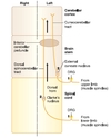
fissures + sulcus

2
Q

SPINAL CORD

anatomy

(gray and white matter)

A

GRAY MATTER

centrally located

butterfly

cell bodies + dendrites + proximal part of axon

WHITE MATTER

surrounds gray matter

tracts or fasciculli

axons

3
Q

SPINAL CORD

components of gray matter

A

dorsal horn (sensory)

ventral horn (motor)

intermediate zone (autonomic)

T1 - L2 + S2 - S4

Clarke’s nucleus (T1 - L2)

4
Q

SPINAL CORD

components of white matter

A

tracts and fasciculli

5
Q

SPINAL CORD

spinal nerves

(31 roots)

A

8 cervical

12 thoracic

5 lumbar

5 sacral

1 coccygeal

6
Q

SPINAL CORD

typical root

(spinal nerve)

A

ventral (motor) and dorsal (sensory) roots

dorsal root ganglion (sensory)

ventral and dorsal ramus (mixed)

7
Q

SPINAL CORD

plexus

A

brachial: C5 - T1

(upper limbs)

lumbarsacral: L2 - S3

(lower limbs)

cervical + lumbar enlargement in spinal cord

8
Q

SPINAL CORD

cauda equina

A

dorsal + ventral roots of

lumbar

sacral

coccygeal

9
Q

SPINAL CORD

conus medullaris

A

caudal end of spinal cord

S3 - S5

adults: L2 vertebra

10
Q

SPINAL CORD

filum terminale

A

slender pial extension

tethers the spinal cord to the bottom of the vertebral column

11
Q

SPINAL CORD

types of nerve fibers

(Erlanger-Gasser)

A

Group A

heavily myelinated

Group B

moderataly myelinated

Group C

unmyelinated

12
Q

SPINAL CORD

Group A nerve fiber

A

150 m/s

somatic fibers (sensory + motor)

subdivision

alpha

beta

gamma

delta

13
Q

SPINAL CORD

Group B nerve fiber

A

15 m/s

sensory and motor autonomic fibers

sensory - general visceral afferent

motor - preganglionic

14
Q

SPINAL CORD

Group C nerve fiber

A

no more than 2 m/s

sensory + motor fibers

sensory - pain + temp

motor - posganglionic (autonomic)

15
Q

SPINAL CORD

other classification of fibers

(functional division)

A

motor fiber

(alfa, beta and gamma)

sensory fiber

(Ia, Ib, II, III and IV)

autonomic

(pre and postganglionic)

16
Q

SPINAL CORD

motor fibers

A

alpha

A-alpha (Erlanger-Gasser)

extrafusal muscle fibers

beta

A-beta (Erlanger-Gasser)

gamma

A-gamma (Erlanger-Gasser)

intrafusal muscle fibers

17
Q

SPINAL CORD

sensory fibers

A

Ia

A-alpha (Erlanger-Gasser)

muscle spindle (primary or annulospiral ending)

Ib

A-alpha (Erlanger-Gasser)

golgi tendon

II

A-beta (Erlinger-Gasser)

muscle spindle (flower-spray ending) + cutaneous mechanoreceptors

III

A-delta (Erlinger-Gasser)

free nerve ending (touch and pressure)

nociceptor (sharp pain)

cold receptors

IV

C (Erlinger-Gasser)

nociceptors (dull pain)

warmth receptors

18
Q

SPINAL CORD

autonomic fibers

A

preganglionic fiber

B (Erlanger-Gasser)

posganglionic fiber

C (Erlanger-Gasser)

(pre is faster than post)

19
Q

GRAY MATTER

rexed laminae

A

dorsal horn

I - VI

intermediate zone

VII

ventral horn

VIII - IX

20
Q

GRAY MATTER

dorsal horn

(general statements)

A

sensory stimulation

(fibers enter the dorsolateral part of spina, via dorsal root)

+

neurons project to higher levels in CNS

+
part of neurons participate in reflexes

21
Q

GRAY MATTER

dorsal horn

(rexed laminae)

A

medial division

proprioception (Ia and Ib, A-alpha fibers)

touch (II, A-beta fibers)

lateral division

sharp pain + cold (III, A-delta fibers)

dull pain, warmth (IV, C fiber)

22
Q

GRAY MATTER

ventral horn

(general statements)

A

innervation of skeletal muscle

alpha + gamma motoneurons

dorsal fibers - flexors

ventral fibers - extensors

medial fibers - proximal musculature

lateral fibers - distal musculature

23
Q

GRAY MATTER

ventral horn

(alpha and gamma motoneurons)

A

alpha motoneurons

A-alpha (Erlanger-Grasser)

extrafusal fibers

neuromuscular junction

gamma motoneurons

A-gamm (Erlanger-Grasser)

intrafusal fibers

muscle spindle (make more sensible to stretch)

24
Q

GRAY MATTER

intermediate zone

A

T1 - L2

contains preganglionic sympathetic neuron cell bodies

+

Clarke nucleus (unconscious proprioception to the cerebellum)

25
NEURAL SYSTEMS concept
systems in spinal cords use neurons in the gray matter and tracts or fasciculi of white matter 3 major (motor, sensory and autonomic) can be found in all levels of CNS
26
MOTOR SYSTEM voluntary innervation of skeletal muscle (basic neural circuit)
**voluntary contraction** basic neural circuit: two motoneurons upper + lower
27
MOTOR SYSTEM voluntary innervation of skeletal muscle (lower motoneuron)
ventral horn (spinal cord) - fibers exit in ventral root and cranial nerve nuclei - fibers exit in cranial nerve **synapse directly in neuromuscular junction** **(motor unit)**
28
MOTOR SYSTEM voluntary innervation of skeletal muscle (upper motoneuron)
cell bodies in brain stem and cerebral cortex axons descend via tract (spinal cord) synapse with lower motoneuron or interneurons
29
MOTOR SYSTEM voluntary innervation of skeletal muscle (upper motoneuron) cell bodies
red nucleus reticular formation lateral vestibular nuclei **cerebral cortex (most important)**
30
MOTOR SYSTEM voluntary innervation of skeletal muscle (upper motoneuron) pathway
note: UMNs have net inhibitory effect on muscle stretch reflexes
31
MOTOR SYSTEM voluntary innervation of skeletal muscle (corticospinal tract) fibers distribution
60% of the fibers precentral gyrus - primary motor cortex and premotor area (Brodmann 4 and 6) 40% of the fibers somatosensory cortical areas (parietal lobe)
32
MOTOR SYSTEM voluntary innervation of skeletal muscle (corticospinal tract) pathway
cerebral cortex internal capsule brain stem (ventral portion) decussation of the pyramids (80-90%) lateral corticospinal tract (contralateral in spinal cord) ventral horn to synapses
33
MOTOR SYSTEM voluntary innervation of skeletal muscle (corticospinal tract) clinical correlate
Lesions above pyramidal decussation **contralateral weakness** Lesions below this level **ipsilateral weakness**
34
MOTOR SYSTEM reflex innervation of skeletal muscle (reflex)
sensory stimulus **(Ia, Ib afferents or pain fibers)** → synapse in spinal cord (mono or polysynapses) → lower motoneuron response (contration or relaxation) **both alpha and gamma**
35
MOTOR SYSTEM reflex innervation of skeletal muscle (flexor withdrawal reflex)
**protective reflex** painful stimulus → spinal cord → withdrawal of stimulated limb crossed extension reflex (to support the body)
36
MOTOR SYSTEM reflex innervation of skeletal muscle (myotatic reflex)
**deep tendon reflex** (monosynaptic and ipsilateral) in response to stretch (**spindles - Ia fibers**) **basic mechanism for regulating muscle tone** **(alpha motoneurons)**
37
MOTOR SYSTEM reflex innervation of skeletal muscle (common myotatic reflexes) 5
38
MOTOR SYSTEM reflex innervation of skeletal muscle (inverse muscle stretch reflex)
collagenous tendon fibers **(golgi tendon organs - Ib fibers)** polysynaptic (**inhibition interneuron**​) facilitate antagonists and inhibit agonist "stretch can be a risc"
39
MOTOR SYSTEM reflex innervation of skeletal muscle (inverse muscle stretch reflex) **mechanism**
golgi tendon organs (Ib fibers) → spinal cord - inhibition interneuron → **direct inhibition action on alpha motoneurons** **+** **gamma motoneurons and upper motoneurons** (regulation of spindles sensitivity and making the alpha motoneurons increase in muscle tone)
40
MOTOR SYSTEM reflex innervation of skeletal muscle (stretch reflex vs inverse reflex)
spindles - in parallel with extrafusal fibers golgi tendon organs - in series with extrafusal fibers
41
MOTOR SYSTEM reflex innervation of skeletal muscle (upper motor neuron function)
**descending control over the reflexes**
42
MOTOR SYSTEM reflex innervation of skeletal muscle (upper motoneurons lesions)
hyperactive muscle streth reflexes + oversensitive golgi tendon organs (increase of muscle tone) = **SPASTIC PARESIS** (ipsilateral - lesion in spinal cord) (contralateral - lesion above the pyramidal decussation)
43
MOTOR SYSTEM reflex innervation of skeletal muscle (lower motoneurons lesions)
hyporactive muscle streth reflexes + golgi tendon organs (reduction of muscle tone) = **FLACCID PARALYSIS** (initially - muscles fasciculations + fibrilations) (after - hypotonia + atrophy) **ipsilateral**
44
MOTOR SYSTEM reflex innervation of skeletal muscle (lower vs upper motoneurons lesions)
45
SENSORY SYSTEM Two ascending pathways with ...
DORSAL COLUMN-MEDIAL LEMNISCAL + ANTEROLATERAL (SPINOTHALAMIC)
46
SENSORY SYSTEM Dorsal column-medial lemniscal system (function) 5
Conscious proprioceptions Fine touch Vibration Pressure Two-point discrimination
47
SENSORY SYSTEM Dorsal column-medial lemniscal system (receptor) **PMM**
Pacinian corpuscle (vibration) Meissner corpuscle (touch) Muscle spindle (proprioception)
48
SENSORY SYSTEM Dorsal column-medial lemniscal system (primary afferent neurons)
Cell bodies in the dorsal root ganglia Enter the cord via class II or A-beta dorsal root fibers **fasciculus gracilis and cuneatus** (dorsal funiculus)
49
SENSORY SYSTEM Dorsal column-medial lemniscal system (fasciculus gracilis vs fasciculus cuneatus)
GRACILIS all spinal cord levels medial lower extremities/trunk CUNEATUS only upper thoracic and cervical levels lateral upper extremities/trunk
50
SENSORY SYSTEM Dorsal column-medial lemniscal system (secondary afferent neuron)
**nucleus gracilis and nucleus cuneatus** (lower part of the medulla - "bulbo") cross the midline (internal arcuate fibers) ascending in **medial lemniscus**
51
SENSORY SYSTEM Dorsal column-medial lemniscal system (thirdary afferent neuron)
Ventral Posterolateral nucleus of the thalamus (**VPL**) Thalamocortical fibers project to the primary somesthesic area (**somatosensory**) Parietal lobe - **postcentral gyrus**
52
SENSORY SYSTEM Dorsal column-medial lemniscal system (lesions)
Astereognosis Negative vibratory testing (128Hz tuning fork) Romberg Ipsi or contralateral
53
SENSORY SYSTEM Anterolateral or spinothalamic tract (function) 3
PAIN TEMPERATURE CRUDE TOUCH
54
SENSORY SYSTEM Anterolateral or spinothalamic tract (primary afferent neuron)
Cell bodies in the dorsal root ganglia Enter the spinal cord via class III or A-delta + class IV Fibers **ascend or descend a couple segments** in the dorsolateral tract of Lissauer
55
SENSORY SYSTEM Anterolateral or spinothalamic tract (secondary afferent neuron)
Cell bodies in the **dorsal horn** gray matter Fibers cross in the **white commissure** **Spinothalamic tract**
56
SENSORY SYSTEM Anterolateral or spinothalamic tract (thirdary afferent neuron)
Ventral Posterolateral nucleus of the thalamus (**VPL**) Thalamocortical fibers project to the primary somesthesic area (**somatosensory**) Parietal lobe - **postcentral gyrus**
57
SENSORY SYSTEM Anterolateral or spinothalamic tract (lesions)
**CONTRALATERAL LOSS OF PAIN AND TEMPERATURE** (unilateral lesions) information crosses almost as soon as it enters anesthesia - 1 to 2 segments below the lesion + everything below that level (Lissauer's tract)
58
SENSORY SYSTEM Spinocerebellar pathways (two major pathways)
DORSAL SPINOCEREBELLAR TRACT (lower extremities and trunk) CUNEOCEREBELLAR TRACT (upper extremities and trunk)
59
SENSORY SYSTEM Spinocerebellar pathways (function)
Unconscious Proprioceptive (muscle spindles + golgi tendon organ) monitor + modulate moviments
60
SENSORY SYSTEM Spinocerebellar pathways (primary afferent neuron)
Cell bodies in the dorsal root ganglia receptor - muscle spindle and goldi tendon organ
61
SENSORY SYSTEM Spinocerebellar pathways (secondary afferent neuron)
Cell bodies * Dorsal spinocerebellar tract - Clarke's nucleus (T1 - L2) * Cuneocerebellar tract - external cuneate nucleus (medulla - bulbo)
62
SENSORY SYSTEM Spinocerebellar pathways (lesions)
uncommon hereditary diseases (Friedreich ataxia - autossomal recessive) ataxia of gait (initial symptom)
63
SPINAL CORD LESIONS Identify cord section (three tips)
Large Ventral Horn Both dorsal columns (gracilis + cuneatus) Lateral Horn
64
SPINAL CORD LESIONS Identify cord section (large ventral horn)
YES C5 - T1 or L2-S2 NO C1 - C4 or T2 - L1
65
SPINAL CORD LESIONS Identify cord section (both dorsal columns)
YES above T5 NO below T5
66
SPINAL CORD LESIONS Identify cord section (lateral horn)
YES T1 - L2 NO C1 - C8 or L3 - S5
67
SPINAL CORD LESIONS Brown-Séquard syndrome
hemisection of the cord 3 main neural systems (corticospinal + dorsal columns + spinothalamic) 2 ipsilateral + 1 contralateral (spinothalamic)
68
SPINAL CORD LESIONS Poliomyelitis
lower motoneurons in ventral horn → flaccid paralysis some patiens recover most function, but others progress to muscle atrophy and permanent disability
69
SPINAL CORD LESIONS Amyotrophic Lateral Sclerosis
pure motor system disease (upper and lower motorneurons) bilateral flaccid weakness - upper limbs + bilateral spastic weakness - lower limbs
70
SPINAL CORD LESIONS Occlusion of the anterior spinal artery
interrupts blood supply to ventrolateral part of spinal cord → corticospinal tract + spinothalamic tract **bilateral loss of pain and temperature and spastic paresis**
71
SPINAL CORD LESIONS Syringomyelia
cavitation of the central canal (cervical spinal cord) early - white comissure (spinothalamic tract - "belt-like") lesion expands - lower motoneurons (ventral horn) late - Horner syndrome (T1 - T4) May be present in hydrocephalus and Arnold-Chiari I
72
SPINAL CORD LESIONS Tabes Dorsalis
Neurosyphilis Bilateral degeneration of dorsal roots + secondary degeneration of dorsal columns high-step stride and Argyll Robertson pupils
73
SPINAL CORD LESIONS Subacute Combined Degeneration
patchy losses of myelin in the dorsal columns and lateral corticospinal tracts
74
SPINAL CORD LESIONS Bladder vs level of the lesion
**Above sacral level** spastic bladder (problems in filing stage - destrusor muscle responds to a minimum amount of stretch) **Sacral level** atonic bladder (loss of pelvic splanchnic motor innervation - continuous dribble of urine)
75
SPINAL CORD LESIONS Brown-Séquard Syndrome - Cervical level