Spinal Disorders Flashcards
(47 cards)
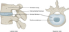
What is the anatomy of the vertebral column?

What is the anatomy of the spinal cord?
- approx 45cm long
- originates from medulla
- through foramen magnum
- ends as conus medullaris (L1-2 in adult)
- cauda equina - motor + sensory nerve roots exiting through lumbar and sacral foramina
- meningeal coverings of cord continuous with brain
- cord closely ensheathed by pia mater
- pia mater forms denticulate ligaments
- space between arachnoid + pia → CSF
- dura forms tough sheath; ends distally at S2

More anatomy of the spinal nerves

Anatomy of the intervertebral disc

Describe the dermatome distribution of the upper and lower limb

What movements are supplied by which nerve roots in the upper limb?

Which are the myotomes of the lower limb?
- Hip flexion → L1-2
- Knee extension → L3
- Ankle dorsiflexion → L4
- Ankle plantarflexion → S1-2
- Foot inversion → L4
- Foot eversion → S1
- Big toe dorsiflexion → L5
What are the ascending (sensory) tracts of the spinal cord?

What are the descending (motor) tracts of the spinal cord?

What does the spinal cord look like on MRI?

What red flags are important to look out for when taking a history for back pain / spinal disorders?
- extremes of age <20 / >50
- Hx of cancer
- unexplained weight loss
- immunosuppression
- IV drugs, steroids
- trauma
- faecal incontinence / loss of anal sphincter tone
- saddle anaesthesia
- globa/progressive weakness in legs
Why is spinal cord compression an emergency?
- requires swift management to prevent irreversible spinal cord injury + long-term disability
- treatment of acute compression → corticosteroids, surgery, radiotherapy
- diagnosis → x-ray or MRI of spine
- spinal cord injury may occur w/ no findings on imaging
- may be acute, sub-acute, chronic
- occurs due to:
- direct cord damage → compression and/or infiltration
- cord vascular supply compromise
Which part of the spinal cord is most commonly injured?
- Cervical (C1-T1) → 55%
- Thoracic (T1-T11) → 15%
- Thoracolumbar (T11-L2) → 15%
- Lumbosacral (L2-S5) → 15% (not cord, cauda equina)
There is a roughly equal distribution between complete and incomplete paraplegia/quadriplegias. Mortality risk is higher in C1-3 compared to C6-8.
What are the causes of spinal cord compression?
- trauma (main cause) → RTAs, falls, sports, knife/gunshot wounds, spontaneous disc protrusions, iatrogenic
- malignancy → extradural, intradural, primary + secondary
- infection → discitis, abscesses, osteomyelitis
- haematoma → AVM, spontaneous, trauma
- cystic lesions → arachnoidal, syringomyelia
- pathological fractures → osteoporosis, steroid therapy
Which tumours are most likely to metastasise to the bone?
‘BLT Plus Ketchup’
- Breast
- Lung
- Thyroid
- Prostate
- Kidney
What are signs and symptoms of spinal cord compression?
- back pain → associated w/ malignancy, compression fractures, infection etc
- sensory → numbness or parasthesia
- motor → weakness or paralysis, hypotonia
- autonomic → bladder or bowel dysfunction
- spasticity → most often w/ malignancy
- neurogenic shock → if lesions in C-spine, hypotension, bradycardia, warm extremities
- Brown-Sequard syndrome

What is the pathophysiology of spinal cord injury?
- injury arises from stretching or from pressure
- injures white matter (myelinated tracts) + grey matter (cell bodies) in cord
- causes loss of some sensory modalities + motor function
- spinal cord nerve roots depend on constant blood supply for appropriate energy stores + substrate, to perform axonal signalling
- conditions that interfere either directly or indirectly w/ blood supply will cause malfunction of transmission pathway
- nerve tracts most vulnerable to mechanical pressure include corticospinal + spinocerebellar tracts, and posterior spinal columns
Spinal cord injury can be classified as complete and incomplete.
What is meant by this?
- INCOMPLETE → any residual motor or sensory function more than 3 segments below level of injury; sensation or voluntary mvmt in legs; sacral sparing; types of lesion = central cord/Brown Sequard/anterior cord/posterior cord
- COMPLETE → no preservation of any motor and/or sensory function more than 3 segments below level of injury; no voluntary anal contraction; no anal sensation; no s4/5 sensation
Complete cord transection syndrome results in a group of symptoms known as spinal shock + when seen at high cervical level → quadriplegia, resp insufficiency, loss of bladder/bowel fxn, anaesthesia below affected level + neurogenic shock.
ASIA classification for pts w/ acute traumatic spinal cord injury.

Transverse myelitis (TM) is a pathogenetically heterogeneous focal inflammatory disorder of the spinal cord. Leads to axonal demyelination. Thought to be viral.
How does it clinically present?
- age 10-10 or 30-39
- motor weakness
- paraesthesias or sensory loss
- bladder → urinary frequency, urgency, incontinence, retention
- bowel → incontinence or constipation
- L’hermitte sign → tingling in limbs on neck flexion
- paroxysmal tonic spasms → painful, involuntary limb spasms
- UMN signs → hyper-reflexia, Babinski +ve, spasticity
- back / trunk / limb pain
What are differentials for the patient “off their legs”?
- Transverse myelitis
- Guillain-Barre syndrome
- HIV-related myelopathy
- Amyotrophic lateral sclerosis
- Multiple Sclerosis
- Diabetic neuropathy
- Polymyositis
- Hereditary muscular dystrophy
- Peripheral neuropathy
- Cerebellar origin (ataxia)
- Spinal cord (stenosis / malignant compression / syringomyelia)
- Degenerative ataxias
- Metabolic/Nutritional (vit B12 def/drugs/dizziness)
What are the investigations for spinal cord compression?
- CT for bony anatomy
- MRI for soft tissue (cord itself)
- CXR, X-ray spine, FBC, U+E, bone profile, ESR, CK, TFTs, plasma electrophoresis, urine (Bence-jones protein), ABPI
Investigate the rest of the body also - is it cancer (primary or mets)? Is it infection (source)? Is it bony degeneration (osteoporosis)? Is it trauma? Any other injuries?
What do MRI findings of spinal cord injury look like?
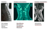
Clinical features depend on the site, level and completeness of the compressive lesion. Any residual motor or sensory function more than 3 segments below the level of injury suggest an incomplete lesion - look for signs of preserved long tract function.
What are 5 types of incomplete injury?
- Central cord syndrome
- Anterior cord syndrome
- Brown-Sequard syndrome
- Posterior cord syndrome
- Cauda Equina syndrome
Whereas, complete lesions show no preservation of motor and/or sensory function more than 3 segments below the level of injury.
With any severe, acute spinal cord lesion there are usually two clinical stages. What are these?
- Spinal shock → initially, there is loss of all reflex activity below level of lesion w/ flaccid limbs, atonic bladder + overflow incontinence, atonic bowel, gastric dilatation and loss of genital reflexes + vasomotor control
- Heightened reflex activity → occurs after about 1-2wks + is associated w/ spasticity of limbs, brisk reflexes and extensor plantar responses. Pts develop a spastic bladder (small capacity w/ urgency and frequency, and autonomic emptying) and hyperactive autonomic function (sweating + vasomotor changes)