Systemic sclerosis Flashcards
(28 cards)
In systemic sclerosis, is it male or female disease? Rough age group? What disease has highest mortality among rheumatic diseases? What used to cause death? What about now?
female to male ratio 3:1 peak age of onset 40-60 Highest mortality among rheumatic disease. Cardiopulmonary disease leading cause of death. Used to be scleroderma renal crisis, now due to ILD and pulmonary HTN.
Classification of systemic sclerosis
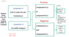
Classification of systemic sclerosis (LeRoy classification) • According to extent of skin involvement • DcSS = skin thickening present proximal to elbows/knees, trunk, in addition to face and distal extremities • LcSS = skin thickening limited to distal to elbows and knees. Can involve the face Sine scleroderma = internal organ involvement with vascular and serological abnormalities, but no detectable skin thickening (<5% of cases)
Describe aetiology of systemic sclerosis Brief description of pathophysiology
Aetiology • Unknown • Genetic predisposition ○ family history important ○ Rare familial cases • Environmental factors ○ Silica ○ Organic solvents ○ Vinyl chloride • 3 pillars to pathogenesis ○ Fibrosis - extensive fibrosis of skin and internal organs. Abnormal production of type 1 collagen. ▪ Skin – scleroderma ▪ Lungs – ILD ▪ Gut – hypomotility and range of consequences ▪ Myocardium ▪ renal arterioles – scleroderma renal crisis ○ Vasculopathy – leads to multiple features of disease including Raynaud’s phenomenon, digital ischaemia and ulcers, pulmonary arterial hypertension, GAVE etc ▪ Raynaud’s phenomenon is one of the first features ▪ over time there is endothelial cell injury, aberrant vascular remodelling, and angiogenesis. This can lead to abnormal blood vessels in the nailfold capillaries, and in the lining of the gut (GAVE) ○ Autoimmunity & inflammation - evidenced by presence of auto-antibodies & pro-fibrotic cytokines. Both cellular and humoral immunity. ▪ clinically, there may be synovitis and myositis
Clinical features of systemic sclerosis
Clinical features • CREST (used to be term to describe lcSS but no longer used as CREST features can be found in both lcSS and dcSS) ○ Calcinosis cutis/subcutaneous calcinosis ▪ More common in long-standing disease. Due to calcium hydroxyapatite deposition. ▪ Small, localised, firm masses on fingers, forearms, and other pressure points. ○ Raynaud’s phenomenon ○ E(o)sopheageal dysmotility ○ Sclerodactyly ○ Telangiectasia • Constitutional symptoms ○ Can occur early ○ Fatigue, arthralgia, myalgias • Skin ○ Puffy fingers (diffuse, non-pitting soft tissue swelling of fingers), hand swelling, foot swelling - common presenting complaint. Can be associated with morning stiffness. ○ Skin thickening of both fingers/hands, extending proximally (sclerodactyly). ○ LcSS = thickening distal to elbows and knees, can involve face (decreases oral aperture). DcSS = in addition to distal/face, also involving proximal to elbows/knees, anterior chest/abdomen. Explosive onset of rapid skin progression after onset of Raynaud’s is characteristic of diffuse disease, which can stabilise and then sometimes regress. ○ May result in loss of function eg, difficulty grasping. Can result in severe contractures. • Telangiectasia - can be found on hands, lips, inside mouth • Abnormal nail fold capillaries - due to changes in micro-vascular that occur even in early disease. ○ Can occur in other diseases eg, dermatomyositis • Raynaud’s phenomenon ○ Another common initial clinical feature ○ Digital ischaemia: fingertip ulcers & infarction, fingertip pitting scars, fingertip pulp loss. Can also occur over bony prominences. • Tendon friction rubs: ○ Palpable, sometimes audible rub, on movement of ankle, wrist, knee, shoulder, or elbow. Caused by irritation of tendon sheath and may be seen early in disease. ○ Usually found in diffuse disease ○ Poor prognostic indicator. Marker for aggressive disease with increased risk for internal organ involvement. • Gastrointestinal ○ GORD - very common (90% of patients) ▪ Oesophageal dysmotility, incompetent lower oesophageal sphincter ▪ Complications: oesophageal strictures, Barrett’s oesophagus, adenocarcinoma, chronic aspiration pnemonitis ○ Bloating - common. Due to decreased stomach motility and small bowel. ○ Constipation - common. Due to decreased bowel motility. ○ Small intestinal bacterial overgrowth - due to decreased bowel motility resulting in bacterial overgrowth. Malabsorptive diarrhoea, secretory diarrhoea (ask about nocturnal diarrhoea), steatorrhoea, weight loss. ○ Faecal incontinence - common. Due to loss of anal sphincter tone, reduced sensation, and constipation with overflow. ○ Gastric antral vascular ectasia (GAVE) - ‘Watermelon stomach’: usually causes chronic GI bleed, rarely acute malaena. Can result in microcytic hypochromic iron deficiency anaemia. ▪ Telengiectasia in GIT can result in chronic GI bleeding as well. • Pulmonary disease - largest cause of mortality ○ Pulmonary arterial hypertension ▪ Can occur anytime in both dcSS and lcSS. Occurs equally (~10%) in both groups. ▪ Typically WHO group I PAH (“pre-capillary”) ▪ Progressive vascular remodelling of small pulmonary arteries –> increased pulm vasc resistance –> RHF ▪ DDx 1) Group 2 Pulm HTN secondary to L heart disease –> *PAWP > 15mmHg 2) Group 3 Pulm HTN in ILD 3) Group 4 Chronic thromboembolic Pulm HTN ® Also look for PEs with V/Q scan as cause of pulmonary HTN ▪ High mortality: □ Untreated 2 year survival <50% □ With new therapies 2 year survival 80% ▪ *Check for iron deficiency –> worse prognosis □ Important to treat ○ Interstitial lung disease ▪ dcSS > lcSS ▪ Usually occurs in first 5 years in dcSS ▪ NSIP >>> UIP on HRCT ▪ Not all patients with ILD progress. ▪ High risk phenotype in ILD: early dcSS, anti-Scl70, elevated CRP, male gender, African-American, extensive involvement on HRCT ○ Chronic aspiration pneumonitis - can occur due to GORD. • Cardiac disease ○ Pulmonary arterial hypertension (see above) ○ Cardiomyopathy of scleroderma - RV or LV diastolic dysfunction. Poor prognostic marker. ○ Myo/pericarditis ○ Arrhythmias due to cardiac fibrosis ○ Increased risk of IHD/coronary artery disease ○ Pericardial effusions 5-10% of patients ○ Cyclophosphamide induced haemorrhagic pericarditis • Scleroderma renal crisis ○ Usually occurs in first 5 years in dcSS. Occurs in 10-20% of dcSS. ○ Due to ischaemic over-activation of RAAS. ○ Triggers: corticosteroids. ○ Risk factors: ▪ Anti-RNA polymerase III ▪ dcSS ▪ Tendon friction rubs ▪ Older age, male gender ▪ Corticosteroids - can be a trigger. Hence recommend <15mg/day especially in dcSS. ○ Clinical features ▪ Abrupt onset moderate-severe hypertension ▪ Progressive renal failure ▪ Bland urinary sediment, or only mild proteinuria with few cells or casts ▪ Microangiopathic haemolytic anaemia in severe cases ▪ Other complications: APO, blurred vision due to retinopathy, headache, encephalopathy ○ Seen in dcSS, usually in early stages. On rare occasions, scleroderma renal crisis can be initial presenting complaint of dcSS. Raynaud’s phenomenon and sclerodactyly may be present but overlooked by the patient. • Synovitis may be present • Muscle disease ○ Inflammatory myositis may be present in subset of scleroderma patients. Usually proximal muscle weakness, elevated CK, +ve EMG/NCS studies, +ve muscle biopsy. ○ Scleroderma myopathy. Elevated muscle enzymes, but no muscle weakness. Also different EMG/NCS findings, and muscle biopsy findings.
What is CREST? Calcinosis cutis -early or late in disease? -what is actually deposited? -where?
• CREST (used to be term to describe lcSS but no longer used as CREST features can be found in both lcSS and dcSS) ○ Calcinosis cutis/subcutaneous calcinosis ▪ More common in long-standing disease. Due to calcium hydroxyapatite deposition. ▪ Small, localised, firm masses on fingers, forearms, and other pressure points. ○ Raynaud’s phenomenon ○ E(o)sopheageal dysmotility ○ Sclerodactyly ○ Telangiectasia
Abnormal nail fold capillaries in systemic sclerosis - do they occur early or late in disease?
typically early in disease
Tendon friction rubs in systemic sclerosis - good or bad prognostic indicator? occurs in dcSS or lcSS?
Poor prognostic indicator. Marker for aggressive disease with increased risk for internal organ involvement. Typically dcSS
Why do you get pulmonary hypertension in systemic sclerosis? what is the % of patients that get pulm HTN? difference between dcSS and lcSS? Which WHO group of pulmonary hypertension? What are the other groups of pulmonary hypertension?
▪ Can occur anytime in both dcSS and lcSS. Occurs equally (~10%) in both groups, but usually occurs in older age in lcSS ▪ Typically WHO group I pulmonary arterial hypertension (“pre-capillary”) Progressive vascular remodelling of small pulmonary arteries –> increased pulm vasc resistance –> RHF ▪ DDx 1) Group 2 Pulm HTN secondary to L heart disease –> *PAWP > 15mmHg 2) Group 3 Pulm HTN in ILD 3) Group 4 Chronic thromboembolic Pulm HTN ® Also look for PEs with V/Q scan as cause of pulmonary HTN
What crucial thing do you have to check for in pulmonary hypertension in systemic sclerosis? why?
▪ *Check for iron deficiency –> worse prognosis □ Important to treat
ILD in systemic sclerosis -more common in dc or lcSS? -when does it typically occur? -what do you see on HRCT? -do all patients with ILD progress? -what is a high risk phenotype?
▪ dcSS > lcSS ▪ Usually occurs in first 5 years in dcSS ▪ NSIP >>> UIP on HRCT ▪ Not all patients with ILD progress. ▪ High risk phenotype in ILD: early dcSS, anti-Scl70, elevated CRP, male gender, African-American, extensive involvement on HRCT
Scleroderma renal crisis -when does it typically occur? dcSS or lcSS? -what’s the pathophysiological mechanism? -risk factors -clinical features
○ Usually occurs in first 5 years in dcSS. Occurs in 10-20% of dcSS. ○ Due to ischaemic over-activation of RAAS. ○ Triggers: corticosteroids. ○ Risk factors: ▪ Anti-RNA polymerase III ▪ dcSS ▪ Tendon friction rubs ▪ Older age, male gender ▪ Corticosteroids - can be a trigger. Hence recommend <15mg/day especially in dcSS. ○ Clinical features ▪ Abrupt onset moderate-severe hypertension ▪ Progressive renal failure ▪ Bland urinary sediment, or only mild proteinuria with few cells or casts ▪ Microangiopathic haemolytic anaemia in severe cases ▪ Other complications: APO, blurred vision due to retinopathy, headache, encephalopathy ○ Seen in dcSS, usually in early stages. On rare occasions, scleroderma renal crisis can be initial presenting complaint of dcSS. Raynaud’s phenomenon and sclerodactyly may be present but overlooked by the patient.
dcSS vs. lcSS?
• Raynaud’s: short history and often same time onset as skin changes vs. long history years before onset of skin changes • Scleroderma involving both distal and proximal to elbows/knees, and trunk, in addition to face vs. skin changes limited to distal limbs and face • Rapid onset skin changes & can form contractures (can plateau and sometimes regress) vs. gradual onset skin changes and contractures less likely • Tendon friction rub vs. none • Constitutional symptoms more likely in dcSS than lcSS • Calcinosis cutis & telengiectasia more likely in lcSS but they do occur in dcSS as well. • Early onset internal organ involvement vs. later onset internal organ involvement ○ ILD more common in dcSS ○ Scleroderma renal crisis more common in dcSS ○ Heart complications more common in dcSS ○ Oesophageal dysmotility & other gut complications (eg, GORD, faecal incontinence) occurs in both subsets of disease equally ○ Pulmonary hypertension equal (~10%) in both subtypes, but usually in older age in lcSS • Auto-antibodies - as described elsewhere
Describe the autoantibodies in systemic sclerosis
○ ANA - ~90% +ve ▪ Centromere pattern – associated with lcSS & better prognosis ▪ Nucleolar pattern - associated with dcSS ▪ Speckled pattern - (associated with RNA polymerase III & topoisomerase I/scl70) ○ Anti-centromere antibody - as above. Centromere pattern on ANA. ○ Anti-topoisomerase I (anti-Scl70) - associated with ILD and diffuse disease. Speckled pattern on ANA. ○ Anti-RNA polymerase III - associated with scleroderma renal crisis & rapidly progressive skin disease. Fine speckled pattern on ANA. ○ Anti-PM/Scl & anti-Sm/RNP - associated with myositis overlaps ○ Anti-U1-RNP - overlap features with MCTD ○ Anti-Th/To - pulmonary hypertension, worse prognosis
What might you see in FBE and blood film in systemic sclerosis?
○ Chronic microcytic hypochromic anaemia - chronic GIT blood loss due to GAVE +/- GIT telengiectasia ○ Microangiopathic haemolytic anaemia in scleroderma renal crisis
ESR and CRP - what do they indicate in systemic sclerosis?
poor prognostic markers
DIfference between inflammatory myositis and scleroderma myopathy in systemic sclerosis?
Elevated CK without weakness seen relatively commonly. Known as scleroderma myopathy. If weakness present, think about inflammatory myositis.
What are the different pulmonary investigations that can be performed in systemic sclerosis?
○ RFT: spirometry, lung volumes, DLCO ▪ Decrease in FVC & DLCO + overall restrictive pattern - interstitial lung disease ▪ Disproportionate drop in DLCO compared with FVC - pulmonary hypertension ○ HRCT ○ TTE ▪ Elevated RVSP - pulmonary hypertension. Refer to right heart catheterisation as TTE findings may not reflect true pulmonary artery pressures. ▪ RV or LV diastolic dysfunction due to cardiomyopathy of scleroderma. ○ Right-heart catheter ▪ In systemic sclerosis, it is WHO group 1 pulmonary hypertension ie, pulmonary arterial hypertension (“pre-capillary”). □ DDx 1) Group 2 Pulm HTN secondary to L heart disease –> *PAWP > 15mmHg 2) Group 3 Pulm HTN in ILD 3) Group 4 Chronic thromboembolic Pulm HTN ® Also look for PEs with V/Q scan as cause of pulmonary HTN
What are the differences in right heart catheterisation findings in:
- pre-capillary pulmonary hypertension
- post-capillary pulmonary hypertension
- mixed pre- and post-capillary pulmonary hypertension

What investigations can you do for GIT findings in systemic sclerosis?
GIT Barium swallow - oesophageal dysmotility, reflux disease
Diminished oesophageal peristalsis
Gastroparesis
Diminished lower oesophageal sphincter tone with evidence of reflux of barium
Strictures
Endoscopy - GAVE, strictures, Barrett’s oesophagus adenocarcinoma
Glucose hydrogen breath test - SIBO
When would you do EMG/NCS and muscle biopsy in systemic sclerosis?
DDx for systemic sclerosis?
Primary Raynaud’s - see Raynaud’s phenomenon page
Localised scleroderma/generalised morphoea
Clinical and histopathological skin indistinguishable from systemic sclerosis
More often seen in children and young adults, rather than older adults.
Does not involve fingers
Lack of Raynaud’s phenomenon, sclerodactyly, or visceral manifestations
Eosinphilic fasciitis
More common in men
Leads to adherence of skin to underlying fascia, usually sparing of hands and feet. Woody induration of extremity or trunk. Peau d’orange (skin pitting like an orange peel) present.
Associated with over-exertion, worse with exercise
Peripheral eosinophilia, fascia biopsy shows eosinophilic inflammatory infiltrate
Scleroedema
Indurated, doughy skin in neck, back, face
Thickened dermis and hyaluronic acid
Associated with diabetes and monoclonal gammopathy
Scleromyxoedema
Waxy, yellow-red papules on head, neck, arms, occurring over thickened and indurated skin
Skin deposition is mucinosis material, not collagen
Associated with amyloidosis, associated with paraproteinaemias
Scleroderma diabeticorum and scleroderma of Buschke
Most patients have diabetes
Skin thickening of neck, upper back, shoulders trunk
Diabetic cheiroarthropathy (“diabetic stiff hand syndrome”)
Associated with diabetes, usually advanced and long-standing.
Thickened skin and contractures of fingers.
Nephrogenic systemic fibrosis
Occurs in usually ESRF patients given gadolinium contrast
Skin changes in distinguishable from scleroderma. Indurated plaques of lower limbs, feet, trunk, hands. However, will not have other systemic features of systemic sclerosis eg, Raynaud’s, pulmonary hypertension etc. Skin biopsy has distinct pathological findings that differentiate it from scleroderma.
May have internal organ fibrosis.
Acrodermatitis chronicum atrophicans
Associated with Borrelia infection
Chronically progressively course leading to skin atrophy, usually lower limbs in symmetric fashion.
Describe autologous haemopoietic stem cell transplant in systemic sclerosis
Autologous HSCT option for early progressive disease at risk of organ failure · Aim: eradicate immune response and re-instate non auto-reactive immune system · Indications: Non-smoker, non-responsive to standard Tx □ AND dSSc within first 5 years with mild-mod organ involvement □ OR LSSc with progressive visceral involvement · Up to 10% mortality overall □ Greater short term mortality with longer term benefit ® With improvement in disease/remission post HSCT □ Mortality largely due to cardiac Cx during and after HSCT ® Scleroderma heart disease ® Radiation induced cardiac injury ® CCF ® Cyclophosphamide induced haemorrhagic pericarditis
Management for skin thickening (scleroderma) of systemic sclerosis
Emollients
Physio
OT
Splinting for hand contractures
Only immunosuppress for progressive skin disease - cyclophosphamide, methotrexate, mycophenolate
Describe management for Raynaud’s phenomenon, critical digital ischaemia, and digital tip ulcers in systemic sclerosis
Raynaud’s phenomenon • Consider DDx of other underlying cause e.g. cryoglobulinaemia and treat • Lifestyle changes- avoid trigger ○ Cold, especially of the digits ○ Caffeine ○ Cease smoking • Vasodilators: ○ 1st line: calcium channel antagonists (nifedipine or amlodipine), ○ 2nd line: ARB (losartan), PDE5 inhibitor (sildenafil), topical GTN/systemic nitrate, alpha-blocker, SSRI (fluoxetine), antiplatelet/ statin • Management of critical digit ischaemia: ○ Requires admission, establish and treat other contributory causes (eg, infection, vasculitis, thromboembolism) ○ IV prostacyclin (iloprost, epoporosteol) ○ Selective Sympathectomy, Botox injection ○ Avoid surgery, unless significant necrotic tissue • Digital tip ulcers ○ Avoid cold, analgesia ○ Treat with § Dressings to remove dead necrotic tissue and allow healing § IV iloprost +/- PDE 5 I (eg sildenafil) § Antibiotics for any infection ○ Consider Bosentan - prevents (though doesn’t treat) ulcers