Test 2 Study Guide/Objectives Flashcards
What are the functions of the skeletal system?
- Support
- Protection
- Assist with Movement
- Mineral Homeostasis
- Blood Cell Production (red bone marrow)
- Triglyceride Storage (yellow bone marrow)
What are the parts of long bones?
- Diaphysis (bone shaft)
- 2 Epiphyses (both ends of the bone at the joints)
- 2 Metaphyses (region between diaphysis and epiphysis)
- Articular Cartilage (covering both epiphysis)
- Periosteum (connective tissue surrounding the diaphysis)
- Medullary Cavity (hollow space within diaphysis)
- Endosteum (thin membrane lining the medullary cavity)

Why is calcium phosphate so important?
- It’s the most abundant mineral salt
- It combines with other mineral salts to calcify bones (harden bones)
What are the 4 types of bone cells?
- Osteoprogenitor cells (bone stem cells able to differentiate into other types of cells)
- Osteoblasts (bone-building cells that secrete matrix)
- Osteocytes (mature bone cells)
- Osteoclasts (remodel bones and cause them to release calcium)
What are some characteristics of compact bone?
- good at providing protection and support
- contain osteons

What are some characteristics of spongy bone?
- Lightweight
- Provides tissue support
- Do not contain osteons
- Always on the interior of the bone
- Irregular thin columns called: trabeculae
- oriented precisely along lines of stress

Why does bone have to form?
During infancy, childhood, and adolescence, bones throughout the body grow in thickness by appositional growth, and long bones lengthen by the addition of bone material on the diaphyseal side of the epiphyseal plate by interstitial growth.
What is Intramembranous Ossification?
- The simpler of the 2 methods of bone development
- Occurs in flat bones when a connective tissue membrane is replaced by bone
- flat bones of skull, facial bones, mandible, medial part of clavical
- Development of ossification center
- Calcification
- Formation of trabeculae
- Development of periosteum

What is Endochondral Ossification?
- Replaces cartilage with bone in the developing embryo and fetus, forms within hyaline cartilage
- Occurs at epiphyseal plates
- Long bones mostly

- Development of the cartilage model made of hyaline cartilage, lined with perichondrium
- Growth of cartilage model
- interstitial growth (from within, increase in length)
- appositional growth (growth of outer surface)
- Development of primary ossification center (central to ends) (inwards)
- Development of medullary cavity (marrow)
- Devlopment of secondary ossification centers (develop around time of birth,outwards from center of epiphysis)
- Formation of articular cartilage and epiphyseal growth plate responsible for lengthwise growth
What is the epiphyseal plate and what are the 4 zones associated with it?
It is the growth plate that lengthens bone. It closes when adolescence ends.
- Zone of Resting Cartilage (cells here do not function in bone growth)
- Zone of Proliferating Cartilage (larger chondrocytes, interstitial growth, divide and secrete extracellular matrix)
- Zone of Hypertrophic Cartilage (large maturing chondrocytes arranged in columns)
- Zone of Calcified Cartilage (final zone, mostly dead chondrocytes)

What is appositional growth?
- At surface periosteal cells differentiate into osteoblasts
- Ridges form around periosteal blood vessel
- Ridges fold together and fuse eventually
- Osteoblasts in endosteum form new concentric lamellae
- Osteoclasts allow for thickening of medullary cavity
What happens during bone growth and remodeling?
- Bone continually renews itself
- Bone resorption (oseoclasts)
- Bone deposition (osteoblasts)
- Interaction between the 2 = remodeling
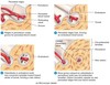
How does a bone repair? What are the phases?
- The healing process involves 3 different phases in 4 steps:
- The reactive phase is an early inflammatory phase
- The reparative phase includes formation of a fibrocartilaginous callus first and a bony callus second
- The bone remodeling phase is the last step as the bony callus is remodeled

Open (Compound) Bone Fracture

Comminuted Bone Fracture

Greenstick Bone Fracture

Impacted Bone Fracture

Pott Bone Fracture

Colles Bone Fracture

What role does calcitonin and parathyroid hormone play in the skeletal system?
What makes up the axial skeleton?
- 80 bones total
- Skull
- Cranium (8)
- Face (14)
- Hyoid bone (1)
- Auditory Ossicles (6)
- Vertebral Column (26)
- Thorax
- Sternum (1)
- Ribs (24)
What makes up the appendicular skeleton?
- 126 bones total
- Pectoral (shoulder) girdles
- Clavicle (2)
- Scapula (2)
- Upper limbs
- Humerus (2)
- Ulna (2)
- Radius (2)
- Carpals (16)
- Metacarpals (10)
- Phalanges (28)
- Radius (2)
- Pelvic (hip) girdles
- Femur (2)
- Patella (2)
- Fibula (2)
- Tibia (2)
- Tarsals (14)
- Metatarsals (10)
- Phalanges (28)
What are some different characteristic types of bones?
- Long (greater seed length than width)
- Short (cube shaped)
- Flat (thin layers of parallel plates)
- Irregular (complex shapes)
- Sesamoid (shaped like a sesame seed)
- Sutural bones are small, extra bone plates located within the sutures of cranial bones
- Sutures are the jointed areas where flat bones come together

What are the different types of bone surface markings?
There are two major types of surface markings:
-
Depressions and openings
- Allow the passage of soft tissues
- Form joints
-
Processes
- Projections or outgrowths that form joints
- Serve as attachment points for ligaments and tendons


