Test 4 Flashcards
1.

Amalgam
Opening or hole in bone located on the external surface of the mandible in the region of the mandibular premolars.
Radiolucent/Radiopaque?
Mental foramen
Radiolucent

1.

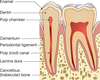
Dental base
Opening or hole in bone located at the midline of the anterior portion of the
hard palate directly posterior to the maxillary central incisors.
Radiolucent/Radiopaque?

Incisive foramen #1
Radiolucent

Identify #1.

Border of maxillary sinus
Linear prominence of bone located on the internal surface of the mandible that extends downward & forward from the ramus?
Radiolucent/Radiopaque?
Internal oblique ridge

Radiopaque
2.

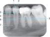
Radiopaque amalgam restorations
1.

PDL space
Identify the air space image #2

nasopharyngeal
Intersection of the maxillary sinus & the nasal cavity
as viewed on a dental radiograph.
Radiolucent/Radiopaque?
Inverted Y
Radiopaque

J or U shape located above the maxillary first molars.
Radiolucent/Radiopaque?

zygomatic process of maxilla #4

Radiopaque
2.

Mylohyoid ridge
1.

Full metal crowns form bridge abutments
Identify the age of this patient

Age 12
Name the classification of dental caries illustrated by 3

C-3 Advanced Caries
Advanced: Lesion that extends to or through the DEJ but does not extend more than half the distance to the pulp
Identify #1.

Border of maxillary sinus
Identify #5.

Lateral pterygoid
Sharp projection of the maxilla located at the
anterior and inferior portion of the nasal cavity.
Radiolucent/Radiopaque?
Nasal Spine
Radiopaque
Identify the age of this patient

Age 8
4
Identify the following:

PDL Space
Rounded prominence of bone that extends
posterior to the third molar region.
Radiolucent/Radiopaque?
Maxillary Tuberosity #2
Radiopaque

Identify the following:
(tube-like passageways through bone supplying blood vessels and nerves to maxillary teeth and bone, appear as narrow bands)

Nutrient Canals in Max Sinus
Identify #3.

Nutrient foramen

6.

Post and core