Test 5 - pulmonary Flashcards

(159 cards)

1
Q

What are the clinical symptoms of brocnhitis? Is fever a common symptom?

A

productive cough

wheezing

fever is unusual

2
Q

How is bronchitis diagnosed?

A

Clinical diagnosis - based on symptoms of cough, wheezing. CXR is only indicated if pneumonia is suspected, and if consolidation is not seen pneumonia can be ruled out.

3
Q

What is pneumonia?

A

Infection of the lung parynchyma (respiratory airways)

4
Q

Pneumonia is categorized into 4 types. Name these 4 types (one type is further divided into two categories)

A

Community acquired (typical and atypical)

Hospital acquired

Health-care associated

Opportunistic

5
Q

What are the clinical symptoms of pneumonia?

A

fever

chest pain

dyspnea

productive cough

6
Q

How is pneumonia diagnosed? Is culture often performed?

A

Diagnosis is made with clinical symptoms and a chest x-ray with focal lung opacities

Culture is not usually performed because patients cannot expectorate sputum and even if they can the culture is contaminated with oropharyngeal bacteria

7
Q

How is pneumonia treated? discuss community-acquired and hospital-acquired treatments

A
8
Q

What is Mycobacterium tuberculosis? (stain, shape, and type of microbe)

A

Tuberculosis is an acid fast rod bacterium

9
Q

How is TB transmitted?

A

air droplets

10
Q

Describe the potential course of TB (primary infection, etc)

A
11
Q

What are the general clinical symptoms of TB?

A

productive cough

fever

malaise

weight loss

+/- hemoptysis

12
Q

If TB becomes systemic, what diseases can the patient develop?

A

Pott’s disease (TB in the vertebrae)

Milliary disease (widespread dissemination)

13
Q

How is active TB diagnosed (there are a few methods you should name)? Can IGRA or a PPD test diagnose active TB?

A

CXR

Sputum smear with acid fast stain (takes 4-6 weeks to culture)

nucleic acid amplification

IGRA and PPD CANNOT diagnose active TB

14
Q

Is PPD specific to Mycobacterium tuberculosis?

A

If the patient recieved a BCG vaccination, PPD will yield a positive result

15
Q

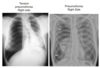
How does primary TB and secondary TB present on CXR?

A

Primary TB has cavitary lesions in the lower/middle lobe(s)

Secondary has cavitary lesions in the upper lobe(s)

Note: hilar LAD may be present in both

16
Q

What are risk factors for developing active TB?

A

Malnutrition

immunocompromized patients (drugs, HIV)

Diabetes

alcoholism

children under 2 years of age

17
Q

How is TB treated?

A
18
Q

What type of infectious agent causes pneumocystis pneumonia and in what patient population is this infection seen in?

A

PCP is a fungal pneumonia (caused by Pneumocystis jirovecii) that is commonly seen in AIDS patients with a CD4 count below 200

19
Q

What are the clinical symptoms of pneumocystis pneumonia?

A

fever

non-productive cough

dyspnea

20
Q

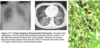
How is pneumocystis diagnosed?

A

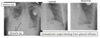
Chest imaging (ground glass opacities) +/- “crushed ping-pong balls” opacities

Bronchoalveolar lavage crushed ping-pong balls morphology

21
Q

What is the treatment for pneumocystis pneumonia?

A

Bactrim (sulfamethoxazole and trimethoprim)

22
Q

What is bronchiolitis?

A

Inflammation of the bronchioles

23
Q

What age is bronchiolitis typically seen in? What are the clinical symptoms of bronchiolitis?

A

Seen in children under 6 months

coryza (stuffy nose)

Rhinorrhea (runny nose)

cough

wheezing

chest wall retractions

respiratory distress

difficulty feeding due to increased work of breathing

24
Q

How is bronchiolitis diagnosed?

A

CXR:

air trapping

peribronchial thickening

subsegmental opacities

25
How is bronchiolitis treated?
Supportive therapy only: fluids, O2, bronchodilators
26
What is laryngeotracheobronchitis?
Croup upper airway obstruction
27
What causes croup?
RSV and parainfluenza
28
What are the clinical symptoms of croup? Is fever common?
Seal bark cough Stridor (inspiratory wheeze) hoarsness fever is not common
29
How do you diagnose croup? Do you use a tongue depressor to examine the pharynx?
Steeple sign on XR DO NOT stick anything in through as the airway could collapse!!
30
How is croup treated?
For kids in respiratory distress - aerosolized racemic epinephrine reduced subglottic edema Systemic steroids
31
What is epiglottitis? What is the most common cause?
inflammation of the epiglottis H. influenzae
32
What are the clinical symptoms of epiglottitis?
fever drooling dysphagia refusal to eat/drink sudden onset of stridor
33
How is epiglottitis diagnosed?
Thumb sign on XR
34
How is epiglottitis treated?
intubation antibiotics
35
Draw the restricive pattern dendritic
Notice that hypersensitivity pneumonitis is under granulomatous diseases and inhaled. Inhalation of particles results in non-caseating granulomas
36
What are the finding in a restricitve pattern? (PFT results)
37
What changes can be seen on a spirogram in restrictive diseases? Are RV and FRC reduced in neuromuscular diseases?
RV is normal/high in neruromuscular disease FRC is normal in neuromuscular disease
38
What changes can be seen on a flow-volume loop in restrictive diseases?
39
What are the 3 categories (heavily focused on in sullabus) of diffuse parenchymal lung disease?
Idiopathic pulmonary fibrosis granulomatous disease inhaled disease
40
What is Idiopathic pulmonary fibrosis?
increased flibroblast growth and collagen synthesis in the alveolar interstitium
41
What are the clinical symptoms of idiopathic pulmonary fibrosis? Is it an acute process? is the prognosis good or bad?
exertional dyspnea (decreaed RBC transit time) dry cough inspiratory crackles at lung bases (velco sounding) digital clubbing It is a slow, insideous process that shows up late in life (poor prognosis)
42
How does IPF change FEV1, FVC, FEV1/FVC, DLCO, A-a O2 difference
reduced FEV1, FVC, DLCO normal FEV1/FVC increased A-a O2 difference
43
How does IPF present on CXR? CT?
CXR low lung volumes, reticulonodular opacities CT honeycombing at lung bases
44
How is IPF diagnosed?
history, PFTs, and imaging biopsy is not always required
45
How is IPF treated?
There are no real treatments for IPF anti-fibrotic agent prifenidone may help to slow down progression
46
What diseases are classified under granulomatous restrictive lung diseases?
Sarcoidosis Hypersensitivity pneumonitis
47
What is sarcoidosis?
inflammation process that results in the formation of non-caseating granulomas in the lungs In severe cases, the disease is systemic
48
Who is at high risk for sarcoidosis? What organ systems are involved in systemic disease?
black women heart, CNS, liver, spleen, skin, eyes
49
What are the clinical symptoms of intrapulmonary sarcoidosis? extrapulmonary?
Intrapulmonary: typically asymptomatic dyspnea nonproductive cough Extrapulmonary: arrhythmias (heart block) uveitis hepatomegaly Lofgren's syndrome (erythemia nodosum, fever, hilar LAD)
50
How is sarcoidosis diagnosed?
symptoms, CXR, and biopsy of non-caseating granulomas
51
How is sarcoidosis treated?
Disease spontaneously resolves in most patients For those with severe pulmonary symptoms, corticosteroids are used
52
What disease closely resembles sarcoidosis?
Berylliosis
53
What is hypersensitivity pneumonitis
parenchymal lung disease characterized by non-caseating granulomas brought on by the inhalation of particles (specific particles)
54
Is HSP reversible?
In most cases, yes
55
What are the clinical symptoms of hypersensitivity pneumonitis?
Presents like pneumonia fever dyspnea cough
56
When do hypersensitivity pneumonitis symptoms present?
4-8 hours after exposure
57
How does HSP present on radiograph? Acute vs Chronic
Acute: ground glass opacities (similar to PCP) with upper lobe predominance Chronic: volume loss reticular opacities honeycombing
58
How is hypersensitivity pneumonitis diagnosed?
re-expose patient to particle and see if symptoms recur
59
How is HSP treated?
Avoid the antigen steroids in acute, severe situations
60
What diseases are classified under inhaled?
Hypersensitivity pneumonitis Asbestosis Silicosis Coal Worker's pneumoconiosis (black lung) Berylliosis
61
What is asbestosis?
A fibrotic lung disease brought on by the inhalation of asbestos
62
Who is at risk for asbestosis?
occupational hazard: shipyard/shipbuilding brak lining work insulation and textiles stone polishing and cutting
63
What is the pathophysiology of asbestos?
Fibers are inhaled —\> lower resp. tract —\> pneumocytes and alveolar macrophages release inflammatory mediators, ROS, free radicals —\> progressive fibrosis
64
How long does asbestosis present after exposure?
many years after exposure
65
How does asbestosis present on CXR?
bilateral patchy reticular opacities w/ associated calcified pleural plaques
66
How is asbestosis diagnosed?
history of exposure, radiology, biopsy (not necessary)
67
How is asbestosis treated?
It isn't. Just monitor for cancer
68
What is silicosis?
Pulmonary disease caused by inhalation of crystalline silica - most abundant mineral on earth found in rocks and sandstone
69
Who is at risk for silicosis?
Occupational: sandblasting masonry polishing mining tunneling
70
What is the pathologic hallmark of silicosis?
silicotic nodule
71
What are the clinical features of silicosis?
dyspnea weight loss fatigue
72
How does silicosis present on CXR?
Acute silicosis: basilar-predominant, alveolar opacities Simple silicosis: small, upper lobe-predominant nodules Chronic silicosis: coalesced nodules w/ upper lobe fibrosis, hilar adenopathy and calcification
73
How is silicosis diagnosed?
history and radiographic findings
74
What is coal worker's pneumoconiosis?
lung disease caused by the inhalation of carbon dust + silica
75
Who is at risk for the black lung?
occupational: coal mining graphite mining milling
76
How does the black lung show up on CXR?
Maybe small upper lobe nodules Chronic: coalescence of nodules
77
How is the black lung diagnosed?
clinical features
78
How is the black lung treated?
supportive therapy
79
Who is at risk for berylliosis?
Occupational metal alloy manufacturing nuclear industry (hanford)
80
Clinically, what other disease does berylliosis resemble?
sarcoidosis
81
What is kyphoscoliosis? What implications does the condition have for pulmonary function?
Lateral deviation + kyphosis (concavity w/ respect to the ventral surface) of the spine. It has the effect of increasing chest wall stiffness, causing restriction.
82
How is kyphoscoliosis diagnosed?
physical exam
83
How is kyphoscoliosis treated?
surgery or braces for young supportive for old
84
What are the three possible sites for neuromuscular dysfunction in restricitve lung diseases?
nervous system neuromuscular junction muscle
85
What are clinical features of neuromuscular diseases?
muscle weakness
86
How do neuromuscular diseases change PFTs? (RV and FRC) What should be done on PFT to assess strength?
RV is normal or high FRC is slightly decreased Max inspriratory and expiratory pressures
87
What is usually the cause diaphragmatic weakness?
weakness usually from phrenic nerve disruption (C3-5)
88
What are clinical features of diaphragmatic weakness?
Unilateral weakness is usually asymptomatic Bilateral weakness results in dyspnea, which is worse when lying down (orthopnea)
89
How is diaphragmatic weakness diagnosed?
upright and supine spirometry (\>20% decrease in FVC when supine) Electromyography unilateral weakness: will show elevated hemidiaphragm in CXR Sniff test to visualize diaphragm under fluoroscopy
90
How is diaphragmatic weakness treated?
Diaphragmatic plication: a surgery that fixes the diaphragm in a flat position Non-invasive ventilation
91
Is it important for the pulmonary vascular system to maintain a low pressure system?
yes
92
How is pulmonary artery pressure related to cardiac output and pulmonary vascular resistance? (equation)
PAP = CO x PVR
93
Describe recruitment and distention
As blood flow increases, intravascular pressures increase which causes initially closed pulmonary vasculature to open (recruitment). In addition, the caliber of already open vessels increase (distention).
94
What is one of the most important vasodilators?
Nitric oxide
95
Define hypoxic vasoconstriction
Hypoxic pulmonary vasoconstriction (HPV) is a vital mechanism that alters pulmonary vascular resistance and redistributes blood to better ventilated areas of the lung.
96
How does alveolar pressure determine blood flow through capillaries? Discuss using the Starling resistor theory (PA, Pa, Pv)
97
Describe how lung volumes affect the caliber of intra and extra-alveolar vessels. Wat is the significance of FCR?
low lung volumes = intra vessels are at their greatest caliber while extra are at their smallest high lung volumes = intra vessels are at their smallest caliber while extra are at their greatest FCR is where the resistance of intra and extra-alveolar vessels are equal
98
Describe the role of bronchial circulation.
Bronchial circulation supplies the lung with oxygen and nutrients and is under systemic pressures becuase it is coming from the systemic circulation
99
Pulmonary hypertension is defined as a mean pulmonary arterial pressure above ___ mmHg.
25 mmHg
100
What can cause pulmonary hypertension? Hint: Mean PAP = (PBF x PVR) + LAP
* Increasing pulmonary vascular resistance (PVR) * Most common * Increasing pulmonary blood flow (PBF) * Only results in hypertension if the effects of recruitment and distention are exhausted * Increasing left atrial pressure (LAP)
101
Name the 5 categories of pulmonary hypertension
Group 1: Pulmonary arterial hypertension Group 2: Left heart disease Group 3: lung diseases Group 4: chronic thromboembolism Group 5: miscellaneous
102
What are some common clinical presentations of pulmonary hypertension?
Loud P2 Elevated JVP Prominent a, c, and v waves Murmur of tricuspid regurgitation Hepatomegaly (severe) Ascites (severe) Lower extremity edema (severe)
103
How does pulmonary hypertension change an ECG?
Tall P waves in lead II (suggestive of right atrial enlargement) Large R wave in V1 (right ventricular hypertrophy) Right axis deviation
104
How does pulmonary hypertension present on radiography?
enlarged pulmonary arteries right heart enlargment
105
How is pulmonary hypertension treated?
treat underlying cause vasodilators: calcium channel blockers, PDE inhibitors, endothelin receptor antagonists
106
What is a venous thromboembolism?
Venous thromboembolism is a combination of two diseases: deep vein thrombosis (DVT) and pulmonary embolism (PE).
107
What causes 90% of PEs?
DVTs
108
How do VTEs affect gas exchange and pulmonary hemodynamics?
Gas exchange: creates dead spaces Blockage sends more blood (increased Q) to other areas creating low VA/Q areas Pulmonary hemodynamics if the block is too big for recruitment and distention mechanisms, PVR will increase
109
What are the 3 major risk factors for VTE?
Stasis Vascular injury Hypercoagulability
110
What are the clinical symptoms of DVT and PE?
DVT swelling, pain, erythema, warmth PE Acute onset of dyspnea, pleuritic chest pain depending on location, cough, hemoptysis, loud P2, tachypnea
111
How is VTE treated?
Anticoagulation with heparin and are put on warfarin or LMWH for 3-6 months depending on their risk factor profile Thrombolytics are reserved for patients with hemodynamic instability and/or cardiac arrest
112
What are arteriovenous malformations?
anatomic shunts Abnormal movement of blood from the systemic venous system to the systemic arterial system without passing through the gas exchanging areas of the lung
113
How do arteriovenous malformations affect gas exchange and pulmonary hemodynamics?
Gas exchange * Because blood bypasses the pulmonary capillaries, PaO2 and SaO2 will decrease. The amount that a decreases depends on the size of the shunt. Pulmonary hemodynamics * PVR is somewhat **_reduced_**, but the amount depends on the size of the shunt. Therefore, a smaller shunt will barely change PVR and a large shunt will have a greater change in PVR
114
What is orthodexia? What causes orthodeoxia?
Shunts located at the base of the lungs lead to orthodeoxia Changing from the supine to the erect posture leads to a redistribution of blood flow which increases the shunt fraction and increases hypoxia
115
What is a paradoxical emboli? Why are patients with an arteriovenous shunt at high risk for one?
A paradoxical embolism is formed in the venous system and can move through a shunt into the arterial circulation
116
What are the causes of arteriovenous malformations?
atrial/ventral septal defects malformation of blood vessels
117
What are the clinical symptoms of AVMs?
Dyspnea Orthodeoxia Platypnea (dyspnea worse in the upright position) Hypoxemia Digital clubbing
118
How are AVMs treated?
surgery embolization of vessels feeding the shunt
119
What is hemoptysis?
Coughing up blood
120
What are the potential sources of hemoptysis? Which is most common?
Bronchial circulation and pulmonary circulation Bronchial circulation is the most common because it is under systemic arterial pressure.
121
What are some causes of bronchial arterial hemorrhage?
Suppurative airway and parenchymal lung diseases Bronchiectasis Tuberculosis Lung abscess Bronchitis Neoplasms in the airway (primary or metastasized)
122
What are some causes of pulmonary circulation hemorrhage?
* Autoimmune disorders * Inflammatory conditions causing vasculitis within the pulmonary circulation causing diffuse alveolar hemorrhage * Vascular disease * Pulmonary embolism * Pulmonary arteriovenous malformation * Pulmonary artery aneurysms
123
What is the Rasmussen's disorder?
In rare cases, TB causes dilation and rupture of a pulmonary artery. This phenomenon is called Rasmussen’s aneurysm
124
What are the clinical symptoms of hemoptysis?
hemoptysis peribronchial cuffing on CXR
125
What is the treatment for hemoptysis?
Massive-hemoptysis (200-600 mL/24 hour) \*\*\*clinical emergency\*\*\* * Localize the source and embolize the vessel * Surgical resection if embolization is not successful Non-massive hemoptysis * Diagnostic workup * Bronchoscopy * CXR * CT
126
What are the 4 causes for non-cardiogenic edema?
High altitude pulmonary edema Re-expansion pulmonary edema Neurogenic pulmonary edema Negative pressure pulmonary edema
127
What is High altitude pulmonary edema
A marked rise in pulmonary artery pressure at high altitude causes fluid to leak into the interstitium
128
What is Re-expansion pulmonary edema?
Overly rapid re-expansion of a collapsed lung during drainage of a pneumothorax or pleural effusion may lead to primarily ipsilateral edema formation
129
What is Neurogenic pulmonary edema?
Following severe CNS injury, large rises in sympathetic outflow increases capillary hydrostatic pressure and permeability
130
What is Negative pressure pulmonary edema
Following resolution of acute upper airway obstruction due to high negative intrathoracic pressures generated while the airway was still obstructed
131
Where is pleural fluid created? How is it absorbed?
Pleural fluid is formed predominantly by the intercostal arteries in the parietal pleura and to a lesser extent the bronchial arteries in the visceral pleura. This fluid is then absorbed predominantly by lymphatics in the parietal pleura
132
What are the 4 causes of pleural effusion?
Increased systemic or pulmonary capillary hydrostatic pressure Decreased colloid osmotic pressure Increased pleural capillary permeability Lymphatic obstruction
133
How does a pleural effusion present on CXR?
positive silhouette sign blunted costophrenic angles meniscus sign
134
What are the 2 types of effusions?
Transudative and exudative
135
What are transudative effusions? Exudative?
Transudative effusions Collections of fluid that accumulate passively as the result of increased hydrostatic pressure or decreased colloid osmotic pressure Exudative effusions Collections of fluid caused by inflammation, increased capillary permeability, or lymphatic obstruction
136
When analyzing pleural fluid studies, what criteria must be met for it to be an exudative effusion?
If **_ANY_** of these criteria are met, the effusion is exudative: Pleural fluid LDH \> ⅔ the upper limit of normal for the serum LDH Pleural fluid LDH:Serum LDH is \> 0.6 Pleural fluid protein:Serum protein is \> 0.5
137
What is a tension pneumothorax?
Creation of a one-way valve that fills the thoracic cavity with each inspiratory breath, but leaves no way for air to escape on exhalation → deviation of mediastinal structures and possible compression of the IVC or SVC
138
What is a pneumothorax?
abnormal collection of air in the pleural space that causes an uncoupling of the lung from the chest wall
139
What are the clinical symptoms of pneumothorax and tension pneumothorax?
Non-traumatic pneumothorax * Dyspnea * Chest pain * Hyperresonance to percussion * Diminished breath sounds Tension pneumothorax * Tachycardia * Hypotension * Elevated JVP * Hyperresonance to percussion
140
How does a tension penumothorax and a pneumothorax present on CXR?
141
Define maximum oxygen uptake (VO2 max)
the amount of oxygen being used at peak exercise
142
What are the 6 factors that determine maximum oxygen uptake?
Ventilation to deliver oxygen to alveoli Gas exchange to move oxygen from the alveoli to the blood Oxygen carriage by hemoglobin Cardiac output to deliver oxygenated blood to the tissues Oxygen delivery to the muscle mitochondria to generate ATP Muscle contraction to perform the sustained exercise
143
Descriebe the role of cardiopulmonary testing (CPET).
In this test, patients exercise on a treadmill or a bicycle. As the exercise progresses, the speed and the incline, or wheel resistance, is increased until the patient can no longer continue. During exercise, the patient's heart rate, oxygen saturation, and ECG are continuously monitored and blood pressure is measured intermittently. In addition, the patient wears a tight fitting mask to allow collection of all exhaled gases to measure minute ventilation, oxygen uptake, and carbon dioxide production. In some cases, blood gases are measured to determine the efficiency of gas exchange and measure dead space
144
Name 4 of the uses of CPET?
Determine a patient’s fitness to undergo a lung resection Follow disease progression Make treatment decisions (like when to list a HF patient for transplant) Monitor training progress in athletes
145
How does maximum exercise affect oxygen uptake?
Oxygen uptake increases linearly until a plateau is reached. This is due to mechanical limitations, a ventilatory threshold.
146
How does maximum exercise affect cardiac output?
Cardiac output increases linearly until a plateau is reached. This is due to mechanical limitations (how fast can the heart beat. The faster it beats, the less time there is to fill the heart. How long increased heart rate be sustained. etc.)
147
How does maximum exercise affect stroke volume?
Stroke volume greatly increases at the onset of exercise due to increased venous return. After that, stroke volume only increases only a little from increased inotropic activity
148
How does maximum exercise affect systemic blood pressure?
Systemic blood pressure progressively increases with exercise. The decreased resistance in exercising muscles is counteracted by the increase in resistance from the renal, splanchnic, and skin beds
149
How does maximum exercise affect pulmonary artery pressure?
Pulmonary artery pressure modestly increases with maximal exercise because recruitment and distention decreases PVR
150
What is the ventilatory threshold?
the ventilatory threshold identifies a level of exertion where blood flow to the exercising muscle is no longer able to completely meet the metabolic demands.
151
Does the ventilatory threshold have a temporal relationship to the lactate threshold?
yes
152
How does maximum exercise affect CO2 tensions? (PACO2 and PaCO2)
PACO2 and PaCO2 both remain pretty constant in early exercise cuz alveolar ventilation increases in proportion to the increased CO2 production Once ventilatory threshold is hit (along w/ lactic acidosis), alveolar ventilation increases a ton and both PACO2 and PaCO2 drop (compensatory respiratory alkalosis)
153
How does maximum exercise affect O2 tensions?
PaO2 is relatively constant during exercise PaO2 does NOT increase after ventilatory threshold, even though minute ventilation and PAO2 have increased (increased A-a O2 difference)
154
How does maximum exercise affect pH?
Arterial pH remains constant until ventilatory threshold, then drops because minute ventilation can’t keep up with the metabolic (lactic) acidosis
155
How does maximum exercise affect VD/VT?
because dead space is largely constant, as VT increases, the VD/VT fraction decreases
156
How does maximum exercise affect the respiratory exchange ratio? R = VCO2/VO2 VA = PIO2 x FIO2 - (PCO2/R)
For the purpose of this class, the value of R is 0.8. At exercise, the use of carbohydrates increases, which increases CO2 production. As CO2 increases, so does R R can rise to levels as high as 1.1 to 1.3
157
What is cardiac limitation? How do CPET data compare to individuals without cardiac limitation?
Cardiac limitation occurs when exercise is limited by the amount of oxygenated blood that can be delivered to the muscles. In other words, the heart reaches limitations before ventilatory or pulmonary systems fail
158
What is ventilatory limitation? How do CPET data compare to individuals without ventilatory limitation?
Ventilatory limitation is when the respiratory system reaches its limits of its ventilatory capacity well before the heart or pulmonary vascular system
159
What is pulmonary vascular limitation? How do CPET data compare to individuals without pulmonary vascular limitation?
pulmonary vascular limitation is inability to recruit and distend underutilized pulmonary vessels in response to increased pulmonary blood flow during exercise