Testicular conditions Flashcards
(47 cards)
Define epididymitis and orchitis
Inflammation of the epididymis (epididymitis) or testes (orchitis)
- 60% of epididymitis is associated with orchitis
- Most cases of orchitis are associated with epididymitis

Explain the aetiology/risk factors of epididymitis and orchitis
Most cases are INFECTIVE in origin
Bacterial
- If < 35 yrs: Chlamydia trachomatis, Neisseria gonorrhoeae, and Mycoplasma genitalium.
-
If > 35 yrs: enteric pathogens- mainly coliforms (e.g. Enterobacter, Klebsiella)
- associated with bladder outlet obstruction, recent instrumentation of the urinary tract, or systemic illness.
- RARE: TB, syphilis
Viral
- Mumps
- Fungal
- Candida if immunocompromised
1/3 are IDIOPATHIC
Explain the risk factors of epididymitis and orchitis
- unprotected sexual intercourse
- bladder outflow obstruction- secondary to bladder neck obstruction, benign prostatic hyperplasia, or urethral stricture
- instrumentation of urinary tract- increased risk of UTI which spreads
- immunosuppression- from a variety of causes, including transplant recipients, HIV, and diabetes
LESS COMMON:
- vasculitis- most commonly Behçet’s syndrome and Henoch-Schönlein purpura.
- amiodarone (drug induced)
- mumps- viral epididymitis is rare in the adult population
- exposure to tuberculosis (TB)
Summarise the epidemiology of epididmytis and orchitis
The most common cause of acute scrotal pain is epididymitis.
May present at any age, with the majority of patients aged 20 to 39 years
Recognise the presenting symptoms of epididymitis and orchitis
Main symptom:
Unilateral scrotal pain and swelling of gradual onset
- Pain and swelling typically develops over the course of a few days
- >6 week’s duration suggests chronic inflammation not epididymitis
- unlike testicular torsion, which is usually of sudden/acute onset
Other symptoms:
- Penile/purulent urethral discharge – found on primary catch urine sample
- Dysuria, frequency
- Pyrexia
- IMPORTANT: ask about sexual history
Recognise the signs of epididymitis and orchitis on physical examination
- Tenderness
- Hot, erythematous, swollen hemiscrotum
- Eliciting a cremasteric reflex may be painful
- Fluctuant swelling or induration of scrotal tissue- may represent a reactive hydrocele or abscess formation.
Wat are the goals of treatment for epididymitis and orchitis?
The general goals in the treatment of acute epididymitis are:
- Symptomatic relief
- Eradication of infection if present
- Prevention of transmission to others (sexually transmitted epididymitis)
- Prevention of potential complications (e.g., abscess formation, infertility, or chronic pain/epididymitis).
Generate a management plan for epididymitis and orchitis
Medical
Antibiotics- for 2-4 weeks
- If <35 yrs, doxycycline (covers chlamydia). If gonorrhoea suspected, add ceftriaxone. Treat sexual partners!
- If >35 yrs (mostly non-STI), associated UTI is common so try ciprofloxacin or ofloxacin
Analgesia + scrotal support
Surgical
- Exploration of testicles if testicular torsion cannot be excluded clinically
- Required if an abscess develops – abscess drainage
Identify possible complications of epididymitis and orchitis
- Pain
- Abscess
- Fournier’s gangrene (if the infection is left untreated and
- spreads)
- Mumps orchitis could cause testicular atrophy and fertility issues
Summarise the prognosis for patients with epididymitis and orchitis
- GOOD if treated
- May take up to 2 months for the swelling to resolve
Define hydrocoele
Collection of serous fluid between the layers of the membrane (tunica vaginalis) that surrounds the testis or along the spermatic cord.
Explain the risk factors of hydrocoeles
- male sex
- prematurity and low birth weight
- infants <6 months of age
- infants whose testes descend relatively late
- increased intraperitoneal fluid or pressure (e.g., following shunts, peritoneal dialysis, or ascites) if there is a patent processus vaginalis.
- Inflammation or injury within the scrotum
- connective tissue disorders

State some causes of hydroceles
Congenital
- most paediatric, resolved within 1 year
Non-communicating hydroceles
found secondary to :
- minor trauma
- infection
- testicular torsion
- epididymitis
- varicocele operation
- testicular tumour
reactive, inflammatory response.
Communicating hydroceles
May occur following:
- increased intra-abdominal fluid or pressure (due to shunts, peritoneal dialysis, or ascites)
- if there is a patent processus vaginalis

Summarise the epidemiology of hydrocoeles
- They are common in male infants and children and in many cases are associated with an indirect inguinal hernia.
- Approximately 1% to 3% of full-term infants
- The incidence in adult men is not known
- Approximately 10% of testicular malignancies are thought to present with hydroceles
Recognise the presenting symptoms of hydrocoeles
-
Variable scrotal swelling/mass
- mass increases in size with activities such as coughing, straining, crying, or raising the arms
- size of the mass will be smaller in the morning than in the evening and after lying down.
- vague sensation of heaviness
- Patients may complain of pain or urinary symptoms due to the underlying cause
Recognise the signs of hydrocoeles on physical examination
Non-tender scrotal mass
- Likely to be soft if the communication is large or tense if it is small.
- It may be restricted to the scrotum or it may extend into the inguinal canal.
Transillumination
- Because of the fluid, most hydroceles are easily transilluminated when a focused beam of light is shone on the scrotum.
in cases of tense hydroceles or thick sacs, the testis may not be palpable.
Identify appropriate investigations for hydrocoeles
Hydroceles are relatively straightforward to diagnose. History and physical examination should be diagnostic and other tests are rarely needed
-
Scrotal USS- if there is:
- inability to palpate the testis
- suggestion of underlying pathology (e.g.fever, GI symptoms such as D+V or shadow on transillumination)
- Raise the suggestion of a different diagnosis or some additional underlying pathology
- Urine - dipstick and MSU for infection
-
Blood - markers of testicular tumours:
- a-fetoprotein
- b-HCG
- Lactate dehydrogenase
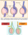
Define varicocoele
An abnormal dilation of the internal spermatic veins and pampiniform plexus that drain blood from the testis, forming a scrotal mass
Explain the pattern of growth of varicocoele
90% of cases on left side; 10% are bilateral because of:
- Lack of effective valves between the left testicular vein and left renal vein- most commonly thought to be caused by incompetent valves within the left internal spermatic vein.
- The angle at which the left testicular vein meets the left renal vein
- The left internal spermatic vein is 8 to 10 cm longer, resulting in increased hydrostatic pressure transmission.
- Increased reflux from compression of the renal vein (between the superior mesenteric artery and the aorta)

Summarise the epidemiology of varicocoele
- Unusual in boys under 10 yrs old
- Incidence increases after puberty
- Incidence: 10-15% in general population
- The majority (>80%) of adult varicoceles are not associated with infertility. However, the prevalence of varicocele is greater in patients with reduced fertility.
Recognise the presenting symptoms of varicocoele
An adult patient with a varicocele is usually asymptomatic and will typically present after failed attempts at conception, seeking an evaluation for infertility.
- 2-10% have symptoms
- Scrotum feels like a bag of worms
- Scrotal heaviness
- Often visible as distended scrotal blood vessels
- May feel dull ache
- Small testicle- larger varicoceles are associated with higher incidence of testicular growth arrest
Recognise the signs of varicocoele on physical examination
Patient must be STANDING for examination
- The side of the scrotum with the varicocoele will hang lower
- The swelling may reduce when lying down
- Valsalva manouevre whilst standing will increase dilatation
- Cough impulse
Identify appropriate investigations for varicocoele
-
scrotal ultrasound with colour flow Doppler imaging
- Used as adjunct to a physical examination to detect varicocele in men with difficult examination
- eg due to small scrotum, or to obesity.
-
semen analysis
- for infertile men with a varicocele, 2 or 3 semen analyses are recommended
- result- variable; reduced sperm count; impaired sperm motility (<50% motile spermatozoa)
-
serum FSH (± GnRH stimulation)
- FSH may be elevated (suggesting testicular dysfunction)
-
serum testosterone
- increasingly recognised as an uncommon cause of decreased testosterone production
Define testicular torsion
Testicular torsion is a urological emergency caused by the twisting of the testicle on the spermatic cord leading to constriction of the vascular supply and time-sensitive ischaemia and/or necrosis of testicular tissue