The Liver Flashcards Preview

Pharmacy; The Healthy Body 2 > The Liver > Flashcards

Flashcards in The Liver Deck (85)
Loading flashcards...
1
Q

THE HEALTHY LIVER

oSynthesis and secretion of _.

oMetabolism

oPlasma proteins– major source of plasma proteins including albumin and _ factors (see blood lectures)

oEndocrine function

oExcretory and degradative functions

oIron storage

A

Bile

Clotting

2
Q

SUMMARY OF LIVER FUNCTIONS

Exocrine (digestive) Functions- synthesis and the secretion of bile for the adequate digestion and absorption of _.

Secretes _ into a bicarbonate rich solution that helps to neutralize acid in the duodenum

Endocrine functions– e.g. Secretes insulin-like growth factor 1 (IGF-1) in response to growth hormone. This promotes cell division in a number of tissues including bones.

Clotting factors- Produces many of the plasma clotting factors including prothrombin an fibrinogen.

Bile salts – essential for the absorption of fat soluble vitamin K that is required for the formation of _ factors in the liver.

Plasma proteins- Synthesizes and secretes proteins including plasma _, acute phase proteins, binding proteins for a variety of hormones and lipoproteins

Metabolism-

  • Coverts plasma glucose to _ and triglycerides
  • Converts plasma amino acids to fatty acids
  • Synthesizes triglycerides and secretes them as lipoproteins
  • Produces glucose from glycogen (_)
  • Converts fatty acids to _ during fasting
  • Produces urea – major end product of amino acid (protein) catabolism and releases into the blood

Cholesterol metabolism-

  • Synthesizes cholesterol and releases into the blood
  • Secretes cholesterol into bile
  • Covers plasma cholesterol into bile salts

Iron and vitamin B12 storage

A

Fats

Bile

Clotting

Albumin

Glycogen

Glyconeogenesis

Ketones

3
Q

ANATOMY

A
4
Q

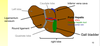
WHAT IS THE LIVER COMPOSED OF?

A

Four incompletely separated lobes that are supported by two ligaments

5
Q

GROSS LIVER ANATOMY

oApprox 1.5 kg (2 % body weight in adults)

oAnatomically four lobes: left, right, caudate and quadrate

oFunctionally considered as two lobes - left and right

oDiaphragmatic surface is the superior upper surface of the liver

oVisceral surface faces adjacent abdominal organs (faces downwards); the porta hepatis and gallbladder are located on this surface.

A
6
Q

WHAT ARE THE FOUR LOBES OF THE LIVER CALLED?

A

Left

Right

Caudate

Quadrate

7
Q

ANATOMY 2

  • Caudate (cauda = tail)
  • Quadrate (quadratus = squre)

The Caudate lobe is next to the _ vena cava

The Quadrate lobe is next to the _ bladder

A

Inferior

Gall

8
Q

WHERE IS THE CAUDATE LOBE OF THE LIVER?

A

Next to the inferior vena cava

9
Q

WHERE IS THE QUADRATE LOBE OF THE LIVER?

A

Next to the gall bladder

10
Q

LIVER STRUCTURE

oFalciform ligament: separates the major right and left lobes and attaches the liver to the _ and anterior abdominal wall

oRound Ligament: Found at the lower edge of the falciform ligament.

oGall bladder – accessory organ – pear shaped sac 7-10 cm long

-Rests in a recess on the inferior, visceral surface of the liver

FACIFORM LIGAMENT – secures the liver to the anterior abdominal wall

Round ligament – remnant of the _ _.

A

Diaphragm

Umbilical cord

11
Q

LIVER LOBES FROM VISCERAL SURFACE

A
12
Q

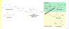
HEPATIC BLOOD CIRCULATION

oThe liver receives blood from two sources; the _ and GI tract.

oHepatic portal vein delivers poorly _ blood from the GI tract.

oHepatic artery delivers oxygenated blood from the heart.

oHepatic portal vein and artery divide into two to supply the left and right side of the liver

A

Heart

Oxygenated

13
Q

WHAT TWO SOURCES DOES THE LIVER RECEIVE BLOOD FROM?

A

Heart

GI Tract

14
Q

HEART STRUCTURE

A
15
Q

HEPATIC CIRCULATION

  • The hepatic portal vein carries blood form the GI tract, _ and pancreas,
  • Rich in nutrients but poor in _.
  • Hepatic artery – branches from _ and eventually splits into right and left hepatic arteries
  • Hepatic vein –at the centre of each lobule is the central vein that drains into the hepatic vein

oAorta supplies oxygenated blood via the hepatic artery

oBlood supplied to the liver via the hepatic portal vein and hepatic artery

oHepatic portal vein drains blood from the capillary beds of the GI tract including spleen and pancreas.

oHepatic vein carries blood away from the liver back to the _.

A

Spleen

Oxygen

Aorta

Heart

16
Q

WHERE DOES THE HEPATIC PORTAL VEIN CARRY BLOOD FROM?

A

Carries blood from the spleen, GI tract and pancreas

17
Q

HOW IS BLOOD SUPPLIED TO THE LIVER?

A

Via the hepatic portal vein and hepatic artery

18
Q

WHAT DOES THE HEPATIC VEIN DO?

A

Carries blood away from the liver back to the heart

19
Q

LIVER HISTOLOGY

o50,000 – 100,000 functional units called _. Hepatic lobules have a small polyhedral shape; these are the functional units of the liver.

oWithin each lobule are cells called _.

oAt the edge of each lobule are portal triads – these are formed by the hepatic portal vein, the hepatic artery and the bile duct

oCentral Vein empties into the hepatic veins and then the vena cava.

A

Lobules

Hepatocytes

20
Q

WHAT ARE PORTAL TRIADS FORMED BY?

A

Formed by the hepatic portal vein, the hepatic artery and the bile duct

21
Q

WHAT ARE THE CELLS INSIDE LIVER LOBULES CALLED?

A

Hepatocytes

22
Q

CLASSIC LIVER LOBULES

Blood flows from the portal triad (contains the portal vein and hepatic artery) towards the central _.

Bile flows in the opposite direction towards the portal triad (contains the bile _)

A

Vein

Duct

23
Q

LIVER LOBULE STRUCTURE

The numerous projections of the wheel spokes are the hepatic sinusoids – thin walled leaky _ where venous and arterial blood mix as they slowly flow through the hepatic lobe towards the central vein.

Hepatocytes absorb nutrients from blood and produce bile that collects in the small bile caniculus.

A

Capillaries

24
Q

WHAT ARE HEPATIC SINUSOIDS?

A

Thin walled leaky capillaries where venous and arterial blood mix as they slowly flow through the hepatic lobe towards the central vein.

25
Q

LIVER LOBULE STRUCTURE 2

A
26
Q

HEPATOCYTES

oLiver cells that separate sinusoidal blood from the canalicular bile.

oPolarized cells

oBasal membrane faces the liver sinusoidal endothelial cells

oApical membrane contributes to bile canaliculi jointly with the directly opposing hepatocytes.

A
27
Q

WHAT ARE HEPATOCYTES?

A

Liver cells that separate sinusoidal blood from the canalicular bile

28
Q

BILE

oExocrine secretory product of the liver

oBile contains HCO3-, cholesterol, lecithin (a phospholipid), bile pigments and bile salts.

oBile salts are important for the absorption of water insoluble _.

oStored and concentrated in the gall _.

oReleased during _.

A

Fats

Bladder

Meals

29
Q

WHAT DOES BILE CONTAIN?

A

HCO3-, cholesterol, lecithin (a phospholipid), bile pigments and bile salts.

30
Q

WHY ARE BILE SALTS IMPORTANT?

A

For the absorption of water insoluble fats

31
Q

LIVER LOBULES

oCounter current flow of _ and bile (opposite directions)

oBile ducts lined by _.

oBlood sinusoid lined by single layer of fenestrated endothelial cells.

oPortal field (portal triad) composed of portal hepatic vein, portal artery and bile duct.

oCentral vein drains to the vena cava.

A

Blood

Hepatocytes

32
Q

WHAT IS THE PORTAL FIELD/PORTAL TRIAD COMPOSED OF?

A

Portal hepatic vein

Portal artery

Bile duct

33
Q

LIVER SINUSOIDAL ENDOTHELIAL CELLS

oFilter between the lumen of the hepatic sinusoid and hepatocytes.

oMinimize any barrier for the bi-directional transfer of small or soluble substrates between blood and the extracellular space of Disse

oFenestrations are approximately 50–150 nm in diameter and most are aggregated into groups of 10–100, so-called liver sieve plates

A
34
Q

KUPFFER CELLS

o80 – 90% tissue macrophages in the liver

oAlso known as stellate macrophages

oDiscovered by pathologist C von Kupffer

oReside in the lumen of the sinusoids of the liver, adherent to endothelial cells

oImportant role in host _.

A

Defence

35
Q

WHAT DO KUPFFER CELLS HAVE AN IMPORTANT ROLE IN?

A

Host defence

36
Q

STELLATE CELLS

oStellate cells are found in the subendothelial space between the basolateral surface of hepatocytes and anti-luminal side of the sinusoidal endothelial cells.

oSpindle-shaped cell bodies with oval or elongated _.

oSingle stellate cell usually surrounds more than two nearby sinusoids.

oThought to be involved in _ formation.

A

Nuclei

Fibrosis

37
Q

WHERE ARE STELLATE CELLS FOUND?

A

Found in the subendothelial space between the basolateral surface of hepatocytes and anti-luminal side of the sinusoidal endothelial cells

38
Q

CYSTIC DUCT

oApprox 4 cm long

oConnects the neck of the gall _ to the common hepatic duct.

oCholangiocytes are epithelial cells that line the intra- and extra-hepatic ducts of the biliary tree

oModifies bile to generate ductal bile.

A

Bladder

39
Q

BILE DUCT CIRCULATION

  1. Bile is transported down Bile canaliculi in the liver.
  2. The bile canaliculi empty into bile ductules and then bile ducts.
  3. Empties into the _ ducts.
  4. Bile then directly enters the _ or diverted to the cystic duct into the gall bladder.
  5. The gall bladder concentrates and stores bile ready for use.
A

Hepatic

Duodenum

40
Q

GALL BLADDER

oPear shaped, 7 -10 cm long

oFundus: wide end of gall bladder and projects from the inferior border of the liver

oBody: contacts the visceral surface of the liver

oNeck: narrow and tappered; makes an S bend into the cystic duct. Internally the mucosa spirals into the spiral fold that keeps the cystic duct open.

A
41
Q

INTRAHEPATIC BILE DUCT ANATOMY

A
42
Q

WHAT ARE CHOLANGIOCYTES?

A

Epithelial cells that line the intra-hepatic and extra-hepatic ducts of the biliary tree

43
Q

WHAT DOES THE CYSTIC DUCT DO?

A

Connects the neck of the gall bladder to the common hepatic duct

44
Q

EXTRAHEPATIC BILE DUCT ANATOMY

A
45
Q

THE IMPORTANCE OF CHOLESTEROL

oSynthesis of cell membranes and contributes to the fluidity of membranes (lipid rafts)

oPrecursor for the synthesis of several molecules including vitamin D, cortisol, aldosterone, progesterone, estrogen, testosterone, bile salts.

A
46
Q

WHY IS CHOLESTEROL IMPORTANT?

A

oSynthesis of cell membranes and contributes to the fluidity of membranes (lipid rafts)

oPrecursor for the synthesis of several molecules including vitamin D, cortisol, aldosterone, progesterone, estrogen, testosterone, bile salts

47
Q

CHOLESTEROL SYNTHESIS

o80 % total daily cholesterol production is found in the liver

oSynthesized from Acetyl Co-A

oMulti-step pathway to form cholesterol from AcCoA that can be divided into 3 major steps.

A
48
Q

WHAT IS CHOLESTEROL SYNTHESISED FROM?

A

Acetyl Co-A

49
Q

CHOLESTEROL SYNTHESIS: STEP 1

A
50
Q

CHOLESTEROL SYNTHESIS: STEP 1 (CONTINUED)

A
51
Q

CHOLESTEROL SYNTHESIS: STEPS 2/3

oSqualene is synthesized from isopentenyl pyrophosphate

oCyclization of squalene to form lanosterol

oLanosterol goes through several steps to eventually form cholesterol.

A
52
Q

CHOLESTEROL TRANSPORT IN THE BODY

o_ molecule and does not dissolve well in aqueous environment

oPackaged with phospholipids and apolipoproteins to form a series of different lipoproteins

oLipoproteins contain a lipid core (contain cholesterol esters and triglyercides) and a hydrophilic outer surface containing phospholipids, free cholesterol and apolipoproteins

oMain cholesterol carrying lipoproteins are low _ and high density lipoproteins (LDL and HDLs)

A

Lipophilic

Density

53
Q

LIVER CHOLESTEROL METABOLISM

oAtherosclerosis is a progressive disease of large arteries and a leading cause of cardiovascular diseases and stroke.

oElevated levels of circulating _ associated with the development of atherosclerosis.

oInhibitors of cholesterol _ used to treat atherosclerosis

oStatins inhibit HMG CoA Reductase in the cholesterol synthesis pathway

o Newer drugs target LDL levels in the body (PCSK9 inhibitors)

A

LDL

Synthesis

54
Q

WHAT IS A COMMON TREATMENT OPTION FOR ATHEROSCLEROSIS?

A

Cholesterol synthesis inhibitors

55
Q

WHAT IS ATHEROSCLEROSIS?

A

A progressive disease of the large arteries, and a leading cause of cardiovascular diseases and stroke

56
Q

BILE SALT PRODUCTION

A
57
Q

BILE SALT SYNTHESIS

Liver receives blood from the gastrointestinal tract via the portal vein and hepatic _.

Blood returns to the system circulation via the hepatic vein into the vena cava.

Liver continuously produces bile into the bile duct that goes to the GI tract or gall _.

A

Artery

Bladder

58
Q

WHAT DOES BILE CONTAIN?

A
  1. Bile salts
  2. Lecithin (a phospholipid)
  3. HCO3-
  4. Cholesterol
  5. Bile pigments
  6. Trace metals
59
Q

BILE PRODUCTION

oHepatocytes secrete hepatic _ into the bile canaliculi

oHepatic bile also contains bile salts, bile pigments, cholesterol and lecithin (a phospholipid)

oEpithelial cells that line the bile ducts secrete a bicarbonate rich fluid that increases the _ of the bile

oLiver produces 600 – 1000 ml bile per day discharged into the duodenum or stored in gall bladder.

A

Bile

Volume

60
Q

CHEMICAL NATURE OF BILE ACIDS

oLiver produces around 500 mg bile salts per day

oPrecursors of bile salts are bile acids and they are synthesized from _.

oPrimary bile acids – cholic acid and chenodeoxycholic acid - are synthesized in the hepatocytes.

oSecondary bile acids – deoxycholic and lithocholic – are formed in the intestine by the dehyroxylating action of bacteria _.

A

Cholesterol

Flora

61
Q

WHAT ARE THE TWO PRIMARY BILE ACIDS AND WHERE ARE THEY SYNTHESISED?

A

Cholic acid

Chenodeoxycholic acid

Synthesised in the hepatocytes

62
Q

WHAT ARE THE TWO SECONDARY BILE ACIDS AND WHERE ARE THEY SYNTHESISED?

A

Deoxycholic

Lithocholic

Formed in the intestine by the dehydroxylating action of bacteria flora

63
Q

BILE SALTS

oPrimary or secondary bile acids are conjugated to _ _ (e.g. glycine) to generate water soluble bile salts.

oExample: cholic acid with glycine forms glycocholate.

oBile salts – hydrophobic and hydrophilic regions that aggregate to form _ at a critical concentration.

oImportant for the emulsification of _.

A

Amino Acids

Micelles

Fats

64
Q

ENTEROHEPATIC CIRCULATION

oThe majority of bile salts are re-absorbed by a _-dependent pathway in the ileum (last segment of small intestine)

oApprox 95% bile salts that enter intestine are recycled back to the _ via the portal vein.

oUptake of bile salts form the portal blood into hepatocytes occurs via a active transport pathways.

oRecycling from the GI Tract to the liver is known as the enterohepatic circulation

A

Sodium

Liver

65
Q

WHAT IS ENTEROHEPATIC CIRCULATION?

A

Recycling from the GI tract to the liver

66
Q

ENTEROHEPATIC CIRCULATION 2

o5% are lost in faeces (but liver makes new bile from cholesterol to replace the lost bile)

oDuring digestion – Bile can be recycled several times via the enterohepatic circulation.

A
67
Q

WHAT IS THE SPHINCTER OF ODDI?

A

A ring of smooth muscle found where the common bile enters the small intestine

68
Q

CHOLECYSTOKININ (CCK)

oPeptide hormone

oProduced by the small intestine

oStimulus for release = amino acid, fatty acids in the intestine

A
69
Q

REGULATION OF BILE SECRETION

A
70
Q

GALL BLADDER

oSphincter of oddi closed -> bile is diverted to the gall bladder.

-Stores bile not required immediately for _ of meals.

oConcentrates the bile by removal of salts and _.

oCan increase concentration of bile salts up to 20 fold.

A

Digestion

Water

71
Q

WHAT HAPPENS TO BILE WHEN THE SPHINCTER OF ODDI IS CLOSED?

A

The bile is directed to the gall bladder, where it is stored

72
Q

EXCRETORY ROLE OF BILE

oBile pigments are breakdown product of heme portion of _ from erythrocytes broken down in the liver and spleen.

oBile contains approx 0.2 % bile pigments

oResponsible for characteristic colour of bile (greenish yellow)

oMajor pigment is bilirubin; found in bile and secreted into duodenum.

A

Haemoglobin

73
Q

WHAT IS THE MAJOR PIGMENT FOUND IN BILE?

A

Bilirubin

74
Q

WHAT ARE BILE PIGMENTS?

A

Breakdown products of heme portion of haemoglobin from erythrocytes broken down in the liver and spleen

75
Q

SYNTHESIS: PLASMA PROTEINS

oLiver secretes large numbers of proteins.

oAlbumin: plasma protein, transport lipids and steroid hormones

oGlobulins: approx 40% total plasma proteins; alpha and beta- globulins also transport lipids and steriod hormones

oClotting factors: most are produced by liver e.g. fibrinogen and prothromin

A
76
Q

WHAT DO ALPHA AND BETA-GLOBULINS DO?

A

They transport lipids and steroid hormones.

77
Q

SYNTHESIS: INSULIN-LIKE GROWTH FACTORS

oInsulin like growth factor (IGF) is an important mediator of growth hormone (GH) action.

oLiver synthesizes IGF-1 and -2 in response to growth hormone

oStructurally similar to pro-insulin

oPotent growth promoting effects.

oIGF-1 levels low in infancy, peak in puberty during growth and declines in adult.

oIGF-2 may be more important in fetal and neonatal growth.

A
78
Q

DRUG METABOLISM

A
79
Q

DRUG METABOLISM: PHASE 1 REACTIONS

oCytochrome P450 enzymes are important

oSuperfamily of related enzymes

oPopulation variation in P450 enzymes are important in terms of therapeutics

oNaturally occurring dietary substances can inhibit (e.g. grapefruit juice) or induce (brussels sprouts) P450 enzymes with impact on drug metabolism

A
80
Q

CLOPIDOGREL METABOLISM

oAnti-thrombotic agent that inhibits _ activation (involved in blood clotting)

oPro-drug that requires oxidation.

oA 2-step metabolism using hepatic P450 enzymes to generate the active thiol metabolite that reacts with platelets

oBinds selectively and irreversibly to P2Y12 receptor on _ membranes.

oThe majority of clopidogrel is also inactivated by blood esterases so only 15% metabolised by hepatic P450 enzymes

oA two step metabolism phase leads to a delayed onset of action for clopidogrel

oGenetic mutations in P450 enzymes are associated with a reduced response to clopidogrel treatment

A

Platelet

Platelet

81
Q

WHAT IS CLOPIDOGREL?

A

Anti-thrombotic agent that inhibits platelet activation (involved in blood clotting)

82
Q

DRUG METABOLISM: PHASE 2 REACTIONS

oPhase 2 is a conjugation step that almost always leads to a pharmacologically inactive or less lipid soluble product that is eliminated in urine or bile.

oGroups most often associated with conjugation are glucuronyl, sulphate, methyl, acetyl, glycyl and glutathione

A
83
Q

PARACETAMOL TOXICITY

oParacetamol is commonly used non-narcotic analgesic-antipyretic agent

oOral drug, peak plasma concentration 30-60 mins and inactivated in liver by conjugation to glucuronide or sulphate

oToxic dose (2-3 x max therapeutic dose) causes hepatotoxicity and potentially renal toxicity

oNormal conjugation pathways become saturated and drug metabolised by mixed function oxidases to form N-acetyl-p-benzoquinone imine (NAPBQI) = very toxic to cells

A
84
Q

IMMUNE SYSTEM: KUPFFER CELLS

oCritical component of the mononuclear phagocytic system

oResident macrophage of the liver

oFound in the hepatic blood sinosoids

oEfficiently phagocytize pathogens entering from the blood circulation

oFirst line of defence against particulates and immunoreactive material passing from the gastrointestinal tract via the portal circulation and may be considered as a final component in gut barrier function

A
85
Q
A