Thorax and Lungs Evaluation Flashcards

(49 cards)

1
Q

Left Upper Lobe Apicoposterior Segment

A

Above the clavicle along the midclavicular line

2
Q

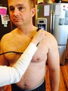
Left Upper Lobe Anterior Segment

A

Somewhere between the 1st and 2nd intercostal space medially

3
Q

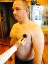
Left Upper Lobe Superior Lingula Segment

A

In the 3rd intercostal space between the midclavicular and axillary lines

4
Q

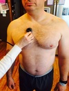
Left Upper Lobe Inferior Lingula Segment

A

Somewhere between the 4th intercostal space and the 5th rib near the apex of the heart

5
Q

Left Lower Lobe Anteromedial Basal Segment

A

Somewhere between the 5th intercostal space and the 6th rib along the midclavicular line

6
Q

Left Lower Lobe Superior Segment

A

Somewhere between 1” below the spine of the scapula and 1” above the inferior angle medially

7
Q

Left Lower Lobe Lateral Basal Segment

A

Somewhere between 1” above/below the inferior angle of the scapula and the 10th intercostal space laterally

8
Q

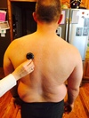
Left Lower Lobe Posterior Basal Segment

A

Somewhere between 1” above the inferior angle of the scapula and the 10th intercostal space medially

9
Q

Right Upper Lobe Apicoposterior Segment

A

Above the clavicle along the midclavicular line

10
Q

Right Upper Lobe Anterior Segment

A

Somewhere between the 1st and 2nd intercostal spaces medially

11
Q

Right Upper Lobe Posterior Segment

A

1” above the spine of the scapula medially

12
Q

Right Middle Lobe Lateral Segment

A

Somewhere between the 3rd intercostal space and the 5th rib laterally

13
Q

Right Middle Lobe Medial Segment

A

Somewhere between the 3rd and 5th intercostal spaces medially

14
Q

Right Lower Lobe Superior Segment

A

Somewhere between 1” below the spine of the scapula and 1” above the inferior angle of the scapula medially

15
Q

Right Lower Lobe Anterior Basal Segment

A

In the 5th intercostal space or 6th rib area between the midclavicular and axillary lines

16
Q

Right Lower Lobe Lateral Basal Segment

A

Somewhere between 1” above/below the inferior angle of the scapula and the 10th intercostal space laterally

17
Q

Right Lower Lobe Posterior Basal Segment

A

Somewhere between 1” above the inferior angle and the 10th rib medially

18
Q

When inspecting rate, rhythm, and depth of breathing you would typically expect RR to be __-__ breaths per minute.

A

12-20

19
Q

What are a few indications of respiratory distress?

A
  • Increased RR
  • Nasal flaring
  • Intercostal and sternal retracftions
  • Visible expression of distress
  • Increased use of neck accessories
  • Paradoxic breathing
20
Q

What are 2 chest abnormalities to look for?

A

Pectus excavatum (funnel chest)

Pectus carinatrum (pigeon chested)

21
Q

Assessment of the neck reveals jugular vein distention, what does this indicate?

A

cor pulmonae (R side heart failure)

22
Q

What things should you look for in regards to chest dimensions?

A
  • scoliosis
  • barrel chest (hyperinflation)
  • kyphosis
  • professorial positioning
23
Q

The normal AP dimension of the chest is how big?

How big is the transverse dimension?

A

1/2 the size of the transverse dimension, which is usually the width of the shoulder

24
Q

Why is important to ask about couch production?

A

Weak and ineffective cough production is indicative ofweak respiratory musculature

25
Why should you assess the digits?
Digital clubbing indicates chronic tissue hypoxia
26
Evaluation of the mediastinum assesses tracheal shift that is due to what?
disproportionate intrathoracic pressures of lung volumes between the 2 sides of the thorax
27
What may be the cause if the contents of the thorax shift toward the affected side?
Lung volume on that side is decreased * lobectomy * peneumonectomy * large degree of atelectasis
28
What may be the cause if the contents of the thorax are shifted towards the unaffected side?
There is increased pressure on the same side * Pleural effusion * Tumor * Untreated Pneumothorax
29
In what position should you palpate the mediastinum for tracheal deviation?
while the patient is sitting upright with the neck flexed slightly to allow the SCM muscles to relax
30
In what position should you palpate the scalenes during quite breathing
31
Describe the positioning to assess motion of the upper, middle, and lower lung lobes
32
In what position should you assess diaphragmatic motion? Describe the process for assessment
In supine, palpate the anterior chest wall with the thumbs over the costal margins and thumb tips meeting at the xyphoid. Instruct the patient to inhale deeply. Assess the movement of the hands; they should travel equally apart with a total circumferential diameter increase of at least 2-3 inches
33
What can be defined as the vibration that is produced by the voice or by the presence of secretions in the airways and is transmitted to the chest wall and palpated by the hand
Fremitus
34
Describe the procedure for assessing fremitus
Place the palms of your hands lightly on the posterior chest wall and instruct the patient to say the word "99"
35
What is indicated if there is increased resonance palpated while testing for resonance?
the presence of an increase in secretions in a particular area (consolidation)
36
What is indicated if there is decreased resonance palpated while testing for resonance?
There is an increase in air in the particular area (air pocketing)
37
In what positon should you perform percussion?
anterior supine, posterior seated
38
Describe the process of percussion Explain the sounds you may hear
* Place the middle finger between 2 ribs, while all other fingers are lifted off the chest * Proceeds in a cephalocaudal direction and back and forth between the left and right sides * Normal sound is when normal lung tissue is percussed and normal resonance is produced * A dull sound is produced with percussion over the liver or other dense tissue and is described as a “thud” * A tympanic sound is loud, long, and hollow and may be heard over an empty stomach or hyperinflated chest
39
Describe the process of determining excursion of the diaphragm
* Position the patient in sitting with the back exposed * Precuss from the apex of the lungs to the bases of the lungs while the patient is quietly breathing * Draw a line at the point where the resonance becomes dull * Then ask the patient to take a maximal inspiration and hold that breath * Percuss caudally until you agian hear the dullness and mark this spot * This distance between the 2 lines represents diaphragmatic excursion
40
What is normal diaphragmatic excursion?
3-5 cm
41
In what patient population will diaphragmatic excursion be reduced?
hyperinflated chests (COPD)
42
What does normal bronchial lung sound sound like? Where can it be heard?
They sound hollow, tubular sounds that are lower pitched. They can be auscultated over the trachea where they are considered normal
43
Describe bronchovesicular lung sounds. Where can they be heard?
Inspiration to expiration periods are equal with bronchovesicular lung sounds. These are normal sounds in the mid-chest area or in the posterior chest between the scapula. They reflect a mixture of the pitch of the bronchial breath sounds heard near the trachea and the alveoli with the vesicular sound.
44
Describe vesicular breath sounds
They are soft and low pitched with a rustling quality during inspiration and are even softer during expiration. These are the most commonly auscultated breath sounds, normally heard over the most of the lung surface
45
Describe what wheezes sound like. What do they indicate?
continuous with a musical quality. Wheezes can be high or low pitched. High pitched wheezes may have an auscultation sound similar to squeaking. Lower pitched wheezes have a snoring or moaning quality narrowing of the airways
46
Describe what crackles sound like What is the difference between fine and coarse crackles?
brief, discontinuous, popping lung sounds that are high-pitched Coarse crackles are louder, lower in pitch and last longer
47
What may crackles be casued by?
restrictive or obstructive respiratory disorders
48
What does pleural rub sound like?
like two pieces of leather or sandpaper rubbing together
49
Where should you assess for pleural rub?
in the lower lateral chest areas