Topic 7 CNS Flashcards

(65 cards)

1
Q

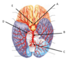
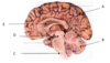
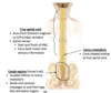
What is the name of the structure labelled E?

A

anterior cerebral artery

2
Q

Describe the three sections of the brain stem.

A
  • As spinal chord enter foramen magnum it is called the brain stem.
  • superiorly it consists of:
    1. medulla oblongata
    2. pons (latin = bridge) - links cerebellum to cerebrum
    3. midbrain
3
Q

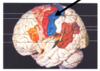
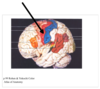
What is the name of the structure labelled D in the diagram?

A

Midbrain

4
Q

What is the innermost layer of the brain and spinal cord meninges called?

A

The pia mater (delicate mother)

Very fine transparant layer that adhers to surface of brain, contains blood vessels.

DUR-AR-PIA

5
Q

What is the true spinal cord?

A
  • true spinal cord runs from the foramen magnum to the 3rd or 4th lumbar vertebra ( at the conus medullaris)
  • part of the CNS - start and finish of the PNS as senosyr and motor nerves travel to and from.
6
Q

What are dural venous sinuses?

A
  • Large dilated blood vessels (spaces) formed between the periosteal and meningeal layer of the dura mater.
  • Contain venous blood that empties into the internal jugular vein
  • Arachnoid granules protrude from the sub-arachnoid space into into them to transfer CSF that has already circulated the CNS
7
Q

What is the ventricular system of the brain comprised of, and what is its function?

A

The brain’s ventricular system is comprised of a series of ventricles and ducts in the brain.

Its function is to produce cerebrospinal fluid (CSF) and circulate it around the tissues of the CNS.

8
Q

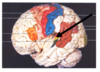
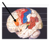
What is the name of the cerebral lobe in cream on the diagram?

A

Temporal lobe

9
Q

Where is the primary sensory cortex located on the diagram below? What is its function?

A
  • posterior to central sulcus
  • recieves sensory information from whole body
10
Q

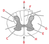
What is the name of the structure labelled M below? What does it contain?

A

Spinal nerve

The dorsal nerve roots (sensory) and ventral nerve roots (motor) combine to form spinal nerves.

11
Q

What is the name of the area of the spinal cord labelled C? What type and part of neurons does it contain?

(i.e. motor/sensory, cell bodies/axons/dendrites)

A

Anterior horns of grey matter

Contains cell bodies of motor neurons.

12
Q

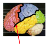
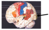
Where is the primary motor cortex of the brain located on the diagram below? What is its function?

A
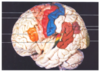
  • Red area in front of central sulcus
  • Sometimes called pre-central gyrus
  • Controls motor function for the whole body
13
Q

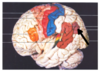
What is the name of the structure labelled A in the diagram?

A

Cerebrum

14
Q

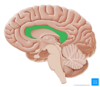
What is the diencephalon and what two important structures does it contain?

A
  • The diencephalon is located between the mid brain and the lateral ventricle.
  • It contains the thalmus (directs traffic from brain stem to correct area of cortex) and the hypothalymus (part of endocrine system that controls homeostasis)
15
Q

What is the name of the structure labelled B in the diagram?

A

Cerebellum

16
Q

What is the conus medullaris?

A

The cone shaped ending of the true spinal cord (around 3rd/4th lumbar vertebra)

17
Q

What is the name of the area of the spinal cord labelled E? What does it contain?

A

Central canal

Contains CSF (cerebrospinal fluid)

18
Q

What are the two layers of the dura mater called and where are they located?

A

The outermost layer is called the periosteal layer

The innermost layer is called the meningeal layer

19
Q

What is the name of the structure labelled C?

A

vertebral artery

20
Q

What is the difference between the distribution of grey matter in the brain versus the spinal cord?

A
  • In the brain the grey matter is located superficial to the white matter
  • in the spinal cord the grey matter is located deep to the white matter (in an ‘H’ shape)
21
Q

Describe the path of arterial blood supply to the posterior brain.

A

► L and R vertebral arteries (travel up brain stem and through foramen magnum)

► Combine to form basilar artery

► branches into:

  1. Left posterior cerebral artery
  2. Right posterior cerebral artery
22
Q

What is the name of the structure labelled H below? What is it carrying?

A

Posterior (dorsal) root

The posterior (dorsal) root is carrying sensory fibres from the body to the cell bodies in the posterior horn of grey matter which then travel up the spine cord.

23
Q

What is the ventricular system in the brain continuous with (down the spinal cord)?

A

Central canal of the spinal cord

24
Q

What is the space between the arachnoid and pia mater meninges called. What does it contain?

A

Sub-arachnoid space

Contains circulating cerebralspinal fluid (CSF)

25
What is the name of the cerebral lobe in **green** on the diagram?
Parietal lobe
26
What is the name of the area of the spinal cord labelled **D**? What type and part of neurons does it contain? ## Footnote *(i.e. motor/sensory, cell bodies/axons/dendrites)*
**Posterior horns of grey matter** Contains **cell bodies** of **sensory neurons**.
27
In which lobe would you find the **primary sensory area**?
parietal lobe
28
Where is the **primary auditory area** located on the diagram below? What is its function?
* deep in temporal lobe * **recieves sensory information about hearing and equilibrium (vestibular area)**
29
What is the name of the structure indicated by the **red arrow**? What is it carrying?
**Anterior (ventral) root** The anterior (ventral) roots consist of axons of **motor fibres** _from_ cell bodies in the anterior horn of grey matter _to_ the muscles in the body (and some glands).
30
In which lobe would you find **Broca's area**?
Frontal lobe
31
How is **CSF reabsorbed** after circulating the brain and spinal cord?
* CSF is circulating in the **subarachnoid space** * It is reabsorbed after circulation through **arachnoid granulations** that protrude from the subarachnoid space into **dural venous sinuses** (between layers of dura mater) * Dural venous sinuses contain venous blood draining into the **internal jugular vein**.
32
In which lobe would you find the **primary motor area**?
frontal lobe
33
Describe the path of arterial blood supply to the **anterior** brain.
**►common carotid artery** ► **internal carotid artery** (enters skull through **carotid canal**) ► branches into: 1. **anterior cerebral artery** (anterior), 2. **posterior communicating artery** (posterior), 3. **middle cerebral artery laterally.**
34
What is the difference between **grey matter** and **white matter** in the brain?
* **Grey matter** contains mostly *cell bodies* of neurons * **White matter** contains mostly *axons/dendrites. A white substance called **myelin** surrounds this part of the cell, appearing white*
35
What is the **cerebellum**? What/how does it attach to?
* The cerebellum recieves sensory information ond then uses this to **coordinate voluntary motor movements** such as posture, balance, speech etc. * It connects to the brain stem via the **cerebellar peduncles**
36
What is the name of the structure labelled **A**?
anterior communicating artery
37
What is the name of the indentation that runs medially between the L and R hemispheres of the brain?
Longitudinal fissure
38
What is the name of the cerebral lobe in **blue** on the diagram?
Frontal lobe
39
Where is the **motor coordination area**?
cerebellum
40
Where is the **anguli gyrus** located on the diagram below? What is its function?
* **comprehension of written material/reading comprehension**
41
In which lobe would you find the **primary auditory cortex**?
Temporal lobe
42
What is **cerebrospinal fluid (CSF)** and what is its function?
* **watery fluid** continuously circulating around brain and spinal cord * provides **cushioning** to protect from jolts and blows * protects against **infection** (also clears infection quickly if occurs)
43
What is the name of the structure labelled **E** in the diagram?
Lateral ventricle
44
What is the name of the cerebral lobe in **red** on the diagram?
Occipital lobe
45
What is the **corpus collosum** and where is it located?
The corpus collosum is a mass of white tissue (axons) that run between the L and R hemisphere and allow them to communicate It is located around the lateral ventricle below the longitudinal fissure.
46
Where is the **pre-motor area/cortex** located on the diagram below? What is its function?
* anterior to primary motor cortex in frontal lobe * **controls repetitive/learned motor skills (e.g. playing a musical instrument)**
47
What feature of the brain is the arrow pointing to?
Lateral sulcus
48
What is the **cauda equina**?
* means *'horses tail'* * located **inferior to conus medullaris** * send and recieves messages from legs, feet and pelvic organs.
49
What are the **ventricles** of the brain called, and where are they located?
1. **Lateral ventricles** - (Ventricles 1&2) found on either side of corpus callosum 2. **Third ventricle** - found on midline, just inferior to lateral ventricles 3. **Fourth ventricle** - found between cerebellum and spinal cord
50
Describe the arterial blood supply to the **spinal cord.**
* the spinal cord gets arterial blood supply from branches of the **vertebral arteries**. * the **anterior** spine has a single artery running down the midline - ***anterior spinal artery.*** * The ***left and right posterior vertebral arteries*** run down the **posterior** surface of the spinal cord.
51
What is the **outermost layer** of the brain and spinal cord meninges called?
**The Dura mater** (*tough mother*) *DUR-AR-PIA*
52
Describe the **circulation of CSF** around the CNS.
1. Mostly **produced in lateral ventricles** 2. Flows down to **3rd ventricle** 3. Travels through **central aqueduct** to **4th ventricle** 4. Either enters **central canal** and travels to base of spine, or enters **subarachnoid space** and flows around brain 5. **Absorbed into the venous blood** at **dural venous sinuses**.
53
Where is **Broca's area** located on the diagram below? What is its function?
* located in **_frontal lobe_** * only found fully developed in dominant hemisphere of brain * **Involved in speech motor planning** ***Brocha's aphasia = normal understanding but reduced length and complexity of speech***
54
What is the name of the duct that **joins the third and fourth ventricle** in the brain called?
**Cerebral aqueduct**
55
Where is the **primary visual area** of the cortex located? What is its function?
* located at back of brain in **_occipital lobe_** * **recieves and processes visual information**
56
What feature of the brain is the arrow pointing to?
Parieto-occipital sulcus
57
What is the name of the structure labelled **D**?
posterior cerebral artery
58
What is the name of the structure labelled **C** in the diagram?
Medulla oblongatta
59
Which major blood vessel supplies branches to the **pons**?
**Basilar artery**
60
Where is **Wernicke's area** located on the diagram below? What is its function?
* located in mostly in **_temporal lobe_** * **Involved in language comprehension** ***Wernicke's aphasia = fluent speech but reduced comprehension***
61
In which lobe would you find **Wernicke's area**?
Temporal lobe
62
What feature of the brain is the arrow pointing to?
Central sulcus
63
What is the the **Circle of Willis**? What arteries is it made up of?
The Circle of willis is a circular region in the middle of the brain formed from the **anastomosis (joining) of the anterior and posterior brain arterial supply.** _Comprised of:_ **L and R anterior cerebral artery** (from internal carotid) **Anterior communicating artery** (joins anterior cerebral arteries) **L and R posterior communicating arteries** (from internal carotid) **L and R posterior cerebral arteries** (from basilar)
64
What is the name of the structure labelled **B**?
basilar artery
65
What is the **middle layer** of the brain and spinal cord meninges called?
**The arachnoid** (*web-like*) *DUR-AR-PIA*