transport in animals? Flashcards
(20 cards)
explain the need for transport systems in multicellular animals in terms of size, level of activity and surface area: volume ratio?

Single vs double circ sytem?

open vs closed circulatory system?

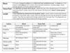
lable this heart?

explain, with the aid of diagrams the differences in the thickness of the walls of the different chambers of the heart in terms of their functions?

describe the cardiac cycle, with reference to the action of the valves in the heart?
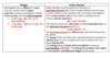
Atrial Systole/ Ventricular Diastole:
Blood enters the atria and the pressure inside the atria increases
The pressure is higher in the atria than the ventricles so the atrioventricular valves open
The atria contract pushing blood into the ventricles
Atrial Diastole/ Ventricular Systole:
4.Blood enters the ventricles and the pressure inside the ventricles increases
The pressure becomes higher in the ventricles than the atria forcing the atrioventricular valves to close to prevent the backflow of blood
The pressure in the ventricles is also higher than in the major arteries (aorta and pulmonary artery), forcing the semi-lunar valves open
The ventricles contract and blood is forced out into the arteries
Atrial Systole/ Ventricular Diastole:
The atria and ventricles are relaxed
The pressure is higher in the major arteries (aorta and pulmonary artery) so the semi-lunar valves close to prevent the backflow of blood
The pressure is slightly higher in the atria than in the ventricles so the atrioventricular valves open
Blood flows passively into ventricles from the atria
) describe how heart action is coordinated with reference to the sinoatrial node (SAN), the atrioventricular node (AVN) and the Purkyne tissue
The cardiac muscle is myogenic – the heart will contract and relax by itself
The SAN initiates a wave of excitation in the right atrium which spreads over the walls of both atria causing atrial systole
The wave of excitation spreads to the AVN, found at the top of the inter-ventricular septum, as there’s a band of fibres between the atria and ventricles which stops the wave of excitation passing to the ventricle walls.
The AVN delays the wave of excitation to allow time for the atria to finish contracting and for blood to flow down the ventricles
The AVN directs the wave of excitation to the Purkyne tissue, which runs down the inter-ventricular septum. The wave of excitation spreads upwards from the apex (base) of the ventricles causing ventricular systole, pushing blood upwards towards the arteries
interpret and explain electrocardiogram (ECG) traces, with reference to normal and abnormal heart activity
Electrocardiograms are used to monitor the electrical activity of the heart by attaching a number of sensors to the skin. Some of the electrical activity generated passes through the tissues next to the heart and then to the skin so that the sensors can pick up the electrical excitation made by the heart and convert this into a trace.

describe, with the aid of diagrams and photographs, the structures and functions of arteries, veins and capillaries?

Differences between arteries vein and capiliiries?

difference beween arteries, veins and caps?

describe the pressure flow from the heart to rest of body?

The pressure in the blood decreases as it moves away from the heart because the blood vessel branchesinto more smaller vessels meaning they have a larger cross sectional area. Also there’s more frictionin the capillaries therefore there is a lower pressure.
It’s important that the pressure is lower by the time the blood reaches the capillaries because they are onlyone cell thick so a high pressure would cause them to burst and become damaged. Also a low pressure means a slow flow rate allowing time for exchange. Furthermore capillaries do not have a lot of elastic tissue.
The pressure in the veins is very low so blood is returned to the heart by the muscle around it contractingand pumping the blood with the help from valves to prevent back-flow of blood.
explain the differences between blood, tissue fluid and lymph
describe how tissue fluid is formed from plasma
At the arterial end of a capillary, the hydrostatic pressure is high, therefore plasma moves out of the capillaries because the pressure is higher in the capillaries than outside, moving down the pressure gradient.
In the capillaries because the plasma proteins are too large to pass through, they stay in the plasma, making the water potential lower than that in the tissue fluid so tissue fluid moves back into the capillaries at the venous end.
describe the role of haemoglobin in carrying oxygen and carbon dioxide?
describe and explain the significance of the dissociation curves of adult oxyhaemoglobin at different carbon dioxide levels (the Bohr effect)?

explaining the o2 dissaosciation curve?
Oxygen Dissociation Curve:
The first oxygen does not attach easily to the haem groups, due to it being in the centre of the haemoglobin molecule
The concentration of oxygen rises making it easier to oxygen to associate with the heam groups.
Once one oxygen molecule binds with a haem group, it causes a conformational change in shape of the haemoglobin molecule – this makes it easier for oxygen molecule to reach the haem group and associate with it
The next two oxygen molecules attach easily to the haem groups
The fourth oxygen molecule finds it difficult to associate with the last haem group. This means that 100% oxygen saturation is difficult even at high pO2 forming a sigmoid curve.

explain the significance of the different affinities of fetal haemoglobin and adult haemoglobin for oxygen
Fetal haemoglobin has a higher affinity for oxygen than adult haemoglobin. This is because the fetal haemoglobin must be able to ‘pick up’ oxygen from the haemoglobin from its mother.
Placenta has low partial pressure of oxygen
At low partial pressure of oxygen, in the placenta, adult haemoglobin will dissociate
Fetal haemoglobin takes up oxygen in lower partial pressure of oxygen

