Vascular System Flashcards
(36 cards)
Subclavian Artery Branches

- vertebral
- internal thoracic
- pericardiophrenic
- (rami perforantes)
- (ant intercostales)
- musculophrenic
- sup epigastric
- post cervical trunk
- intercostalis superior
- cervicalis profunda
- thyrocervical trunk
- inferior thyroid
- ascending cervical
- transverse cervical
- superficial cervical
- suprascapular
- dorsal scapular (sometimes a branch of trans cervical)
Continues on to become axillary artery
Axillary Artery Branches

- sup thoracic
- thoracoacromial:
- rami pectoralis, deltoid, acromial
- subscapular
- thoracodoraslis and circumflexa scapulae
- circumflexa posterii humerii
- circumflexa anterior humerii
- thoracolateralis
BRACHIAL
- branches
- profunda brachii
- sup ulnar collateral
- inf ulnar collateral

Thoracic Aorta Branches
Visceral and Parietal Branches:
- bronchial (v)
- pericardial (v)
- oesophageal (v)
- mediastinal (v)
- post intercostal (3 - 11) (p)
- sup phrenic (p)
- subcostal (p)
External Carotid Artery

- sup thyroid
- lingual
- facial
- (sternocleidomastoid)
- ascending pharyngeal
- occipital
- post auricular
- maxillary
- superficial temporal
Opthalmic Artery
- lacrimalis
- centrales retinae
- ciliares longis i brevis
- ciliares anteriores
- rami musculares
- supraorbitalis
- palpabralis mediales
- ethmoidal anteriores
- ethmoidal posteriores
- dorsalis
- frontalis
Abdominal Aorta
Visceral Paired
- suprarenalis media
- renalis
- ovaria / testicularis
Abdominal Aorta
Visceral Unpaired
- celiac trunk
- superior mesenteric
- inferior mesenteric
Abdominal Aorta
Parietal Branches
- aa lumbales
- a phrenic inf
- median sacral
Coeliac Trunk
T12 level

- splenic:
- left gastroepiloic
- panceatic branches
- short gastric arteries (to fundus)
- common hepatic:
- proper hepatic, left and right hepatic, cystic from right
- right gastric
- gastroduodenal; right gastroepiploic and pancreaticoduodenalis sup
- left gastric

Superior Mesenteric Artery
L2
- pancreaticoduodenalis inf
- jejunalis et ilei
- iliocolica - appendicularis
- colica dextra
- colica media
Inferior Mesenteric Artery
L3
- colica sinsistra
- sigmoidei
- rectalis sup

Inetrnal Iliac
Visceral Branches
- umbilicales - vesicales sup
- vesicales inf
- rectalis media
- pudenda interna - uterina - vaginalis, tubarius, ovaricus
- ductus deferens
Internal Iliac
Parietal Branches
- iliolumbar
- sacralis lateralis
- gluteal (sup et inf)
- pudenda interna (both parietal and visceral)
- obturator
IMA Anastamoses
There are two major anastamoses of the IMA, both involving a union with branches of thesuperior mesenteric artery:
Marginal artery (of Drummond) – forms a continuous arterial circle along the inner border of the colon. Straight vessels (vasa recta) arise from the artery to supply the colon. It is formed by the union of several branches; the ileocolic, right colic and middle colic of the SMA and left colic and sigmoid branches of the IMA.
Arc of Riolan – anastamosis between the middle colic branch of SMA and the left colic branch of IMA. It is less common than the marginal artery, and indeed its existence has been questioned by some surgeons.

External Iliac
The common iliac artery bifurcates into the internal iliac artery and external iliac artery at the level of the pelvic brim anterior to the sacroiliac joint.
The external iliac artery courses medially along the iliopsoas muscle . After it enters the thigh under the inguinal ligament, it changes name and continues as the femoral artery, supplying the lower limb.
Branches
inferior epigastric artery
deep circumflex iliac artery
Femoral Artery
The main artery of the lower limb is femoral artery. It is a continuation of the external iliac artery (terminal branch of the abdominal aorta). The external iliac becomes the femoral artery when it crosses under the inguinal ligament and enters the femoral triangle.
In the femoral triangle, the profunda femoris artery arises from the posterolateral aspect of the femoral artery. It travels posteriorly and distally, giving off three main branches:
Perforating branches – Consists of three or four arteries that perforate the adductor magnus, contributing to the supply of the muscles in the medial and posterior thigh.
Lateral femoral circumflex artery – Wraps round the anterior, lateral side of the femur, supplying some of the muscles in the lateral side of the thigh.
Medial femoral circumflex artery – Wraps round the posterior side of the femur, supplying the neck and head of the femur. In a fracture of the femoral neck, this artery can easily be damaged, and avascular necrosis of the femur head can occur.

Obturator Artery
The obturator artery arises from internal iliac artery in the pelvic region. It descends via the obturator canal to enter the medial thigh, bifurcating into two branches:
Anterior branch – This supplies the pectineus, obturator externus, adductor muscles and gracilis.
Posterior branch – This supplies some of the deep gluteal muscles.
Gluteal Arteries
The gluteal region is largely supplied by the superior and inferior gluteal arteries. These arteries also arise from the internal iliac artery, entering the gluteal region via the greater sciatic foramen.
The superior gluteal artery leaves the foramen above the piriformis muscle, the inferior below the muscle. In addition to the gluteal muscles, the inferior gluteal artery also contributes towards the vasculature of the posterior thigh.

Popliteal Artery
The popliteal artery descends down the posterior thigh, giving off genicular branches that supply the knee joint. It moves through the popliteal fossa, exiting sandwiched between the gastrocnemius and popliteus muscles. At the lower border of thepopliteus, the popliteal artery terminates by dividing into anterior and posterior tibial arteries.
The branches of the popliteal artery are:
anterior tibial artery
posterior tibial artery
sural artery
medial superior genicular artery
lateral superior genicular artery
middle genicular artery
lateral inferior genicular artery
medial inferior genicular artery
Posterior Tibial Artery
The posterior tibial artery continues inferiorly, along the surface of the deep muscles (such as tibialis posterior). It accompanies the tibial nerve in entering the sole of the foot via the tarsal tunnel. During the descent of the posterior tibial artery in the leg, the fibular artery arises. This artery moves laterally, penetrating the lateral compartment of the leg. It supplies muscles in the lateral compartment, and adjacent muscles in the posterior compartment.
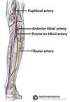
Anterior Tibial Artery
The other division of the popliteal artery, the anterior tibial artery, passes anteriorly between the tibia and fibula, through a gap in the interosseous membrane. It then moves inferiorly down the leg. It runs down the entire length of the leg, and into the foot, where it becomes the dorsalis pedis artery.

Arteries in the Foot
Arterial supply to the foot is delivered via two arteries:
Dorsalis pedis (a continuation of the anterior tibial artery)
Posterior tibial
The dorsalis pedis artery begins as the anterior tibial artery enters the foot. It passes over thedorsal aspect of the tarsal bones, then moves inferiorly, towards the sole of the foot. It thenanastamoses with the lateral plantar artery to form the deep plantar arch. The dorsalis pedis artery supplies the tarsal bones and the dorsal aspect of the metatarsals. Via the deep plantar arch, it also contributes to the supply of the toes.
The posterior tibial artery enters the sole of the foot through the tarsal tunnel. It then splits into the lateral and medial plantar arteries. These arteries supply the plantar side of the foot, and contributes to the supply of the toes via the deep plantar arch.
Branches of Pudenda Interna
- inf rectal
- perineal
- posterior labial/scrotal
- bulb of vestibule/penis
- dorsal artery of clitoris/penis
- deep artery of clitoris/penis
Veins Superior to Heart

- empty into SVC
- azygous (from below)
- brachiocephalic
- internal jugular
- vertebral
- external jugular
- subclavian
- axillary
- cephalic
- brachial


