Venous Disease Flashcards
(21 cards)
List the main superficial veins of the legs.
- Great Saphenous Vein 2. Small saphenous vein 3. Anterior accessory saphenous vein (AASV) 4. Vein of Giacomini 5. Sapheno-femoral junction (SFJ) 6. Sapheno-popliteal junction (SPJ)
What is the pathway of the great saphenous vein?
- Originates in Medial Foot as part of dorsal venous arch - Continues proximally , along the medial aspect of the foot as the medial marginal vein of the foot - GSV then passes anterior to the medial malleolus - Ascends along the tibial edge of the medial calf to cross the knee. - Lies within a fascial compartment (not as large and well defined as in the thigh) - From the upper calf to the groin, the GSV lies within a very clearly defined fascial compartment (superficial and deep fascial fascia) known as the “saphenous eye”’
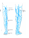
What is the pathway of the small saphenous vein?
- Drains the Postero-Lateral Aspect of the Leg and lateral aspect of the foot. - Originates in the lateral foot as part of the dorsal venous arch. - Ascends proximally behind the lateral malleolus as continuation of lateral marginal vein of foot - Frequently terminates at the popliteal vein, but this may vary. - The SSV lies for its entire length in an inter-fascial compartment defined by the deep muscular fascia and superficial fascia

What are perforators?
- Perforators act as alternative pathways from superficial to deep - They usually contain one way bicuspid valves that allow blood flow from superficial to deep - All perforators are accompanied by an artery

Pathway of deep veins (distal to proximal)
- Popliteal vein 2. Femoral Vein (not to be referred to as the Superficial Femoral Vein) 3. Deep Femoral Vein (aka Profunda Femoris Vein) 4. Common Femoral Vein 5. External Iliac Vein

List the two theories of the pathophysiology behind chronic venous insufficiency leading to ulcers?
- White cell trapping hypothesis 2. Fibrin cuff hypothesis
Explain the white cell trapping hypothesis behind venous insufficiency to ulceration.
-WBC’s are larger and less deformable than RBC’s - When perfusion pressure is reduced by venous hypertension, - WBC plug capillaries and RBC build up behind - WBC activation occurs - Endothelial adhesion by WBC releases proteolytic enzymes and oxygen free radicals causing endothelial and tissue damage
Explain the fibrin cuff hypothesis behind venous insufficiency to ulceration.
- increased venous pressure is directly transmitted to capillaries resulting in capillary elongation and increased endothelial permeability - larger molecules such as fibrinogen becomes deposited into tissues => fibrin - accumulation of fibrin acts as barrier to oxygen => tissue hypoxia => ulceration
What classification is used for venous disease?
CEAP: clinical, etiology, anatomic, pathophysiological
List the clinical classification (C) options for CEAP.

List the etiological (E) options for CEAP.

List the anatomical (A) options for CEAP.

List the pathophysiology options for CEAP.

Define varicose veins
= elongated, tortuous, dilated veins

What is primary varicose veins and secondary varicose veins?
Primary: affects the superficial veins or perforators in the absence of deep incompetence
Secondary (post-thrombotic): deep vein incompetence usually due to venous obstruction that destroys the valves
e.g. recanalisation of previous DVT (venous obstruction)
Clinical presentation of varicose veins
a. Cosmetic issue: telangiectasia (small dilated BV), reticular veins (dilated medium veins), varices (dilated vessel with tortuousity)
b. Pain:
- general leg ache, or heaviness
- venous claudication (no relief with rest; requires elevation 10-20 min)
c. Swelling: ankle pitting oedema
d. Skin changes:
- varicose eczema
- pigmentation
e. Lipodermatosclerosis: inverted champagne bottle
f. Atrophie blanche: confluence of white and depressed scars
g. Ulceration
What is the pathophysiology behind lipodermatosclerosis?
Increased blood pressure in the veins (venous hypertension) can cause diffusion of substances, including fibrin, out of capillaries. Fibrotic tissue may predispose the tissue to ulceration. Recurrent ulceration and fat necrosis is associated with lipodermatosclerosis. In advanced lipodermatosclerosis the proximal leg swells from chronic venous obstruction and the lower leg shrinks from chronic ulceration and fat necrosis resulting in the inverted coke bottle appearance of the lower leg
Ix of varicose veins
- Venous duplex
- Descending venography
Conservative Rx for varicose veins
- Conservative:
- Elevate legs
- Avoid standing still
- Dressings for ulceration
- Graduated compression stockings (increase in class, for more severe)
Non-conservative Rx of varicose veins
- Sclerotherapy: scars the vein so it sticks together
- Open surgery
- Endovenous laser therapy (EVLT): ablat eportino of the vein
- Radiofrequency ablation (RFA)
