Weak 1: Brain Flashcards

(244 cards)

1
Q

What are the 3 main parts of the brain?

A
  1. cerebrum
  2. cerebellum
  3. brainstem
2
Q

What are the 3 main parts of the brainstem?

A
  1. midbrain
  2. medulla
  3. pons
3
Q

What are the 2 main components of the cerebrum?

A
  1. cerebral hemisphere
  2. diencephalon
4
Q

What are the 2 main parts of the diencephalon?

A
  1. thalamus (relay to cortex)
  2. hypothalamus (control of autonomics)
5
Q

What are the 4 main parts of the cerebral hemisphere?

A
  1. cerebral cortex
  2. basal ganglia
  3. hippocampus
  4. amygdala
6
Q

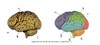
List the 5 lobes of the cerebral cortex?

A
  • frontal (Motor cortex)
  • parietal (Somatosensory cortex)
  • occipital (Visual cortex)
  • temporal (Auditory cortex)
  • limbic (Drives, emotions, memory)
7
Q

basal ganglia divides into what 2 parts?

A
  1. caudate nucleus
  2. lenticular nucleus
8
Q

What is the BASIC function of the cerebellum?

A

coordination

9
Q

What is the BASIC function of the hypothalamus?

A

autonomics + homeostasis

10
Q

What is the BASIC function of the thalamus?

A

relay to cortex

11
Q

What is the BASIC function of the basal ganglia?

A

movement control

12
Q

What is the BASIC function of the frontal lobe?

A

motor cortex

13
Q

What is the BASIC function of the parietal lobe?

A

somatosensory cortex

14
Q

What is the BASIC function of the occipital lobe?

A

visual cortex

15
Q

what is the BASIC function of the temporal lobe?

A

auditory cortex

16
Q

What is the BASIC function of the limbic lobe?

A

drives, emotions and memory

17
Q

What is the BASIC function of the hippocampus/ amygdala?

A

limbic structures; associated with drives, emoitions and memory

18
Q

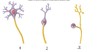
What are the 2 classes of cells in the nervous system?

A
  1. neurons
  2. neuroglial cells
19
Q

What are the main functions of neurons?

A

Neurons are a functional unit of the NS that recieve and transmit neural signal in the form of action potentials.

20
Q

(True or false) cerebral hemisphere includes structures deep to those on surface.

A

true

21
Q

anterior is also known as ______

A

ventral

22
Q

posterior is also known as ______

A

dorsal

23
Q

the top of the brain would be known as _______

A

dorsal

24
Q

What would the bottom of brain be known as? why?

A

ventral. dorsal and ventral are homologous to posterior and anterior and used synonymously in most cases, but as the brain started off as a rod like structure - the dorsal (posterior) aspect grew out to be on top. Now it is just the nomenclature

25
What does rostral mean?
toward head (superior)
26
what does caudal mean?
toward tail (inferior)
27
what are the 4 types of neuroglial cells? what is their collective function?
Collective function: To support and protect neurons. 1. Astrocytes (maintain BBB) 2. Oligodendrocytes (prod + maintain myelin) 3. Microglial cells (phagocytic) 4. Ependymal cells (prod CSF)
28
What is the basic function of astrocytes?
located between neurons and help support their activity. Involved with the secretion or absorption of neural transmitters and maintenance of the blood–brain barrier (funnily enough recent studies have actually shown astrocytes dont even have that much to do with the BBB but we will forget about that cause amandas lectures only mention "BBB" association so we will run with that)
29
What is the basic function of microglial cells?
phagocytic: part of the immune system within brain tissue helping clear dead cells and other debris.
30
What is the basic function of oligodendrocytes?
Produce myelin: produce and maintain the myelin sheath that coats and insulates the axon making electrical conduction more efficient.
31
What is the basic function of ependymal cells?
Produce CSF
32
Explain the following part of a neuron: Dendrites
receive impulses, short and often highly branched dendritic spine.
33
Explain the following part of a neuron: axons
transmit impulse away, can branch to form 'collaterals'
34
What is found within the cell body of a neuron?
nucleus + organelles which produce neurotransmitters
35
Explain the following part of a neuron: presynaptic terminals (terminal of boutons)
store synaptic vesicles containing neurotransmitters
36
Explain the different functional classifications of neurons.
Sensory (afferent): AP's towards CNS Motor (Efferent): AP's away from CNS Interneurons (Association neurons): Transmit signals from one neuron to another.
37
Explain multipolar neurons. Which neurons are multipolar?
Many dendrites, one axon. Most neurons of CNS and motor neurons are multipolar.
38
Explain unipolar neurons. Which neurons are unipolar?
appears to have only axon, no dendrites. Makes up all/most sensory neurons except for the sensory neurons in the retina of the eye and nose.
39
Explain bipolar neurons. Which neurons are bipolar?
A dendrite and an axon. Sensory neurons in retina of the eye and nose.
40
Briefly explain the functional advantage of myelination of axons. Are these axons 'continuous'?
- myelin protects and insulates axons from one another - Speeds up transmission - Functions in repair of axons - not continuous (contain nodes of ranvier)
41
What do unmyelinated axons 'rest against'? How does the speed of conduction in unmyelinated axons compare to those which are myelinated?
- Rest against oligodendrocytes - no myelin sheath wrapping - conduction of nerve impulse slower
42
where is gray matter found? (generally)
In non myelinated areas
43
where is white matter found?
axons which are mostly myelinated
44
explain what is meant by the 'nuclei within the brain"
dense areas deep in brain or brain stem where there is a collection of cell bodies. Usually functionally related. This is GREY matter -deep neuron cell bodies -synapses -synaptic terminals
45
what is the composition of the cortex of the brain?
gray matter; neuron cell bodies, synapses, synaptic terminals
46
t/f: location of gray and white matter is reversed in cortex and spinal cord
true
47
What makes up the cerebrum?
- left and right cerebral hemispheres - diencephalon (thalamus, hypothalamus, subthalamus, epithalamus)
48
what are the main components of the cerebral hemispheres?
frontal, parietal, temporal, occipital and limbic lobes along with the insular cortex, basal ganglia structures, and other limbic system structures. (does not include the central (midline) diencephalic structures)
49
the area of the brain other then the brainstem and the cerebellum is known as the \_\_\_\_\_\_\_\_\_\_
cerebrum
50
Consider the cerebral cortex. How thick is it? What time of matter? Approximately how many neurons? What are the gyri, sulci and fissures found on it?
- 2-4mm thick - Convoluted gray matter - approx 25 billion neurons - 2/3 cortex hidden from surface - forms folds (gyri) and grooves (sulci or deep fiscures)
51
what is the proportional weight of the cerebral cortex?
1/2 of brains
52
what is the fissure that seperates left and right sides of brain into cerebral hemispheres?
longitudinal fissure
53
is the insula considered a cerebral lobe?
no
54
is the insula visible from surface
no
55
where does the insula lie
deep to lateral fissure
56
brodmanns areas divides cerebral hemispheres into __ regions. Each region can be associated with a _____ \_\_\_\_\_
44, particular function
57
what's more common in the NS, neurons or neuroglial cells?
neuroglial cells far more common (counter-intuitive)
58
schwann cells myelinate axons in the \_\_\_, whereas oligodendrocytes myelinate axons in the \_\_\_\_
PNS, CNS
59
Explain myelin
A layer of fatty tissue segmentally encasing the fibers of many neurons; enables vastly greater transmission speed of neural impulses as the impulse hops from one node to the next.
60
explain the grey/white matter distribution of the brain (out to in)
grey white grey
61
precentral gyrus is part of the ___ lobe
frontal
62
where is the cingulate sulcus located?
superior border of limbic lobe
63
where is the calcarine sulcus
through occipital lobe
64
which regions of the brain have both primary and secondary association regions
auditory (temporal) somatosensory (parietal) visual (occipital)
65
Sensory input is directed to the ____________ 1st for initial analysis
primary cortical region (sensory)
66
what do secondary SENSORY association areas do?
integrate information from multiple sources and make sense of it, and then make decisions based on the processing.
67
The frontal lobe contains ______ and \_\_\_\_\_\_\_cortical regions
primary and secondary
68
primary motor cortex... function? location?
- easiest motor area to stimulate - controls specific muscles (particularly distal limb muscles) - at precentral gyrus - particularly controls distal limb muscles
69
secondary motor areas... function? based upon input from where? can motor output be stimulated from these areas?
- planning movt/ making overall decision of what movt should be - based upon input from basal ganglia, cerebellum and other cerebral areas - motor outpup can also be generated from these regions
70
what is the role of the auditory association area?
comprehension of speech
71
what is the role of the gnostic area
integrates senses
72
what is the location of the primary motor cortex?
precentral gyrus
73
What is the difference between the frontal cortex and the prefrontal cortex?
prefrontal is anterior and ventral (inferior) to frontal prefrontal doesnt include the motor part of the frontal cortex, it refers to just the personality/ decision making etc
74
\_\_\_\_\_\_\_ is the area that is often referred to as the thinking part of the brain. Spefically, this area helps with
prefrontal and frontal cortex -decision making, planning, motivation, self awareness, and social behaviour
75
Cerebral white matter contains 3 neuron fibre types depending on _______________ . These 3 are...
where they travel 1. projection fibres 2. association fibres 3. commissural fibres
76
explain: projection fibres
Consists of neurons ascending or descending to or from cerebral cortex through brainstem and spinal cord. The main projection fibres are collectively known as the internal capsule. Essentially projection fibres form ascending and descending tracts.
77
explain association fibres
they run between gyri of same hemisphere. They do this in a kind of "UUUUU" shape, in between the gyri dipping to go around the sulci.
78
explain commissural fibres
they run from one hemisphere to another, the main commisural fibre is the corpus callosum.
79
somatosensation is refering to......
taste, touch, pressure, vibration, pain, and temperature
80
what is the homonculus
a rendering of the body in which each part is shown in proportion to show how much of the somatosensory cortex is devoted to it. Essentially its the mapping of which parts of the body are connected to which part of the pre/ postcentral gyrus - it is somatotopically organised
81
The precentral gyrus contains the \_\_\_\_\_\_\_\_\_\_\_\_\_\_\_\_\_\_, whilst the postcentral gyrus contains the \_\_\_\_\_\_\_\_\_\_\_\_\_\_\_\_\_\_
Motor cortex Somatic sensory cortex
82
why are some structures of the brain C shaped
due to expansion of cerebral cortex
83
What are the 5 C-shaped structures? Why are they C-shaped?
1. cerebrum 2. caudate nucleus (basal ganglia structure) 3. corpus callosum 4. lateral ventricle 5. limbic structures (CCCLL) Due to expansion of cerebral cortex being disproportionate to other deeper telencephalic structures.
84
Explain 3 functions of the limbic system
- modulates processes to do with hypothalamus, such as... 1. feeding behaviour 2. endocrine activity 3. sexual behaviour 4. autonomic control - affect short term immediate response to stimuli (euphoria, happiness, anger) - central regulation of MOTIVATION, LEARNING and MEMORY
85
Give the structure and function of the basal ganglia.
group of structures found deep in cortex and midbrain that influence the thalamus which provides feedback to the cerebral cortex for initiation and control of motor responses and reduces muscle tone.
86
what is the only part of the diencephalon which can be seen on an intact brain? where is it seen?
hypothalamus; on ventral surface of cerebrum, inferior and anterior to thalamus
87
the hypothalamus forms the ___ \_\_\_ and ___ of the third ventricle
lateral wall; floor
88
the third ventricle divides ..............
the hypothalamus into left and right halves which are continous anteriorly
89
what are the mian functions of the hypothalamus? (7) (SEW FACT)
- energy homeostasis regulation (feeding, growth, metabolism) - water balance (drinking, water reabsorption) - stress (adrenal glands -\> cortisol) - thermoregulation - fever initiation and immune responses - circadian rhythms - arousal and reproduction
90
what is meant by: circadian rhythms?
sleep/ wake cycles
91
Explain the structure of the thalamus
- left and right thalami (seperated 3rd ventricle) - Two egg shaped strucutures connected medially to each other in 70-80% of people as the intralaminar adhesions - divide into anterior/medial/ lateral regions - each of these regions contain groups of nuclei with specific functions
92
explain the function of the thalamus (pre long answer grr) (SAM MAC)
- **sensory relay station** (visual, auditory, somatic) - **cognitive functions** associated with visual and auditory stimuli - **motor control** via feedback from basal ganglia to motor cortex - mediates **emotional** behaviour/ memory/ cortical activation - helps form **complex behavioural patterns** based on incoming sensory input - recieves information about **waking/ arousal states**
93
the hippocampus is found deep in the \_\_\_\_\_\_\_\_\_\_\_\_\_
temporal lobe
94
from **inferior** to **superior** list the parts of the brainstem.
medulla pons midbrain
95
how many cranial nervesare there? how many of these emerge out of the brainstem
12 10
96
ascending and descending tracts travel out/ in the brain via the \_\_\_\_\_
brainstem
97
the brainstem connects the ______ to other areas of the brain and spinal chord
cerebellum
98
brainstem contains nuclei which control.....
respiration, CVS, consciousness, sleep and alertness
99
10 out of the 12 cranial nerves emerge out of the brainstem. Specifically, how many in the pons, medulla and midbrain?
2 from the midbrain 3 from the pons 1 between pons and medulla 4 from medulla
100
explain the structure of the cerebellum
- lies posterior to pons and medulla - connected to brainstem via cerebellar peduncles - consists of two hemispheres (right and left) seperated by the 'vermis' - like the cerebrum has superficial cortex, white matter and deep nuclei
101
how is the cerebellum connected to the brainstem
via cerebellar peduncles
102
explain the cerebellar function.
1. planning programming and monitoring of voluntary movt 2. postural adjustments 3. coordination of eye and head movements
103
The cingulate gyrus is part of the ___ lobe
limbic
104
basal ganglia are ____ to diencephalic structures, they are not midline.
lateral
105
basal ganglia controls the amount of ____ \_\_\_\_from the thalamus, which in turn provides feedback to the cerebral cortex for the initiation and control of ___ \_\_\_
excitatory output motor responses + muscle tone
106
all sensory information apart from olfactory must first pass through the \_\_\_\_\_, which integrates the information and relays it to other areas of the brain
thalamus
107
midbrian divides into a roof (\_\_\_\_\_\_\_) and a floor (\_\_\_\_\_\_\_)
roof = tectum floor = tegmentum
108
what structure divides the midbrain into roof and floor
cerebral aqueduct
109
2 out of 12 cranial nerves dont pass through brainstem. Which ones are these?
1. olfactory 2. optic
110
A (directional)
Dorsal
111
B (directional)
Posterior
112
C (Directional)
Ventral
113
D (Directional)
Anterior
114
1
Multipolar neuron
115
2
Bipolar neuron
116
3
pseudo-unipolar neuron
117
1
multipolar neuron: Dendrite
118
2
multipolar neuron: Cell body
119
3
multipolar neuron: Dendrite
120
4
multipolar neuron: Axon
121
A
multipolar
122
D
Multipolar
123
F
bipolar neuron
124
G
Unipolar neuron
125
E
Multipolar neuron
126
A
Gyrus
127
B
Sulcus
128
C
Cerebral cortex
129
D
Cerebral white matter
130
E
Fissure
131
1
Central culcus (of Rolando)
132
2
Lateral sulcus (sylvian fissure)
133
3
Pre-occipital notch
134
4
Frontal lobe
135
5
Temporal lobe
136
6
Parietal lobe
137
7
Occipital lobe
138
8
Brainstem
139
9
Cerebellum
140
1
Cingulate gryus
141
2
Top of central sulcus
142
3
Parietoocipital sulcus
143
4
Intraventricular foramen
144
5
Cerebral aquaduct
145
6
Fourth ventricle
146
7
calcarine sulcus
147
8
diencephalon
148
9
Frontal lobe
149
10
limbic lobe
150
11
parietal lobe
151
12
occipital lobe
152
13
temporal lobe
153
14
brainstem : Midbrain
154
15
Brainstem: Pons
155
16
Brainstem : Medulla
156
17
cerebellum (Vermis)
157
18
cerebellum (Hemisphere)
158
part in blue
insula
159
1
Precentral sulcus
160
2
central sulcus
161
3
post central sulcus
162
4
Lateral sulcus
163
1
Subpariertal sulcus
164
2
Central sulcus
165
3
Cingulate sulcus
166
4
Callosal sulcus
167
5
Parietooccipital sulcus
168
6
Calcarine Sulcus
169
7
Occipitotemporal sulcus
170
8
Collateral sulcus
171
Brodmanns 1,2,3
Primary somatosensory area within the postcentral gyrus
172
Brodmanns 17
Primary visual area
173
Brodmanns 41 and 42
Primary auditory area
174
Brodmanns 43
Primary gustatory area
175
Brodmanns 5 and 7
Somatosensory (integrate and interperate)
176
Brodmanns 18 and 19
Visual association area (interpret)
177
Brodmanns 22
Auditory association area (wernicke's) - (comprehension of speech)
178
Brodmanns 5, 7, 39 and 40
Gnostic area (integrates senses)
179
Brodmanns 4
Primary motor area (precentral gyrus)
180
Brodmanns 6
premotor area
181
Brodmanns 44 and 45
Motor speech area (broca's area) - Production of speech, control of tongue and airway.
182
Brodmanns 8
frontal eye field
183
Brodmanns 4, 6, 8, 44, 45 (collectively)
part of frontal cortex and related to motor control
184
Brodmanns 11 and 47 (collectively)
Prefrontal cortex (thinking part of brain)
185
1
Association fibers
186
2
Projection fibers
187
3
Thalamus and internal capsule
188
4
Corpus callosum (commissural fibers)
189
5
Projection fibers (internal capsule)
190
1
Corpus callosum (main connection between left and right hemispheres for commissural fibers)
191
What are the red arrows pointing to?
Projection fibers (specifically the internal capsule)
192
1
lateral ventricle
193
2
Fornix
194
3
Third ventricle
195
4
Fourth Ventricle
196
5
Hippocampus
197
6
Cingulate gyrus
198
7
Parahippocampal gyrus
199
1
Cingulate gyrus
200
2
Hypothalamus
201
3
olfactory bulb
202
4
mammillary body
203
5
hippocampus
204
6
Corpus callosum
205
7
fornix
206
8
amygdala
207
9
parahipocampal gyrus
208
1
hippocampus (deep in temporal lobe)
209
2
Fornix
210
1
Body of fornix
211
2
Crura of fornix
212
3
hippocampus
213
4
amygdaloid bodies
214
5
mamillary bodies
215
6
columns of fornix
216
1
Body of fornix
217
2
Hippocampus
218
3
parahippocampus
219
1 (specifically)
Cingulate gyrus
220
1
head of caudate nucleus
221
2
body of caudate nucleus
222
3
tail of caudate nucleus
223
4
putamen
224
1
Thalamus
225
2
Epithalamus
226
3
Subthalamus
227
4
Hypothalamus
228
what is being highlighted in red
Thalamus
229
1
rostral midbrain
230
2
caudal midbrain
231
3
rostral pons
232
4
Caudal pons
233
5
rostral medulla
234
6
caudal medulla
235
1
Cerebral aqueduct
236
2
pineal gland
237
3
superior colliculus
238
4
inferior colliculus
239
5
cerebellum (vermis)
240
6
Cerebellum (hemisphere)
241
7
medulla
242
8
fourth ventricle
243
9
basal pons
244
10
tegmentum