Week 1 Flashcards
(40 cards)
Describe the types of membrane proteins
The types of membrane proteins found in the membrane depend on the cell type. Hay:
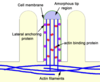
Structural proteins: Proteins which attach cytoskeletal filaments to cell membrane. Adhesion molecules which attach cells to extracellular matrix
Transport proteins: eg carrier, pump and channel proteins.
Receptors: for chemical signalling between cells
Proteins with enzymatic activity
Receptor proteins are more likely to be glycoproteins
Proteins can be embedded in the membrane or associated with the membrane surface.
What are conjugated proteins?
have a chemical group associated w/ their polypep chain. Eg haemoglobin has an Iron containing heme group.
What are carrier proteins?
facilitate diffusion of large polar molecules.
A molecule/ ion combines with the binding site of a carrier protein.
ATP then transfers a phosphate group to the carrier protein on the inside of the membrane.
The carrier has a shape change, carrying and then releasing the molecule in the membrane before reverting back to its original shape.
Describe Cholesterol
Cholesterol occurs dentro the membrane and limits movement of the phospholipids, making the membrane more rigid. The organic lipid fits between the phospholipids, maintaining membrane strength.
At higher temps it stabilises cell membrane structure, and at lower temps maintains fluidity. The more unsaturated the lipids, the more fluid it will be.
Describe glycolipids
Glycolipids, occur on the outer surface of the membrane with their associated sugars. They may be involved in intercellular communication.
Describe centrioles
Each centriole is composed of nine sets of 3 microtubules.
In a pair of centrioles, the individual centrioles are arranged perpendicular to each other.
Centrioles occur in pairs and are found in an area of the cell near the nucleus called the centrosome (cell center).
The centrioles organise the microtubular network within the cell. And organises the development of the microtubules in the cilia
Describe ribosomes
Ribosomes consist of 2 subunits.
Site of mRNA translation for protein synthesis. Occurs either in cytosol (cell protein synthesis) or attached to ER (membrane or secretion protein synthesis)
Describe the rER
A network of tubules, vesicles and flattened cisternae continuous with the nuclear envelope
Function: protein synthesis in conjunction with ribosomes and modification of newly synthesised protein by glycosylation and structural changes
Describe the smooth ER
An irregular network of tubes and vesicles continuous with the rough ER
Lipid and steroid biosynthesis, protein processing and intracellular transport by packaging of products into vesicles
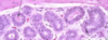
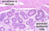
Describe the GApp
Vesicles from the ER fuse to form flattened, membrane bound sacs.
The GA modifies and packages proteins in vesicles for transport
Hay transport vesicles, which move within the cell, and secretory vesicles which move molecules out of the cell, (exocytosis).
The Golgi apparatus has 3 main roles:
Modification of proteins by addition of sugars
Proteolysis, activation of peptides
Sorting and packaging of macromolecules into vesicles for transport within cell and for secretion
Describe exocytosis of proteins
Transcription forms mRNA, which leaves the nucleus and joins onto a ribosome on the rough ER.
Protein moves through the ER assuming its 3-D shape en route.
Vesicles containing the protein are pinched off the rER.
These pinched off vesicles fuse to form flattened sacs of the Golgi apparatus. Proteins are modified within the Golgi apparatus.
Vesicles containing the MODIFIED protein are pinched off the G. ap.
Vesicle fuses with the cell membrane and releases the protein.
Describe microtubules
Microtubules are hollow fibres present in all cells except RBCs. Formed from 2 protein subunits, α and ß tubulin, which join alternatively to form protofilament chains. These arrange into groups of 13, forming the microtubule.
Microtubules are constantly forming and unforming w/in the cell. They grow out of the centrosome and are stabilised by associating w various proteins.
Microtubules have many functions:
Intracellular transport
Present in cilia and flagella
Form cell cytoskeleton
Form filaments of the mitotic spindle
Form centrioles and basal bodies

What are intermediate filaments?
Intermediate filaments are intermediate in size between microtubules and microfilaments. They vary slightly according to which cell type they appear in.
For example cells showing muscle differentiation contain the intermediate filament desmin and epithelial cells all contain cytokeratin.
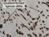
How can intermediate filaments be used to detect breast cancer?
The cells contain cytokeratin, the epithelial type of intermediate filaments
All the cancer cells have stained black/brown with immunoperoxidase using an antibody which reacts with human cytokeratin.
The connective tissue around the cancer does not stain as these cells contain a DIFF type of intermediate filament, which doesn’t react w the cytokeratin antibody.
What is the cytoskeleton?
The cytoskeleton is made out of proteins like microfilaments, microtubules, and intermediate filaments, which all provide structural stability.
The cytoskeleton is v dynamic, allowing the cell to change shape by selectively contracting and extending filaments. This is important in some cell functions like muscle contraction, cell division, cell movement
The cytoskeleton also helps structures within the cell move from one area to another.
What are microfilaments?
Microfilaments are composed of actin and are the smallest proteins of the cytoskeleton. Various isoforms of actin occur in various amounts in diff cell types.
The majority of the microfilaments occur just below the cell membrane where they form a crosslinked mesh which provides mechanical support to the cell membrane.
Actin molecules can form bundles which protrude the cell membrane to form microvilli.
In some cells actin interacts with Myosin to generate motion.

What are microvilli? Draw a diagram in your answer
Microvilli are small finger like projections found on the apical surface of most epithelial cells.
The no and shape of the microvilli correlates w the absorptive capacity of the cell. Mircovilli increase SA of the cell.
What is the endosymbiotic theory?
Mitochondria are believed to have evolved in eukaryote cells as a result of symbiotic relationship with a mitochondrion-like prokaryote organism.
In support of this theory mitochondria have their own DNA and can synthesise protein.
Draw and describe mitochomdria
Mitochondria provide energy to cells through oxidative phosphorylation. They are present in all cells except red blood cells. Their morphology varies with cell type.

What is oxidative phosphorylation and the ETC?
NADH gives its e- to complex I, getting oxidised back to NAD+. FADH2 gives its e- to Complex II. E-s from the complexes flow down a chain of e- carriers in the mitochondrial membrane.
Energy is released as electrons pass along the ETC. This energy is used to move H+ ions from the mitochondrial matrix into the intermembrane space, creating a steep electrochem gradient across the inner membrane.
H+ diffuse down this gradient via protein channels joined to ATP synthase. H+ ions change the enzyme a.s shape, allowing ADP and phosphate ions to bind. ADP is phosphorylated to ATP, catalysed by ATPsynthase
W/in the matrix, the H+ and e-s recombine to form H2. These combine with 02 to form 2H20. 02= final e acceptor.
Describe the nucleus and nucleolus
The nucleus contains DNA and is the site of RNA transcription.
The nucleolus is a compartment within the nucleus. It is the site of transcription and processing of ribosomal RNAs and their assembly into ribosomal subnunits before export to the cytoplasm.
The nucleolus varies in size depending on how metabolically active the cell is.
The cell DNA is organised into linear molecules called chromosomes. During metaphase the chomosomes are fully condensed and can be visualised on metaphase spreads for karyotype analysis.
What is a karyotype/ karyotyping?
Karyotype: the number and visual appearance of the chromosomes in the cell nuclei of an organism or species.
During metaphase the chromosomes condense and become distinguishable. Metaphase chromosomes are used during the karyotyping procedure that is used to look for chromosomal abnormalities
What is epithelia?
Epithelia are sheets of epithelial cells covering the outside of the body, line many hollow organs and can have secretory and absorptive functions.
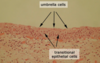
Describe squamous epithelium
Squamous (flat) cells form a squamous epithelium e.g. skin and the lining of some hollow organs. The epithelium can either be simple (made up of a single layer of cells) or stratified (many layers of squamous cells)
Formed of multiple layers of squamous cells on top of each other. They’re joined juntos by specialised junctions called desmosomes.
The upper part of some squamous epithelia is made of squamous cells filled with a large amount of keratin (intermediate filament), which makes the surface hard-wearing, like in skin.
Squamous epithelium can also be found in organ linings like the oral cavity (mouth and pharynx), oesophagus, anal canal and the vagina.
The epidermis is a stratified squamous epithelium (i.e. made up of many layers). The underlying dermis is made of fibrous connective tissue.