Week 1 Flashcards

1
Q

What are the components of…

  • the upper urinary tract
  • the lower urinary tract?
A

Upper - left and right kidneys, left and right ureters

Lower - bladder and urethra

2
Q

What part of the urinary tract is included in the following areas?

  • abdomen
  • pelvis
  • perineum
A

Abdomen - kidneys, proximal ureters

Pelvis - distal ureters, bladder, proximal urethra

Perineum - distal urethra

3
Q

The kidneys are intra/retroperitoneal

The great vessels (IVC and aorta) are intra/retroperitoneal

A

Kidneys are retroperitoneal

Great vessels are also retroperitoneal

4
Q

What are the three structures of the renal hilum? Which of these structures always sits most anteriorly?

A

Renal artery

Renal vein - always sits anteriorly

Ureter

5
Q

From superficial to deep, what layers surround the kidney to the peritoneum? (5)

A

Visceral peritoneum

Paranephric fat

Renal (deep) fascia

Perinephric fat

Renal capsule

6
Q

What muscle sits medially to the kidney?

What muscle sits posteriorly to the kidney?

A

The Psoas major sits medially

The Quadratus lumborum sits posteriorly

7
Q

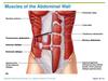
What are the three layers of abdominal wall muscle?

A

External oblique

Internal oblique

Transversus abdominis

8
Q

Where do the following drain to…

  • Renal artery
  • Renal vein
A

Artery - abdominal aorta

Vein - IVC

9
Q

At what vertebral level are the kindeys (right and left are at different levels!)

A

Right - L1-L3

Left - T12-L2

10
Q

When attempting to ballot the kidneys, what should you ask the patient to do? Why?

A

Ask the patient to breathe in.

As the liver and spleen lie in direct contact with the diaphragm, when the patient breathes in this expands the lungs and pushes these organs down. The liver and spleen are also in contact with the kindeys, so on inspiration they too are pushed down and may be “trapped” on balloting

11
Q

What is the name of the recess where fluid would collect if a patient was supine?

A

The hepatorenal recess between the kidney and the liver

12
Q

At what level does the abdominal aorta bifurcate?

What happens to the relationship between the arteries and veins at this point?

A

The abdominal aorta bifurcates at the level of the umbilicus (L3-L4)

While the renal arteries are posterior to the renal veins, the common iliac arteries are anterior to the common iliac veins

13
Q

Where does the lymph from the a) kidneys and b) ureters drain to?

A

a) lymph from the kidneys drains to the lumbar nodes (located around the great vessels)
b) lymph from the ureters drains to both the lumbar and the iliac nodes

14
Q

What’s the difference between a supra-renal and infra-renal AAA?

A

Supra-renal AAAs include the renal arteries, and renal artery stenosis is a result of the AAA (aneurysm narrows and occludes artery)

Infra-renal AAAs are below the renal arteries, and renal artery stenosis may be combined with the AAA (both caused by atherosclerosis)

15
Q

What anatomical variations may be seen in kidney development?

A

Bifid renal pelvis

Bifid ureter and unilateral duplicated ureter

Retrocaval ureter

Horseshoe kidney

Ectopic pelvic kidney

16
Q

What are the two main parts of the kidney contained within the renal capsule?

Where are the nephrons contained?

A
  • The cortex
  • The medulla

Nephrons are contained in renal pyramids, which are contained within the medulla

17
Q

Proximally to distally, what are the various structures of a nephron?

A

Glomerulus

Proximal convoluted tubule

Loop of Henle

Distal convoluted tubule

Collecting duct

Minor calyx

18
Q

Describe the drainage of urine from the kidney to the ureter.

At what point is the first constriction in this pathway?

A

Nephron collecting ducts > minor calyx > major calyx > renal pelvis > ureter

The diameter of these structures continues to increase until the pelviureteric junction (where the renal pelvis becomes the ureter)

19
Q

What are the three sites of ureteric constriction?

Why are these clinically important?

A
  1. Pelviureteric junction
  2. Ureter crossing the anterior aspect of the common iliac artery
  3. Ureteric orifice into the bladder (corner of the trigone)

Clinical importance - this is where kidney stones will most likely cause issue

20
Q

Why is there a colicky pain associated with kidney stones?

A

The ureters have a peristaltic motion. Upon obstruction, there is increased peristalsis proximally to the site of the blockage

21
Q

What is hydronephrosis? What causes it?

A

“Water inside the kidney”, caused by back pressure of urine into calyces, which compresses the nephrons and results in renal failure.

Acutely painful

22
Q

What are the different areas of the pelvic cavity? What structures form the boundaries between these areas?

A

False pelvis - from the iliac crests to the pelvic inlet, contains abdominal organs

True pelvis - pelvic inlet to pelvic floor, contains pelvic viscera

Perineum

False pelvis and True pelvis are separated by the pelvic rim (inlet), True pelvis and Perineum are separated by the pelvic floor, specifically the levator ani

23
Q

What structures pass through the pelvic floor?

A

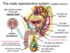
Distal parts of the alimentary (rectum), renal (bladder) and reproductive tracts

24
Q

Describe the path of the ureters from the abdominal cavity to the bladder. At what point to they turn medially?

A

Leave the kidney, travel inferiorly and pass anteriorly to the common iliac vessels to enter the pelvis

Run anteriorly, along the lateral walls of the pelvis

At the level of the ischial spine the ureters turn medially to enter the posterior aspect of the bladder

25
What mechanisms help to prevent reflux of urine back into the ureters when the bladder contracts?
Entry of the ureters into the bladder is **inferomedial** **Detrusor muscle fibres** encircle the ureteric orifices and tighten when the bladder contracts
26
In the male, when standing up, what is the most dependent part of the peritoneal cavity?
The **rectovesicle pouch** - the space between the bladder and rectum
27
What are the two recesses in the peritoneum in the female? Which of these is the most dependent?
The **vesico-uterine pouch** - between the bladder and uterus The **rectouterine pouch (pouch of Douglas)** - between the uterus and the rectum. **This is the most inferior part of the female peritoneal cavity**
28
How does the ureter run in relation to the uterine tube and uterine artery? How does the ureter run in relation to the vas deferens?
The ureter runs **inferiorly to these structures** (water under the bridge)
29
What larger artery gives off the majority of arteries supplying the pelvis? Name some of these smaller arteries
**Internal iliac artery** Female * Vesicular arteries (to bladder) * Uterine artery * Middle rectal artery * Vaginal artery Male * Vesicular arteries * Middle rectal artery * Prostatic arteries (often branches of the vesical arteries)
30
What structures form the three corners of the trigone?
2 ureteric orifices Internal urethral orifice
31
What is the name of the muscle that forms the main bulk of the bladder wall? What does this muscle allow in the male, and for what purpose?
The **detrusor muscle** Forms a smooth muscle sphincter, the **internal urethral sphincter muscle**, which contracts during ejaculation to prevent retrograde passage of semen into the ureters
32
Where does the bladder lie when it is a) empty and b) full? What are the 2 routes of catheterising a patient?
a) the bladder lies within the pelvis b) the bladder may extend out of the pelvis Two routes of catheterisation * **urethral** - more common * **suprapubic** - through the anterior abdominal wall, need to be sure to avoid the peritoneal cavity however as this could cause peritonitis. Easier to do if the bladder is full.
33
Again, what feature separates the pelvis and the perineum?
The **levator ani**
34
The external urethral orifice is under voluntary/involuntary control
Voluntary
35
Describe the passage of sperm
Synthesised in the **seminiferous tubules of the testes**, then stored in the **epididymis**. Passed along the **vas deferens**, which passes anteriorly and then superiorly to the bladder before joining the **seminal gland**. Then passes into the **ejaculatory duct** which passes through the prostate, then the spongy urethra and finally exits out of the external urethral orifice
36
Describe the passage of urine in the male
Ureters into bladder, then through internal urethral sphincter and into the **prostatic urethra** Then passes through the external urethral sphincter into the **spongy urethra** and exits out of the **external urethral orifice**
37
What structures are contained within the spermatic cord? (3)
Vas deferens Testicular artery Pampiniform venous plexus
38
What is the name of the protective sac surrounding the testes?
**Tunica vaginalis**
39
What is the term for when there is excessive fluid accumulating within the tunica vaginalis?
Hydrocele
40
Where do the left and right testicular veins drain into?
Left - left renal vein then IVC Right - directly into IVC
41
What region of the prostate most commonly becomes cancerous? Why is this beneficial to clinicians?
The peripheral zone is where most prostate cancers begin. This is useful as it means prostate cancers can be detected early via palpation
42
What are the three cylinders within the penis that become engorged with blood during erection?
Right and left **corpus cavernosum** ## Footnote **Corpus spongiosum**
43
What is the blood supply to a) the penis and b) the scrotum?
a) internal iliac \> internal pudendal \> deep arteries of the penis b) internal iliac \> internal pudendal AND external iliac
44
Describe lymph drainage from a) the scrotum and penis and b) the testes
a) lymph from scrotum and penis drains to the **superficial inguinal lymph nodes** b) lymph from the testes drains to the **lumbar nodes** around the aorta (due to embyrological development)
45
From what embryonic layer do the kidneys arise?
The **mesoderm**
46
How many mesonephric units are produced initially? What do they regress to and when?
**40** units are initially produced craniocaudally, then regress at **the end of week 5 to 20 pairs** These then differentiate into **excretory units** which function between **weeks 6-10**, and then regress
47
What structure appears mid week 5, begins to function between weeks 9-11 and becomes the kidney permanent? What does it form from?
**Metanephros** Forms from the **ureteric bud** and the **metanephric blastema**
48
What do the following structures develop into? - ureteric bud - metanephric blastema
Ureteric bud develops into **collecting ducts** Metanephric blastema develops into **nephrons**
49
Where do the kidneys initially develop, and what direction do they travel in? When does this happen?
Kidneys initially develop in the **pelvis** and **ascend upwards** into the abdomen, attaining their adult position by around **week 9**
50
What are the 5 Rs for prescribing fluids?
Resuscitation Replacement Routine maintenance Redistribution Reassessment
51
Where are errors in prescribing IV fluids particularly likely?
Emergency departments Acute admission units General medical and surgical wards
52
How much fluid can be given to a patient in resuscitation before it needs to be escalated?
Up to 3 litres
53
For maintenance IV fluids, how much should be given for the following... - water - sodium, potassium, chloride - glucose?
Water - **25-30ml/kg a day** Sodium, Potassium and Chloride - **1 mmol/kg a day** Glucose - **50-100g** **a day** Point is: it is NOT a one-size-fits-all, and needs to be calculated **based on patient's weight**
54
What does NICE recommend to be the best form of fluid replacement in a resuscitation setting? Why? What are the exceptions to this?
NICE typically recommends **balanced** **crystalloid solutions**, such as Hartmann's, lactated Ringer's or Plasma-Lyte 148. This is because excessive amounts of 0.9% Sodium Chloride can potentially result in **hyperchloraemic metabolic acidosis** Exceptions to the above includes **rhabdomyolysis** (and AKI/CKD) - in this instance 0.9% NaCl would actually be preferred initially because of the risk of **hyperkalaemia** (these conditions result in raised serum potassium)
55
How do the guidelines for using fluid in Resuscitation vs Replacement vary?
They don't! Still recommend **Balanced crystalloid solutions**
56
What mix of fliuds is generally used for Routine Maintenance?
0. 18% NaCl/4% dextrose 0. 45% NaCl 5% dextrose Point is - a Saline/Dextrose mix is typically used in **maintenance**, but remember that **glucose is of no use in a resuscitation context**
57
In giving maintenance IV fluids, the total amount per day increases as weight of the patient increases. What else increases with patient weight?
The **Rate at which the fluid is given**, from 50ml/hour in patients weighing 35-44 kg to 100ml/hour (max) in patients weighing 75kg or more
58
When giving fluids in a resus scenario, why after half an hour or so does the patient's BP start to drop and their HR start to rise again?
Of the fluids given, only approx 18% remain within the intravascular tree
59
When _might_ human albumin solution (HAS) be _considered_ for fluid resuscitation, according to NICE? In practice, for what other conditions can it be used?
Only conisdered in patients with **severe sepsis**, according to NICE 20% HAS can be used in **large volume paracentesis** e.g. draining fluid from a patient with ascites, and also in **Hepatorenal Syndrome** 4.5% HAS can be used as exchange fluid for **Therapeutic Plasma Exchange** - as done in numerous autoimmune conditions that feature detectable auto-antibodies e.g. various types of vasculitis, Goodpasture's syndrome etc.
60
Name some blood products. Usually only one or two of these isolated components of full blood is given to a patient. Why? Under what circumstances is the whole thing given?
- Packed Red Cells - Platelets - Fresh Frozen Plasma - Cryoprecipitate (e.g. clotting factors) Full blood isn't usually given because the **white cells contained in whole blood can elicit an immune response**. The exception is in the event of **massive haemorrhage**
61
What is omsolarity? What two things are needed to be able to calculate it?
Concentration of osmotically active particles present in a solution Can be calculated if the following are known: - molar concentration of the solution - the number of osmotically active particles
62
What will be the effect on a cell if it is placed in the following tonicity of solutions? - isotonic - hypotonic - hypertonic
Isotonic - no change Hypotonic (mroe water) - increase in cell volume Hypertonic (less water, concentrated salt solution) - decrease in cell volume
63
What are the two major compartments in which total body water exists in the human body?
Intracellular fluid (67% tbw) Extracellular fluid (33% tbw)
64
What are the components of extracellular fluid (ECF)?
Plasma - 20% Interstitial fluid - 80% Lymph and transcellular fluid - negligible
65
Give some examples of tracers, along with the fluid volume the can be used to measure
**3H2O** - used to measure total body water **Inulin** - used to measure ECF **Labelled albumin** - used to measure plasma
66
What is the difference between sensible and insensible losses of water? How is most water lost from the body?
Sensible - through means that we can control e.g. sweat, urine, faeces Insensible - through means that we cannot control e.g. lungs and skin Most water is lost from the body via **urine**
67
Are the following ions present in a greater amount in the ICF or ECF? - Na+ - Cl- - K+ - HCO3-
Na+ - greater in **ECF** (140 mM vs 10 mM) Cl- - greater in **ECF** (115 mM vs 7 mM) K+ - greater in **ICF** (140 mM vs 4.5 mM) HCO3- - greater in **ECF** (28 mM vs 10 mM)
68
In terms of ionic makeup, how do plasma and interstitial fluid compare?
They are essentially the same! Except that plasma also has a small amount of **protein anions**
69
What are the main ions in... - ECF - ICF And how do their osmolarities compare?
ECF - Na+, Cl-, HCO3- ICF - K+, Mg+ and negatively charged proteins ECF and ICF osmolarities are **usually identical** (approx 300 mosmol/l), despite the fact that cell membranes are selectively permeable
70
What would happen to ECF and ICF volumes in the following situations... - if the osmotic conc. of ECF increases (e.g. dehydration) - if the osmotic conc. of ECF decreases (e.g. fluid excess)
ECF osmotic conc. increases - ECF becomes hypertonic compared to ICF, resulting in a **decrease in ICF volume** and **increase in ECF volume** ECF osmotic conc. decreases - ECF becomes hypotonic compared to ICF, resulting in an **increase in ICF volume** and **decrease in ECF volume**
71
What is potassium important for, and what is the effect if potassium leaks from ICF to ECF?
K+ plays a key role in establishing **membrane potential**, especially in cardiac and skeletal muscle cells If K+ leaks out of the ICF, this can cause **muscle weakness and paralysis** (skeletal muscle) and **cardiac irregularities and cardiac arrest** (cardiac muscle)
72
What % of total cardiac output do the kidneys receive?
Despite their relatively small size, the kidneys receive **25% of total cardiac output**
73
Describe the blood supply to the nephron
Artery \> afferent arteriole \> glomerulus \> efferent arteriole \> peritubular capillaries \> vein
74
What are the two types of nephron in the kidney? Which are more numerous? What are the 2 main differences between these two types of nephron?
Juxtamedullary (20%) and Cortical (80%) In the juxtamedullary nephron, the **Loop of Henle is much longer** and there are not peritubular capillaries, instead there is a **single vasa recta**
75
What are podocytes?
Cells on the inner lining of Bowman's capsule Have "foot-like" extensions that mesh together with each other to form filtration slits
76
What cell type in the kidney produces and secretes renin? What cell type in the kidney is sensitive to changes in NaCl? Where are both of these cell types found?
Renin is produced and secreted by **granular cells** NaCl in tubular fluid is sensed and monitored by **macula densa cells** Both of these cell types are found at the **juxtaglomerular apparatus**, where the afferent and efferent arterioles fork around the **distal tubule**
77
Of the plasma that enters the glomerulus, how much is filtered and how much is unfiltered and passed into the efferent arteriole? Once filtered, what happens to the plasma in the kidney tubule?
Only **20%** is filtered and the remaining 80% is passed into the efferent arteriole The tubule acts as a **conveyer belt** and both tubular absorption and secretion with the peritubular capillaries occurs along the length of the tubule
78
How is rate of filtration calculated?
Rate of flitration of X = [X]plasma x GFR
79
How is rate of excretion of a substance calculated? How is rate of reabsorption of a substance calculated? What about rate of secretion?
Rate of excretion of X = [X]urine x urine production rate If rate of filtration \> rate of excretion then **net reabsorption has occurred** - Rate of reabsorption of X = rate of filtration of X - rate of excretion of X If rate of filtration \< rate of excretion then **net secretion has occurred** - rate of secretion = rate of secretion - rate of filtration
80
Nerve revision - what are the 5 types of nerve fibre?
_Sensory_ * **somatic sensory** - sensations from the body wall i.e. external environment * **visceral afferent** - sensation from our organs i.e. internal environment _Motor_ * Autonomic Nervous System - motor responses to our organs i.e. internal environment, stimulate smooth (involuntary) muscle muscle, cardiac muscle or glands * **Sympathetic** * **Parasympathetic** * **somatic motor** - motor responses to our body wall i.e. external environment, stimulate skeletal (voluntary) muscle to contract
81
What nerve types are involved in normal renal system motor function?
Ureteric peristalsis and bladder contraction - **sympathetic and parasympathetic** Urethral sphincter control - **sympathetic and parasympathetic** (internal sphincter), as well as **somatic motor** (external sphincter and levator ani) as these structures are in contact with the perineum (body wall)
82
What nerve types are involved in sensory pain from the renal system?
Pain from the kidneys, ureters and bladder - **visceral afferent** Pain from the urethra - **visceral afferent** in pelvis, **somatic sensory** in perineum Pain from the testis - **visceral afferent**, but also **somatic sensory** due to the close proximity to the scrotum
83
What nerve types are involved in motor and sensory control of urinary continence?
Voluntary control of the elimination of urine from the bladder - **sympathetic, parasympathetic and somatic motor** (motor), and **visceral afferent** (sensory)
84
What nerve types are found in the lumbar and sacral plexus (motor and sensory)?
**Somatic sensory** **Somatic motor**
85
How many pairs of spinal nerves are there?
31
86
Between what spinal cord levels do sympathetic nerve fibres leave the CNS via spinal nerves?
T1-L2 - **thoracolumbar outflow**
87
How do sympathetic nerve fibres reach... - the smooth muscle/glands of the body wall - the smooth muscle/glands of the body (other than the body wall)?
Smooth muscle/glands of the body wall - reached within **spinal nerves** Smooth muscle/glands of the body (other than the body wall) - reached within groupings of nerves called **splanchnic nerves** - **cardiopulmonary** and **abdominopelvic**
88
What are the two groups of splanchnic nerves called?
**Cardiopulmonary** **Abdominopelvic**
89
How do spinal nerve fibres get from the CNS to the kidneys, ureter an bladder?
Leave the spinal cord approximately between **T10-L2** and enter the **sympathetic chains but do not synapse** Instead, they leave the sympathetic chains within the **abdominopelvic splanchnic nerves** and synapse at the **abdominal sympathetic ganglia** which are located around the abdominal aorta Postsynaptically, the nerves then **follow the arteries**
90
What is the name given to the collection of post-synaptic nerve fibres found clustered on the outside of arteries? Other than sympathetic nerve fibres, what other types of nerve fibre take part in these collections?
**Periarterial plexuses** Also found in these plexuses are **para****sympathetic**and**visceral afferent** nerve fibres (all going to/coming from the same organs)
91
How do parasympathetic nerves leave the CNS? Specifically, how do parasympathetic fibres reach the smooth muscle/glands of the hindgut and pelvic organs?
Only within **4 cranial nerves** (CN III, CN VII, CN IX and CN X) and the **sacral spinal nerves** - **craniosacral outflow** Smooth muscle/glands of the hindgut and pelvic organs are reached via **pelvic splanchnic nerves**
92
Parasympathetic nerve fibres innervating the following structures are carried in what nerves? - Kidneys and ureters - Bladder
Kidneys and ureter - carried within the **Vagus nerve (CN X)** Bladder - carried within the **pelvic splanchnic nerves (S2-S4)** NB - anything after the splenic flexure of the colon is supplied by S2-S4
93
What are the only parts of the renal system that **somatic motor nerve fibres** go to?
Those **within the perineum** e.g. the urethra and its sphincter Remember - somatic motor nerve fibres don't supply organs, they only supply **body wall structures**
94
How might pain present in the following parts of the renal system? - Kidneys - Ureters - Bladder - Urethra
Kidneys - pain is felt in the **loin** Ureters - pain is felt **from loin to groin** Bladder - pain is felt **suprapubically** Urethra - pain is felt in **the distal perineum**
95
How do **visceral afferent** nerve fibres get from the kidneys back to the CNS?
Run alongside the **sympathetic nerve fibres**, back to the spinal cord, entering between **T11-L1**
96
Okay, so someone presents with loin pain and you think it could be from the kidneys! But wait, what are the differentials of loin pain?
- skin origin (e.g. herpes zoster) - muscular pain - vertebrae - spinal nerve root compression - lower lobe pneumonia
97
Visceral afferent nerve fibres supplying the ureters re-enter the spinal cord at what level? With what nerve fibre type do they run alongside? What are some differentials for groin pain?
Re-enter with **sympathetic nerve fibres between T11-L2** Differentials for groin pain * hernias (inguinal or femoral) * lymphadenopathy * testicular pathology
98
Visceral afferent nerve fibres supplying the bladder re-enter the spinal cord at what level? With what nerve fibre type? What are some differentials for suprapubic pain?
Parts of the bladder touching the peritoneum run alongside **sympathetic fibres** and enter the spinal cord between **T11-L2** Parts of the bladder not touching the peritoneum run alongside **parasympathetic fibres** and enter the spinal cord between **S2-S4** (pelvic splanchnic nerves) Differentials * hindgut organ pathology e.g. sigmoid diverticula * other single, midline pelvic organs whose superior aspect touches the peritoneum e.g. uterus etc.
99
Visceral afferent nerve fibres supplying the urethra re-enter the spinal cord at what level? With what nerve fibre type? What are some differentials for perineal pain?
Proximal urethra - visceral afferents run alongside **parasympathetic nerve fibres** back to spinal cord levels **S2-S4** **Somatic sensory** **nerve fibres** from the remaining urethra are carried within the **pudendal nerve**, also back to levels **S2-S4** Differentials * Vaginal tear * Anal canal fissure * perineal genital ulcers
100
Visceral afferent nerve fibres supplying the testis re-enter the spinal cord at what level? With what nerve fibre type? Differentials for scrotal pain?
Due to embryological origin, visceral afferent nerve fibres run alongside **sympathetic fibres** back to the spinal cord at levels **T10-T11** However, due to close proximity to scrotum, pain from the testis can also present localised to the scrotum and/or groin (**L1**) Differentials * skin lesions * strangulated inguinal hernia
101
What nerves are importanty in controlling urine flow? From what spinal level do they leave?
**Pelvic splanchnic (S2-S4)** **Visceral afferent fibres (S2-S4)** **Pudendal nerve (S2-S4)**
102
The anterior/posterior rami of spinal nerves can form plexuses, from which named nerves emerge. Name the nerves of the lumbar plexus, and give their spinal nerve level
**Anterior rami** of spinal nerves form plexuses Lumbar Plexus * (Subcostal) * Iliohypogastric (L1) * Ilioinguinal (L1) * Lateral Cutaneous Nerve of the Thigh (L2, L3) * Genitofemoral (L1, L2) * Femoral (L2-L4) - **L****ateral to Psoas major** * Obturator (L2-L4) - **Medial to Psoas major**
103
What are the four forces that comprise net filtration pressure? Which of these is the largest contributor, and therefore most important force?
1. **Glomerular capillary blood pressure - 55 mm Hg** 2. Bowman's Capsule hydrostatic pressure - 15 mm Hg 3. Capillary oncotic pressure - 30 mm Hg 4. Bowman's Capsule oncotic pressure - 0 mm Hg
104
# Define GFR What will happen to the GFR in the following scenarios... - kidney stone - diarrhoea - severe burns
**Rate** at which **protein-free plasma** is filtered from the glomeruli into the Bowman's Capsule **per unit time** Kidney stone - increase in Bowman's capsule fluid pressure resulting in a **decrease in GFR** Diarrhoea - increase in capillary oncotic pressure (plasma proteins retained) resulting in a **decrease in GFR** Severe burns - decrease in capillary oncotic pressure (plasma proteins lost) resulting in an **increase in GFR**
105
What substance can be used to measure GFR clinically? Why is it useful? What molecule produced naturally by the body can be used in place of this?
**Inulin** Useful because... * freely filtered at the glomerulus * neither absorbed nor secreted * not metabolised by the kidneys * not toxic * easily measured in urine and blood **Creatinine** can be used instead
106
What substance can be used to calculate renal plasma flow? What makes a good marker of renal plasma flow?
**Para-amino hippuric acid** (PAH) A good marker of RPF should be **filtered and completely secreted**