Week 1 Flashcards

(163 cards)

1
Q

Define the components of the nervous system

A

CNS - brain, spinal cord

PNS - 12 pairs of cranial nerves, 31 pairs of spinal nerves and their branches

Enteric nervous system - found in the digestive system from oesophagus to rectum, neurones of which are found largely in one of two plexuses in the walls of the gut - myenteric plexus and the submucosal plexus

2
Q

What is the most numerous cell in the CNS?

Name the 4 types that there are of this cell within the CNS

A

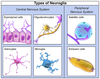
Glial cells are the most numerous cell type in the CNS

4 major types are astrocytes, oligodendrocytes, microglia and ependymal cells

3
Q

What are astrocytes and what do they do?

A

Type of glial cell in the CNS

Has many roles, including providing physical support and repair, maintaining the BBB and environmental homeostasis, including the removal of excess potassium ions

4
Q

What are oligodendrocytes and what do they do?

What condition is are they characteristically affected in?

A

Type of glial cell, produce myelin in the CNS

Characteristically affected in multiple sclerosis

5
Q

What are microglia and what do they do?

A

Type of glial cell. Specialised CNS phagocytes that have a similar lineage to macrophages. Have both immune monitoring and antigen-presenting functions.

They are haemopoietic and migrate to the CNS

6
Q

What are ependymal cells and what do they do?

A

Type of glial cell. Ciliated cuboidal/columnar epithelial cells that line the ventricles

7
Q

Again, list the different types of glial cells found in the CNS.

What types of glial cells exist in the PNS?

A

CNS - astrocytes, microglia, ependymal cells and oligodendrocytes

PNS - Schwann cells and satellite cells

8
Q

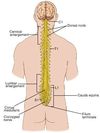
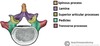
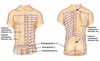
Describe the distribution of white and grey matter in a) the brain and b) the spinal cord

A

Brain - WHITE matter is on the INSIDE and is made up mostly of myelinated axons and their support cells. GREY matter is on the OUTSIDE and consists of a huge number of neurones, cell processes, synapses and support cells

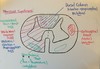
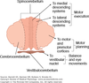
Spinal cord - WHITE matter is on the OUTSIDE while GREY matter is on the INSIDE

9
Q

Sensation arrives in the anterior/posterior horns of the grey matter of the spinal cord

Motor signals leave the anterior/posteroior horns of the grey matter of the spinal cord

A

Sensation arrives in the POSTERIOR (dorsal) grey matter horns

Motor signals leave the ANTERIOR (ventral) grey matter horns

10
Q

The cerebral hemispheres in the adult are derived from what embryonic structure?

A

Telencephalon

11
Q

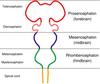
The thalamus and hypothalamus in the adult are derived from what embryonic structure?

A

Diencephalon

12
Q

The midbrain in the adult is derived from what embryonic structure?

A

Mesencephalon

13
Q

The pons and cerebellum in the adult are derived from what embryonic structure?

A

Metencephalon

14
Q

The medulla oblongata in the adult is derived from what embryonic structure?

A

Myelencephalon

15
Q

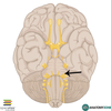
What is the corpus callosum and what processes is it involved in?

A

Large white matter tract that allows communication between the left and right hemispheres

Surrounded by the cingulate gyrus

Involved in memory and motion (among other things)

16
Q

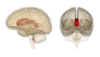
What is the lentiform nucleus? What structure is it next to and what is it made up of?

A

Triangle of grey matter sitting next to the thalamus and is just lateral to the internal capsule

Made up from the putamen and the globus pallidus

17
Q

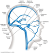
What arteries make up the Circle of Willis?

A

The vertebral arteries coming together to form the basilar artery

The internal carotid arteries

(The middle cerebral arteries are not actually considered part of the circle!)

The anterior cerebral arteries (+anterior communicating artery)

The posterior cerebral arteries (+posterior communicating arteries)

18
Q

Which parts of the brain do the following arteries supply?

  • anterior cerebral
  • middle cerebral
  • posterior cerebral
A

Anterior cerebral supplies mainly the superior and anterior portions of the brain (in a mohawk distribution)

Middle cerebral supplies the majority of the temporal and parietal lobes

Posterior cerebral supplies most of the occipital lobe

19
Q

What is a collection of nerve cell bodies called when it is in a) the CNS and b) the PNS?

A

CNS - collection of nerve cell bodies is called a nucleus

PNS - collection of nerve cell bodies is called a ganglion

20
Q

The cell bodies of multipolar neurones are found in the CNS/PNS and the cell bodies of unipolar neruones are found in the CNS/PNS

A

Multipolar neurone cell bodes are found in the CNS

Unipolar neurone cell bodies are found in the PNS

21
Q

Motor (efferent) fibres move in what direction?

Sensory (afferent) fibres move in what direction?

A

Motor (efferent) - move towards the body wall/cavity or organ

Sensory (afferent) - move towards the brain

22
Q

CN I - name, sensory or motor, where does it connect to?

A

CN I - olfactory

Sensory (special)

Connects to the forebrain

23
Q

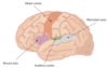
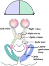
CN II - name, sensory or motor, where does it connect to?

A

Optic nerve

Sensory (special)

Connects to the forebrain

24
Q

CN III - name, sensory or motor, where does it connect to?

A

Oculomotor

Motor

Connects to midbrain

25
CN IV - name, sensory or motor, where does it connect to?
**Trochlear** **Motor** Connects to the **midbrain**
26
CN V - name, sensory or motor, where does it connect to?
**Trigeminal** **Both** sensory and motor Connects to the **pons**
27
CN VI - name, sensory or motor, where does it connect to?
**Abducens** **Motor** Connects to the junction between the pons and the medulla (**pontomedullary junction**)
28
CN VII - name, sensory or motor, where does it connect to?
**Facial** **Both** sensory and motor Junction between the pons and the medulla (**pontomedullary junction**)
29
CN VIII - name, sensory or motor, where does it connect to?
**Vestibulocochlear** **Sensory** Junction between the pons and the medulla (**pontomedullary junction**)
30
CN IX - name, sensory or motor, where does it connect to?
**Glossopharyngeal** **Both** sensory and motor Connects at the **medulla**
31
CN X - name, sensory or motor, where does it connect to?
**Vagus** **Both** sensory and motor Connects to the **medulla**
32
CN XI - name, sensory or motor, where does it connect to?
**Spinal accessory** **Motor** Connects to the **spinal cord**
33
CN XII - name, sensory or motor, where does it connect to?
**Hypoglossal** **Motor** Connects to the **medulla**
34
How many spinal nerves are there? How many are in each group?
**31 pairs** of spinal nerves 8 cervical 12 thoracic 5 lumbar 5 saccral 1 coccygeal
35
Spinal nerves are very long/short Where are they found?
Spinal nerves are in fact very **short!** They are only found in the **intervertebral foramina**
36
After leaving the intervertebral foramina, what do spinal nerves become? Which is bigger - anterior or posterior? And what do they supply?
After leaving the intervertebral foramina the spinal nerves split into the **anterior and posterior rami** to connect to structures of the soma Posterior rami are **smaller and supply the posterior body wall** Anterior rami are **larger and supply the anterolateral body wall**
37
Which of these is correct? Anterior/posterior rami \> spinal nerves \> rootlets \> roots \> spinal cord Anterior/posterior rami \> rootlets \> roots \> spinal nerves \> spinal cord Anterior/posterior rami \> spinal nerves \> roots \> rootlets \> spinal cord Anterior/posterior rami \> roots \> spinal nerves \> rootlets \> spinal cord
Anterior/posterior rami \> spinal nerves \> roots \> rootlets \> spinal cord
38
What does each spinal nerve supply a body segment with?
**General sensory supply** to all structures **Somatic motor supply** to skeletal muscles **Sympathetic nerve supply** to the skin and smooth muscle of the arterioles
39
# Define the following terms... Dermatome Myotome
Dermatome - **area of skin supplied with SENSORY innervation by a single spinal nerve**. Also supplies sensory innervation to deeper structures. There is also some degree of overlap with adjacent spinal nerves Myotome - **skeletal muscles supplies with MOTOR innervation from a single spinal nerve**. May be deep to the dermatome of the same number, but not always!
40
What are the dermatomes associated with the following structures? - male nipple - umbilicus - deltoid - posterior neck, scalp and shoulder - upper limb - lower limb, gluteal region and perinuem
Male nipple - T4 Umbilicus - T10 Deltoid - C4 Posterior neck, scalp and shoulder - C2, C3 and C4 Upper limb - C5-T1 Lower limb - L2-Coccygeal 1
41
What are the 4 bilateral plexuses of the body? What are plexuses and what levels are these 4 at?
Cervical plexus - C1-C4 Brachial plexus - C5-T1 Lumbar plexus - L1-L4 Saccral plexus - L5-S4 Plexuses are the **intermingled ANTERIOR RAMI of adjacent spinal nerves**
42
Explain the anatomy of the sympathetic nerve supply to the soma and the routes sympathetic nerves take on leaving the CNS to reach target organs
Originates in the brain and travels down the spinal cord **Thoracolumbar outflow** (leaving the spinal cord between T1 and L2) - these segments of the spinal cord have **lateral horns** Presynaptic sympathetic axon delivers motor innervation therefore passes through the **anterior rootlets/roots**, then takes 1 of 4 paths 1) ascends and synapses 2) synapses at the same level 3) descends then synapses 4) passes through the sympathetic trunk without synapsing to enter an **abdominopelvic splanchnic nerve**
43
What is the name of the tapered cone shape that the spinal cord ends in? What does it then continue as?
Spinal cord ends at the **conus medullaris** This then continues as a thin strip of connective tissue called the **filum terminale**
44
Describe the distribution of white and grey matter in the spinal cord in comparison to the brain. How is white matter typically divided structurally in the spinal cord?
In the brain - grey matter on the outside with white matter in the middle In the spinal cord - grey matter in the middle forming an H-shape. white matter on the outside (consisting of axons, glial cells and blood vessels) White matter in the spinal cord is typically divided into 4 separate columns - 1 anterior, 1 posterior, 2 lateral
45
What special feature can be noted in the grey matter of the spinal cord between the levels of T1 and L2? Why is this present?
Grey matter has **small lateral horns on either side**. These contain the preganglionic sympathetic neurones (which have thoracolumbar outflow)
46
When rootlets come together to form roots, what do they pass through when on their way to the intervertebral foramina where they become spinal nerves?
They pass through the **subarachnoid space**
47
Describe the arterial blood supply of the spinal cord
3 major **longitudinal arteries** - 1 anterior and 2 posterior, originating from the vertebral arteries and running the whole length of the spinal cord **Segmental arteries** derived from vertebral, intercostal and lumbar arteries **Radicular arteries** which travel along the dorsal and ventral roots
48
Describe the venous drainage of the spinal cord
**Epidural space** - space between the dura and the bone - contains adipose tissue and the **anterior and posterior epidural venous plexuses**
49
How does white matter vary from the top of the spinal cord to the bottom?
There is a **greater amount of white matter at the TOP** and decreases as the spinal cord descends
50
Describe the Dorsal Column/Medial Lemniscus (DCML) system
Provides **proprioception, fine touch and vibration** (especially in the upper limbs) Fibres in the DCML system **cross in the** **medulla** in a 3-neurone relay. E.g. if something is sensed with the fingers of the right hand, fibres on that same side will translate the signals up until the medulla, at which point they will pass over onto the left hand side and go on to the **thalamus**, where they will then be passed onto the cortex Fibres from the thalamus have to pass through the internal capsule in order to reach the cortex, hence why a lesion here can affect movement
51
Describe the spinothalamic pathway
Carries **pain, temperature and deep pressure** Fibres cross **segmentally** i.e. synapse immediately and travel up the anterior spinal cord on the opposite side (as opposed to the DCML which travels up the dorsal column) Fibres **do NOT synapse in the brainstem**, but instead travel **straight to the thalamus** and synapse there
52
What is the primary somatosensory cortex?
Where sensory information first arrives. By the time information arrives here, it has already crossed the midline meaning that **information from the LEFT hand side of the body is represented on the RIGHT hand side of the cortex**, and vice versa Located in the **POST-central gyrus**
53
Describe the corticospinal tract
Provides **fine, precise movements**, especially of the distal limbs Form visible ridges, referred to as "**pyramids**", on the anterior surface of the medulla (hence the alternative name - the pyramidal tract) Approx. 85% of fibres decussate at the **CAUDAL MEDULLA** (**decussation of the pyramids**) but importantly not all fibres! **CROSSED** fibres form the **LATERAL corticospinal tract** **UN-CROSSED** fibres from the **VENTRAL(ANTERIOR) corticospinal tract**, which crosses segmentally
54
How might an infarct of the internal capsule present clinically?
Can result in a lack of descending motor control via the corticospinal tract, resulting in **spastic paralysis w/ hyperreflexion of the upper limbs** ("decorticate posturing") Other signs and symptoms * Weakness of the face, arm and/or leg (caused by lacunar infarcts in the internal capsule, this presentation is known as a "pure motor stroke" * Babinski positive * Clonus * Possible weakness and sensory loss in the contralateral side, if affecting the posterior limb of the internal capsule ("mixed sensorimotor stroke")
55
Where are the synaptic connections in the following pathways... - DCML pathway - Spinothalamic pathway - Corticospinal pathway
DCML - 1st order neurone runs up the **ipsilateral** spinal column, synapses with 2nd order in **lower medulla** at either the gracile fasciculus or the cuneate fasciculus. Then synapses with 3rd order neurone in **thalamus** Spinothalamic - 1st and 2nd order neurones synapse (decussate) at the **level of the spinal cord**. Run up the **contralateral side**. Then synapses with 3rd order neurone in **thalamus** Corticospinal - **descending tract.** Most fibres decussate at the level of the **medulla** and travel down the spinal cord in the **lateral corticospinal tract**. Those that don't travel down in the **anterior corticospinal tract**
56
How is body location represented in the primary motor cortex?
Medially is foot, leg and hip, and moving laterally it becomes trunk, arm, hand and finally the face, tongue and larynx
57
What are the 3 major extrapyramidal motor tracts?
Tectospinal tract Reticulospinal tract Vestibulospinal tract
58
Describe Brown-Sequard Syndrome in terms of cranial nerves+motor system, reflexes and sensation
**Lateral hemisection** of the spinal cord Cranial nerves + motor system - **ipsilateral paralysis** (corticospinal tract) Reflexes - **ipsilateral hyperflexion** and **contralateral extensor plantar reflex** Sensation - **ipsilateral loss of vibration sense and proprioception** (medial lemniscus of DCML), **contralateral loss of pain and temperature sense** (spinothalamic)
59
Describe the structure of the cerebellum - how many lobes and fissures of note?
Anterior lobe Posterior lobe (largest) Flocculus (part of the flocculonodular lobe) Primary fissure - separates the anterior and posterior lobes Horizontal fissure Posterolateral fissure - separates the posterior and flocculonodular lobes
60
What 3 layers is the cerebellar cortex divided into?
Molecular layer (outer) Purkinje layer (middle) Granular layer (inner)
61
How is the cerebellum attached to the brainstem?
Via 3 separate thick bundles of nervous tissue called **peduncles** Superior cerebellar peduncle Middle cerebellar peduncle - by far the largest Inferior cerebellar peduncle
62
What are the 3 functional divisions of the cerebellum? What would happen if a lesion occurred in each of these divisions?
Vestibulocerebellum - lesion can lead to disturbance of balance Spinocerebellum - lesions can result in disturbance of postural control Cerebrocerebellum
63
What is the main role of the cerebellum?
**Co-ordination** of skeletal muscle contraction - balance, posture etc.
64
Cerebellar hemispheres influence the ipsilateral/contralateral side of the body How does this translate to lesions affecting the cerebellum, in contrast to the cerebrum?
Cerebellar hemispheres influence the **ipsilateral** side of the body This means that a lesion in the cerebellum will affected **the same side of the body**, as opposed to in the cerebrum where a lesion would affect the contralateral side
65
What symptoms would a unilateral hemispheric lesion in the cerebellum cause?
Disturbance of coordination of limbs, resulting in intention tremor and unsteady gait
66
What symptoms would a bilateral cerebellar dysfunction cause? How is this commonly caused temporarily?
Slowed, slurred speech Bilateral loss of coordination of the arms Staggering, wide-based gait (cerebellar ataxia) **Acute alcohol exposure** results in bilateral cerebellar dysfunction
67
What is the basal ganglia made up of?
The basal ganglia are a group of structures found deep within the cerebral hemispheres and include... - **caudate nucleus** (cerebrum) **- putamen** ​(cerebrum) **- globus pallidus** ​(cerebrum) **- substantia nigra** (found in the midbrain) **- subthalamic nucleus** (found in the diencephalon)
68
What two components make up the "striatum"? What three components make up the "Corpus striatum"? What two components make up the "lenticular nucleus"? NB - all of these are classed within the basal ganglia
Striatum * The caudate nucleus * The putamen Corpus striatum * The caudate nucleus * The putamen * The globus pallidus Lenticular nucleus * The putamen * The globus pallidus
69
Broadly speaking, what are the functions of the basal ganglia?
FASCILITATION of **purposeful movement** INHIBITION of **unwanted movement** Also has a role in posture and muscle tone
70
Degeneration of what is the classic pathology seen in Parkinson's Disease?
Degeneration of the **dopaminergic neurones of the substantia nigra**
71
How does the "direct pathway" of the basal ganglia function? Damage to this pathway is a characteristic feature of Parkinson's Disease/Huntington's Disease
Direct pathway - enhances outflow of the thalamus, **enhancing the desired movement** Damage to this pathway is a characteristic feature of **Parkinson's Disease​** (unable to initiate movement)
72
How does the "indirect pathway" of the basal ganglia function? Damage to this pathway is a characteristic feature of Parkinson's Disease/Huntington's Disease
Indirect pathway - **inhibits outflow from the thalamus**, suppressing unwanted movement Damage to this pathway is a characteristic feature of **Huntington's Disease** (loss of inhibition, resulting in involuntary movements)
73
Unilateral lesions of the basal ganglia affect the ipsilateral/contralateral side of the body What symptoms do these lesions result in?
Unilateral lesions of the basal ganglia affect the **contralateral** side of the body Symptoms * changes in muscle tone * dyskinesias (abnormal, involuntary movements) * tremor * chorea (rapid, asymmetrical movements usually affecting distal limb muscles) * myoclonus (muscle jerks)
74
What are the 3 cardinal symptoms of Parkinson's Disease? What other symptoms might present?
1. **Akinesia/bradykinesia (slowness of movement)** 2. **Rigidity** 3. **Resting tremor** Others - **anosmia**, balance issues, nerve pain, urinary problems, constipation, erectile dysfunction/sexual dysfunction, dizziness, excessive sweating etc. Cognitive/Psychiatric - depression/anxiety, mild cognitive impairment, dementia
75
What are the extrinsic muscles of the back?
Levator scapulae Rhomboids Trapezius Latissimus dorsi
76
What are the two groups of intrinsic muscles of the back?
**Erector spinae** (superficial) - Ileocostalis, Longissimus, Spinalis **Transversospinalis** (deep)
77
Describe the typical features of a vertebra
Spinous process x1 Transverse process x2 Articular processes - x2 superior, x2 inferior Vertebral arch - x2 pedicles, x2 laminae
78
What are the 5 main ligaments associated with the vertebral column? Where is each found?
**Ligamentum flavum** - short, connects adjacent laminae of vertebrae, posterior to the spinal cord **Posterior longitudinal ligament** - narrow and weak, found between the spinal cord and the vertebral body. Prevents over-flexion of the spine **Anterior longitudinal ligament** - broad and strong, found on the anterior surface of the vertebral body. Prevents over-extension of the spine **Suprasinous ligament** - strong and fibrous, connects the tips of the spinous processes **Interspinous ligament** - weak and membranous, connects the superior and inferior surfaces of adjacent spinous processes
79
What is the difference between the sacral canal and sacral hiatus? What is the clinical relevance of the sacral hiatus?
Sacral canal - superior end of the sacrum, continuation of the spinal canal and houses the sacral nerves Sacral hiatus - **palpable** area at the inferior end of the sacrum, **site of injection with local anasthetic** when performing caudal anaesthesia to numb the sacral spinal nerve roots
80
Where does the spinal cord a) begin and b) end? What is the name of the end point?
a) spinal cord begins at the **foramen magnum** and is continuous with the medulla b) spinal cord ends at **level L1/L2** (usually, but can vary between T12-L3) The end point is called the **conus medullaris**
81
How might a lumbar puncture result in an epidural haematoma?
The spinal cord is surrounded by meninges, but also a layer of epidural fat which contains venous plexuses. If damaged during an LP, this could cause a haematoma, which in turn could result in compression of the spinal cord
82
Where are the following nerve dermatomes? - L1 - L2 - L3 - L4 - L5
L1 - groin (pocket region) L2 - anterior thigh L3 - anterior knee L4 - medial lower leg (inc. malleolus) L5 - lateral lower leg (inc. dorsum of foot)
83
Where are the following nerve dermatomes? - S1 - S2 - S3 - S4 - S5
S1 - heel and outside of the foot S2 - posterior knee and thigh S3 - buttock S4 - perineum S5 - perianal skin
84
Describe the path an action potential takes when testing a dermatomal sensory function
E.g. T10 function AP generated in the axons of the stimulated receptors at T10 then conduct centrally along the same axons within the anterior ramus. AP conducted via the same axons then pass into the T10 spinal nerve, then into the T10 posterior root, and then into the T10 posterior rootlets. Finally the AP arrives at the posterior horn of the T10 spinal cord segment
85
Give an example of a nerve that has more than one spinal nerve root, and therefore overlaps more than one 'true' dermatome
**Femoral nerve** - spinal nerve root levels are L2, L3 and L4 The area of cutaneous innervation of the femoral nerve therefore crosses 3 true dermatomes - those of the L2, L3 and L4 spinal nerves
86
Describe the cutaneous nerve innervation of the neck, both posteriorly and anterolaterally What rhyme can be used to remember the anterolateral sensory branches of the cervical plexus?
Posteriorly - mainly the posterior rami of spinal nerves C2-C8 Anterolaterally - sensory branches of the cervical plexus. **Lesser occipital** (L2), **Great auricular nerve** (C2, C3), **Transverse cervical** (C2, C3) and **Supraclavicular nerves** (C3, C4) **"Little Goats Treat Softly"**
87
What is "Erb's Point"?
A.k.a. the "nerve point" of the neck Point at which the 4 superficial branches of the cervical plexus (Lesser occipital, Great auricular nerve, Transverse cervical and Supraclavicular nerves, Little Goats Tread Softly) emerge from behind muscle The muscle in question is the **posterior border of the sternocleidomastoid**
88
Describe the cutaneous nerve innervation of the trunk, both anterolaterally and posteriorly
Anterolaterally - anterior rami of intercostal nerves **T2-T12** (nb - T12 is a subcostal nerve). After this it is done by the **iliohypogastric and ilioinguinal nevres** (bifurcation of anterior ramus of L1) Posteriorly - posterior rami of spinal nerves **T2-L3**
89
Why might pain from an MI refer to the arm?
Because of the T1 and T2 anterior rami spreading across the chest
90
Describe the peripheral cutaneous nerve innervation of the upper limb
Firstly know the dermatomes of the upper limb * C4 - shoulder * C5 - lateral arm * C6 - lateral forearm, thumb and lateral half of index finger * C7 - medial half of index finger, middle finger, lateral half of ring finger * C8 - medial half of ring finger, pinkie and medial wrist * T1 - medial forearm Then know that these areas are supplied by named nerves, i.e. axons from multiple segments of the spinal cord e.g. C3,C4. Important ones * **supraclavicular nerves** (C3, C4) * **axillary nerve** (C5, C6) - "badge patch" region * **cutaneous branch of musculocutaneous nerve** (C5, C6, C7) * **radial nerve** (C5, C6, C7, C8, T1) * **median nerve** (C5, C6, C7, C8, T1) * **ulnar nerve** (C7, C8, T1)
91
The anatomical snuff box is supplied by the cutaneous branches of what named nerve? When relocating a dislocated shoulder, sensation in what part of the arm should be tested following relocation?
The **radial nerve** Sensation in the **badge patch** area should be tested following relocation of a shoulder, as this is supplied by the **axillary nerve** and is in close proximity to the joint capsule
92
Describe the peripheral cutaneous nerve innervation of the lower limb (thigh first)
Again, first know dermatomes Anteriorly * **Ilioinguinal nerve** (L1) - inside of proximal thigh * **Genitofemoral nerve** (L1, L2) - anterior proximal thigh * **Lateral cutaneous nerve of the thigh** (L2, L3) - lateral thigh * **Obturator nerve** (L2, L3, L4) - inner thigh, below area supplied by ilioinguinal nerve * **Femoral nerve** (L2, L3, L4) - anterior thigh Posteriorly * **Posterior cutaneous nerve of the thigh** (S1, S2, S3) - popliteal fossa and posterior thigh * **Posterior rami a.k.a. cluneal nerve** (S1, S2, S3) - proximal thigh and gluteal region
93
Describe the peripheral cutaneous nerve innervation of the leg
Anterior * **Saphenous nerve** (from femoral, L3, L4) - anterior and medial leg * **Sural nerve** (from tibial and common fibular, S1, S2) - lateral leg * **Superficial fibular nerve** (L4, L5, S1) - dorsum of foot EXCEPT space between first two toes * **Deep fibular nerve** (L5) - space between first two toes Posterior * **Calcaneal nerves** (of the tibial nerves, L4-S3) - heel * **Medial and plantar nerves** (of the tibial nerves, L4-S3) - plantar and lateral foot * **Sural nerve** (from tibial and common fibular, S1, S2) - lateral and posterior leg * **Saphenous nerve** (from femoral, L3, L4) - medial posterior leg
94
Clinical testing of myotomes - shoulder Which muscle and myotome causes a) abduction and b) adduction?
a) abduction - **deltoid, C5** b) adduction - **pectoralis major and latissimus dorsi, C7**
95
Clinical testing of myotomes - elbow joint Which muscle and myotome causes a) flexion and b) extension?
a) flexion - **biceps brachii, C5 and C6** b) extension - **triceps brachii, C7 and C8**
96
Clinical testing of myotomes - finger joints Which muscle and myotome causes a) flexion, b) extension, c) abduction and d) adduction?
a) flexion **- digit flexors, C8** b) extension - **digit extensors, C7** c) abduction - **dorsal interossei, T1** (DAB) d) - adduction - **palmar interossei, T1** (PAD)
97
Clinical testing of myotomes - wrist joint Which muscle and myotome causes a) flexion and b) extension?
a) flexion - **carpal flexors, C6, C7** b) extension - **carpal extensors, C7, C8**
98
Clinical testing of myotomes - hip joint Which muscle and myotome causes a) flexion and b) extension?
a) flexion - **psoas major, L2, L3** b) extension - **gluteus max and hamstrings, L5, S1**
99
Clinical testing of myotomes - knee joint Which muscle and myotome causes a) flexion and b) extension?
a) flexion - **hamstrings, L5, S1** b) extension - **quadriceps, L3, L4**
100
Clinical testing of myotomes - anklejoint Which muscle and myotome causes a) dorsiflexion, b) plantar flexion, c) inversion and d) eversion?
a) dorsiflexion - **tibialis anterior, L4, L5** b) plantar flexion - **gastrocnemius and soleus, S1, S2** c) inversion - **tibialis anterior and posterior, L4** d) eversion - **fibularis longus and brevis, L5, S1**
101
What effect do a) UMN lesions and b) LMN lesions have on muscle contraction?
a) UMN lesions cause muscle **spasticity** b) LMN lesions cause muscle **flaccidity**
102
What nerve roots are associated with the following reflexes? - biceps brachii
Biceps brachii - **C5 (C6)**
103
What nerve roots are associated with the following reflexes? - triceps brachii
Triceps - **C7**
104
What nerve roots are associated with the following reflexes? - supinator/brachioradialis
Supinator/brachioradialis - **C6**
105
What nerve roots are associated with the following reflexes? - ankle
Ankle - **S1**
106
What nerve roots are associated with the following reflexes? - knee
Knee - **L4**
107
All cranial nerves exit **anteriorly** except CN IV which exits ___ and CN ___ which exits laterally
CN IV exits **posteriorly** **CN VIII** exits laterally
108
Which special sensory cranial nerve is the only sensory modality that does NOT synapse in the thalamus prior to reaching the cortex?
The **Olfactory Nerve (CN I)**
109
Why is CN XI different to the other cranial nerves, in terms of where it exits the brainstem?
CN XI **arises from the cervical spinal cord** (mostly, partly also from the nucleus ambiguus in the medulla
110
What are the 3 sensory nuclei associated with the trigeminal nerve? Bonus points - what sense(s) is each associated with?
**Mesencephalic nucleus** - proprioceptive info from chewing muscles **Pontine trigeminal nucleus** - discriminitive touch, vibration **Spinal nucleus** - pain, temperature
111
What are the functions of CN VII?
1. motor - muscles of facial expression, stapedius 2. parasympathetic - innervation to pterygopalatine and submandibular ganglia 3. Taste - anterior 2/3rds of tongue via the chorda tympani
112
What are the functions of CN IX?
1. Tactile sense, pain and temp from the posterior tongue 2. Taste - posterior 1/3rd of tongue 3. Parasympathetic - fibres to the otic ganglion (parotid gland) 4. Motor - stylopharyngeus
113
What are the functions of CN X?
1. Tactile sense, pain, temp from the pharynx, larynx, trachea, oesophagus and thoracic and abdominal viscera 2. Taste - epiglottis 3. Parasympathetic - innervation to ganglia serving thoracic and abdominal viscera 4. Motor (striated muscle of the pharynx and larynx)
114
What muscles are supplied by the Hypoglossal nerve (CN XII)?
Genioglossus Hyoglossus Styloglossus Intrinsic tongue muscles
115
What are the 4 nuclei associated with the facial nerve (CN VII)?
**Facial motor nucleus** - muscles of facial expression **Salivatory nucleus** - parasympathetic innervation of submandibular and pterygopalatine ganglion **Solitary nucleus** - taste in the anterior 2/3rds of the tongue **Spinal trigeminal nucleus** - small region of somatosensation associated with the ear
116
What are the 4 nuclei associated with the glossopharyngeal nerve (CN IX)?
**Solitary nucleus** **Spinal trigeminal nucleus** - small region of somatosensation associated with the ear **Inferior salivatory nucleus** **Nucleus ambiguus** - supplies the stylopharyngeus muscle
117
What are the 4 nuclei associated with the vagus nerve (CN X)?
**Dorsal (motor) nucleus** **Solitary nucleus** **Spinal trigeminal nucleus** **Nucleus ambiguus**
118
Which cranial nerves are associated with the solitary nucleus (taste and visceral sensory info)?
CN VII CN IX CN X
119
Which cranial nerves are associated with the superior and inferior salivatory nuclei?
CN VII CN IX
120
Which cranial nerves are associated with the nucleus ambiguus (motor efferents to muscles of pharynx, larynx and upper oesophagus)?
CN IX CN X (cranial part of CN XI)
121
The solitary nucleus (taste and general visceral sensations) is in a V shape, and is composed of what two nuclei?
**Gustatory nucleus** (taste) **Commissural nucleus** (visceral afferents)
122
Which tract is the part of the pyramidal tract that is motor to cranial nerves? This includes fibres to the motor nuclei of which cranial nerves? What is interesting about the input to the facial nucleus?
Which tract - **corticobulbar tract** Which cranial nerves - **CN V, VII, X and XII** Input to the facial nucleus is **only bilateral to the parts that control the upper face** i.e. supply to the lower half of the face is CONTRALATERAL, while supply to the upper half of the face is BILATERAL
123
What is the 'reticular formation'? What are the functions of some of its centres?
Reticular formation is a network of loosely aggregated cells w/ cell bodies, axons and dentrites that intermingle in the central core of the brainstem. Distinct from the long pathways (e.g. DCML, corticospinal tract) Functions * participation in the conduction and modulation of pain * integration of some basic functions e.g. respiration, sleep etc. * integration of cranial nerve reflexes * influence voluntary movement * regulate autonomic activity
124
What are the 5 layers of the SCALP?
S - skin C - connective tissue (**contains arterial blood supply**) A - aponeurosis L - loose connective tissue P - periosteum
125
What 4 bones make up the pterion?
Frontal Parietal Temporal Sphenoid
126
What are the two layers of dura mater? What can be found between these two layers?
Periosteal layer Meningeal layer **Dural venous sinuses** are found between the two
127
# Define the following features... - diaphragm sellae - tentorium cerebelli - falx cerebri
Diaphragm sellae - tough sheet of dura mater that forms a roof over the pituitary Tentorium cerebelli - tent of dura mater that separates the cerebellum from the cerebrum Falx cerebri - midline structure of dura mater that separates the left and right hemispheres
128
Which spinal nerve mainly provides sensory innervation to the dura mater?
Mainly the **trigeminal nerve (CN V)** (also branches of the vagus nerve and small contributions from the upper cervical spinal nerves)
129
What is the purpose of cranial sutures?
Fibrous joints that help to prevent skull fractures from spreading (minimise propagation)
130
Describe the venous drainage of the brain - what are the sinuses involved and where do they drain to?
Blood is drained from the brain via the **cerebral veins** into the dural venous sinuses Anterior * **Cavernous sinuses** (either side of the sphenoid bone) Midline * **Superior sagittal sinus** (ultimately drains to the confluence of sinuses) * **Inferior sagittal sinus** (ultimately drains to the confluence of sinuses) Posterior * Occipital sinus (drains to the confluence of sinuses) * **Confluence of sinuses** (drains to the right and left transverse sinuses, which drain to the internal jugular vein) Lateral * Transverse sinuses * **Sigmoid sinuses** (drains into the internal jugular vein at the jugular foramen)
131
What is the "danger triangle" of venous drainage of the brain and why is it clinically relevant?
**Facial veins**, which drain to the **ophthalmic veins**, which drain to the **cavernous sinuses** There are **no valves** present in the facial or ophthalmic veins, meaning that infection could spread along these veins into the dural venous sinuses (e.g. from squeezing a spot!)
132
How can CSF be sampled?
Lumbar puncture at **L3/L4** or **L4/L5**
133
Describe the flow of CSF in the brain through the various ventricles
Left and right **lateral ventricles** (via **interventricular foramina of Monro**) To the **third ventricle** (via **cerebral aqueduct** a.k.a. aqueduct of Sylvius) to the **fourth ventricle** (via the **median aperture** (x1) or **lateral apertures** (x2)) to the **subarachnoid space**
134
Where do the following occur? - extradural (a.k.a. epidural) haemorrhage - subdural haemorrhage - subarachnoid haemorrhage
Extradural - between the **bone and the dura**, typically due to a ruptured middle meningeal artery as a result of trauma to the pterion Subdural - separates the **dura from the arachnoid**, due to torn cerebral veins and associated with falls in the elderly + people w/ drinking problems Subarachnoid - bleed is into the **CSF of the subarachnoid space**, due to a ruptured Berry aneurysm (in the Circle of Willis) or congenital aneurysm
135
Brain injury associated with falls in elderly and drunk people. Appears like a banana on CT
**Subdural** haemorrhage
136
Brain injury associated with trauma to the pterion. Appears as a lemon shape on CT
**Epidural** haemorrhage
137
What layers does a needle providing epidural anaesthesia pass through? What further layers are passed through when performing a lumbar puncture?
Skin Superficial and Deep fascia **Supraspinous** then **interspinous ligaments** **Ligamentum flavum** and into the epidural space (containing fat and veins) In a LP, the needle will continue through the **dura mater and arachnoid mater** into the subarachnoid space
138
What potential complication could arise as a result of a lumbar puncture?
If the extradural venous plexus is damaged by the needle on insertion, this could result in an **epidural haematoma** developing which could compress the spinal cord or cause cauda equina
139
CN I - test(s)
Ask the patient to occlude one nostril and test a familiar smell in the other
140
CN II - test(s)
Acuity (Snellen chart) Colour (Ishihara plates) Fields (4 quadrants) Reflexes (pupillary light reflex) Fundoscopy
141
CN III - test(s)
Like CN II, use a light to test for pupillary constriction
142
CN IV - test(s)
'H' test of eye movement
143
CN V - test(s)
Sensory * ask patient to close their eyes and gently brush each of the 3 dermatomes * ask the patient to tell you when they feel the stimulus * remember to do both sides! Motor * palpate strength of masseter and temporalis contraction by asking patient to clench teeth * ask patient to open jaw against resistance
144
CN VI - test(s)
'H' test of eye movement
145
CN VII - test(s)
Raise eyebrows (frontalis) Close eyes tightly (orbicularis oculi) Puff out cheecks (orbicularis oris) Smile (elevators of lips)
146
CN VIII - test(s)
Rinnie and Weber tests
147
CN IX - test(s)
Ask patient to say "aaah" - tests muscles of the palate. If pathology, uvula will point towards the **contralateral side** Ask the patient to swallow a small amount of water - tests laryngeal muscles, a swallow lasting more than 1 second is pathological Listen to the patient's speech - hoarseness may suggest pathology of the larynx (RL nerve)
148
CN X - test(s)
N/A
149
CN XI - test(s)
Ask patient to shrug shoulders Ask patient to turn their head one way and then the other
150
CN XII - test(s)
Ask patient to stick out tongue - if pathology, tongue will point **towards the affected side**
151
What is the name of the structure in the inner ear that transduces chemical energy into electrical energy?
**Organ of Corti**
152
What are the superior olivary nucleus and the nucleus of lateral lemniscus important for, respectively?
Superior olivary nucleus - important for **sound localisation** Nucleus of lateral lemniscus - important for **relays for stapedial and tensor tympany reflexes**
153
What are the two important auditory areas of the brain? Which lobe are they found in?
Primary Auditory Cortex Auditory Association Cortex (Wernicke's Area) Both are found in the **temporal lobe (left side)**
154
Fibres carrying information regarding LOW frequency sound end in the _____ part of the auditory cortex Fibres carrying information regarding HIGH frequency sound end in the _____ part of the auditory cortex
LOW frequency = **anterolateral part** HIGH frequency = **posteromedial part**
155
How would a patient with damage to Broca's area present?
Patients have difficulty **producing language** Often only use simple words, and the most important words in the sentence Patients tend NOT to have difficulty comprehending language This is termed **expressive aphasia**
156
How would a patient with damage to Wernicke's area present?
Patients have difficulty **comprehending language** Can range from saying words out of order to completely meaningless words This is termed **sensory** or **receptive aphasia**
157
What are the components of the visual pathway?
Optic nerve Optic chiasm Optic tract Lateral geniculate nucleus (of the thalamus) Optic radiation Visual cortex
158
Where are the visual areas of the brain?
**Primary visual cortex** (Calcarine sulcus runs through here) **Visual association cortex** Both of which are in the **occipital lobe**
159
What is Meyer's Loop?
Fibres of the geniculocalcarine tract (lateral geniculate body to visual cortex) initially form part of the internal capsule Those fibres carrying visual information from the UPPER HALF of the field of vision first loop anteriorly around the temporal part of the lateral ventricle - this is Meyer's Loop (green lines in the diagram)
160
The visual cortex allows for ____ eye movements The frontal eye fields allow for ____ eye movements
Visual cortex - **tracking eye movements**, tend to be smooth Frontal eye fields - **movements of command** i.e. independent of moving visual stimuli, tend to be "jumpy" (saccadic)
161
What reflexive movements are looked for in assession vision?
Direct light reflex Consentual light reflex (due to pretectal fibres projecting bilaterally to the Edinger-Westphal nucleus) Accommodation reflex (convergence of gaze, contraction of ciliary muscles, pupillary constriction)
162
# Define the following terms... Homonymous hemianopia Heteronymous hemianopia
Hemianopia = blindness for half the visual field in one or both eyes Homonymous = vision is lost in the same visual field in both eyes Heteronymous = vision is lost in opposing visual fields in both eyes
163
What are the 3 types of fibres found within the white matter of the cerebral hemispheres?
**Association fibres** - connect cortical sites lying in the same hemisphere **Commissural fibres** - connect one hemisphere to the other, usually connecting areas of similar function **Projection fibres** - connect hemispheres to deeper structures including thalamus, corpus striatum, brain stem and spinal cord