Week 1 Flashcards

(125 cards)

1
Q

What are the sites of haematopoesis (production of blood cells) at the following points in development?

  • Embryo
  • Birth
  • Birth to maturity
  • Adult
A

Embryo - yolk sac, then liver, then marrow. 3rd-7th month - spleen

Birth - mostly bone marrow, liver and spleen when needed

Birth to maturity - number of active sites in bone marrow decreases but ability for haematopoiesis is retained

Adult - bone marrow of skull, ribs, sternum, pelvis and proximal ends of femur

2
Q

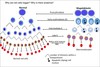
Describe the haemopoietic tree

A

Long-term haematopoietic stem cells (self-renewing) become short term haematopoietic stem cells, which then become multipotent progenitor (MPP) cells

These then differentiate into either common myeloid progenitor (CMP) cells or common lymphoid progenitor (CLP) cells

CMP

  • megakaryocyte-erythrocyte progenitors (MEP)
    • erythrocytes
    • platelets
  • granulocyte-monocyte progenitors (GMP)
    • granulocytes
    • macrophages
  • Pro-DC
    • dendritic cells

CLP

  • Pro-DC
    • dendritic cells
  • Pro-T
    • T cells
  • Pro-NK
    • NK cells
  • Pro-B
    • B cells
3
Q

Describe the steps from the Common Myeloid Progenitor (CMP) cell to the mature neutrophil (granulopoiesis)

A

CMP > myeloblast

Myeloblast > promyelocyte

Promyelocyte > myelocyte

Myelocyte > metamyelocyte > N. band forms >…

Neutrophil

4
Q

Describe the steps from the Common Myeloid Progenitor (CMP) cell to mature red cell/erythrocyte (erythropoiesis)

What changes occur in the cell morphology, and at what point?

A

CMP > pronormoblast

Pronormoblast > basophilic/early normoblast (ribosome synthesis)

basophilic/early normoblast > polychromatophilic/intermediate normoblast (haemoglobin accumulation, nucleus condenses)

polychromatophilic/intermediate normoblast > orthochromatic/late normoblast

Nucleus is then lost, some RNA retained

Reticulocyte > Mature red cell/erythrocyte (remaining RNA is completely lost over ~7 day period)

5
Q

Describe the steps from the Common Myeloid Progenitor (CMP) to platelet

A

CMP > megakaryoblast

Megakaryoblast > promegakaryocyte

Promegakaryocyte > megakaryocyte

Platelets form on the periphery of the megakaryocyte and bud off

6
Q

What are the types of granulocyte?

A

Eosinophils

Basophils

Neutrophils

7
Q

Describe the structure and function of neutrophils

A

Structure

  • Segmented (polymorphic) nucleus
  • Stains neutrally (hence the name)

Functions

  • short life in circulation (transit into tissues)
  • phagocytoses invading molecules
  • kills invaders with granule contents, killing itself in the process
  • in doing so, attracts other cells
  • numbers are increased by body stress e.g. infection, trauma etc.
8
Q

Describe the structure and function of Eosinophils

A

Structure

  • bi-lobed (usually)
  • bright orange/red granules

Function

  • fight parasitic infections
  • involved in hypersensitivity reactions and often elevated in patients with allergic conditions
9
Q

Describe the structure and function of Basophils

A

Structure

  • infrequent in circulation
  • large, deep purple granules, obscuring the nucleus

Function

  • circulating version of tissue mast cell
  • mediates hypersensitivity reactions
  • Fc Receptors bind IgE
  • granules contain histamine
10
Q

Describe the structure and function of monocytes (macrophages)

A

Structure

  • large, singular nucleus
  • faintly staining granules

Function

  • circulate for a week, then enter tissues to become macrophages
  • phagocytose invaders, killing them and presenting antigens to lymphocytes
  • attract other cells
  • more long-lived than neutrophils
11
Q

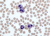
How does a lymphocyte change in appearance when it becomes activated (a.k.a. from mature to atypical)?

A

Mature - small, with condensed nucleus and a rim of cytoplasm (B)

Activated - large, with plentiful blue cytoplasm extending around neighouring red cells (A)

12
Q

Effector cells are easily identifiable, but how are the precursors identified?

A

Immunophenotyping - examine the expression profile of proteins (antigens) on the surface of the cells. Done with mAbs w/ fluorescent tags

13
Q

What is a common site for bone marrow aspiration?

A

Posterior iliac crests

14
Q

Describe some of the key features of a typical red blood cell

A

Full of Hb

No nucleus - can’t divide or replace damaged proteins meaning lifespan is limited (approx 120 days)

No mitochondria - limited to glycolysis for energy generation

Flexible - requires specialised membrane

High surface area/volume ratio

15
Q

What structures in the membrane of a RBC allow it to be flexible (like a hiking tent!)?

A

Protein spars’ - alpha and beta spectrin

16
Q

Describe the structure and function of haemoglobin

A

Structure

  • Tetrameric globular protein made up of 2 alpha and 2 beta chains (in adults)
  • Haem group Fe2+ is found in the centre of this protein in a flat porphyrin ring

Function

  • Oxygen delivery
  • Acts as a buffer for H+
  • Involved in CO2 transport
17
Q

Describe the process of RBC destruction

A

Usually occurs in the spleen

Old RBCs are taken out of the circulation by macrophages and red cell contents are recycled

  • globin chains are broken down into amino acids
  • haem group is broken down into iron and bilirubin
18
Q

Briefly describe the regulation of RBC production

A

Hypoxia sensed by kidneys

Erythropoietin is produced which stimulates RBC production in the bone marrow

More RBCs are released, and EPO levels subsequently drop

19
Q

How does a RBC a) generate ATP and b) prevent Fe2+ from becoming Fe3+ (oxidation)?

A

ATP is generated via glycolysis or Embden-Meyerhof pathway which yields a net generation of ATP and NADH

NADH prevents the oxidation of Fe2+ to Fe3+

20
Q

What protects our RBCs from reactive oxygen species such as hydrogen peroxide?

What is the rate limiting enzyme in the recycling of this molecule?

A

Glutathione (GSH) reacts with hydrogen peroxide to form water and an oxidised glutathione product (GSSG). This is then replenished by NADPH.

The rate-limiting enzyme in this process is glucose-6-phosphate dehydrogenase (G6PD), a deficiency of which can potentially result in anaemia in individuals that are unable to compensate by producing more reticulocytes e.g. in conditions of high oxidative stress, such as infection

21
Q

How is CO2 transported to the lungs?

A

60% transported as bicarbonate (RBCs are important in generation)

30% transported bound directly to haemoglobin - carbamino-Hb

10% is dissolved in solution

22
Q

Describe the dissociation curve for oxygen and why it is unusual

A

Unusual as it doesn’t follow Michaelis-Menten kinetics. Dissociation curve is sigmoidal

As one O2 molecule binds to a subunit, the Hb shape changes (allosteric effect) to increase its affinity for binding further O2 molecules

23
Q

What does a shift to either the right or the left in the oxygen dissociation curve mean?

What factors may cause this to happen?

A

Right shift indicates decreased oxygen affinity of Hb allowing more oxygen to be available to tissues

Left shift indicates increased oxygen affinity of Hb, allowing less oxygen to be available to tissues

Left Shift

  • increased pH
  • decreased CO2
  • decreased temperature
  • decreased 2,3-DPG
  • foetal haemoglobin
  • myoglobin

Right Shift (Bohr Effect)

  • decreased pH
  • increased CO2
  • increased temperature
  • increased 2,3-DPG
24
Q

Anaemia is defined as a reduced total red cell mass, however this is not easy to measure in routine practice. What two surrogate markers are used to assess anaemia, and how are they measured?

A

Haemoglobin concentration - spectrophotometer

Haematocrit - ratio of whole blood that is red cells if a sample was allowed to settle, normally about 50%

25
In what situations are the two surrogate markers of red cell mass not a reliable indicator of anaemia?
Hb/hct are not a good marker of anaemia if... there has been rapid blood loss of 50% blood volume with subsequent fluid replacement i.e. plasma expansion (Hb will increase by haematocrit will remain at the same level, meaning the ratio will be lowered) Or in the case of haemodilution (again, plasma expansion)
26
How do reticulocytes compare to normal mature red cells?
Slightly larger than average red cells Still have remnants of RNA and stain purple/deeper red as a consequence Blood film appears **polychromatic**
27
Regarding red cell values in a blood test, which are measured and which are calculated?
Measured * Hb concentration * Mean Cell Volume (MCV) * Number of red cells (concentration) Calculated * Haematocrit * Mean cell haemoglobin
28
Anaemia can be pathophysiologically classified into either being due to decreased production (low reticulocytes) or increased loss/destruction (high reticulocytes). What are the causes of both?
Decreased production (low reticulocyte count) * Hypoproliferative - **reduced amount** of erythropoiesis * Maturation abnormality - erythropoiesis is present but **ineffective** * **cytoplasmic** defects are due to impaired **haemoglobinisation** * **nuclear** defects are due to impaired **cell division** Increased loss/destruction (high reticulocyte count) * Bleeding * Haemolysis
29
What red cell parameter is useful in distinguishing cytoplasmic vs nuclear defects (both resulting in decreased production/low reticulocyte count)?
**Mean Cell Volume** If MCV is low (**microcytic anaemia**) then consider problems with **haemoglobinisation** If MCV is high (**macrocytic anaemia**) then consider problems with maturation (NB - cells haven't increased in size, they just remain large due to not dividing)
30
Where does haemoglobin synthesis occur?
In the cytoplasm - defects result in small cells (microcytic anaemia)
31
How would an anaemia due to a deficiency in haemoglobin synthesis appear on a blood film? What are some of the causes of this type of anaemia?
Red cells would appear **hypochromic** (lacking in colour) and **microcytic** (small) Causes * Haem deficiency * Lack of iron for erythropoiesis * **Iron deficiency anaemia** - most common cause worldwide * Chronic disease * Problems with porphyrin synthesis * lead poisoning * pyridoxine responsive anaemias * Globin deficiency * **Thalassaemia** - second most common worldwide * (Congenital Sideroblastic Anaemia - v. rare)
32
What molecule transports Iron from stores to make red cells? How much iron is in the circulation at any one time?
**Transferrin** Only a tiny amount of iron is actually present in the circulation as it moves to and from storage sites (mostly bone marrow)
33
Iron turnover is fast/slow Where is most of the iron in the body found?
Iron turnover is **fast** (4mg in plasma pool and 20mg is moved a day) Most of the iron is found in **haemoglobin** (approx. 2500mg, vs 4mg in plasma, 500mg in macrophages, 500mg in parenchymal tissues...)
34
What molecule binds to iron when it is stored in the liver?
**Ferritin**
35
What molecule best represents the body's iron supply? What molecule best represents the body's iron storage?
Supply - **transferrin** Storage - **ferritin**
36
What two indicators can confirm iron deficiency when used together?
Anaemia (decreased functional iron) Reduced storage iron (low serum ferritin)
37
What are some of the causes of iron deficiency?
Inadequate dietary intake * Relative deficiency - esp. women of child bearing age and children * Absolute deficiency - e.g. vegetarian diets Excessive loss * Blood loss, usually GI in nature * tumours * ulcers * NSAIDs Malabsorption * Coeliac disease * Achlorhydria
38
What is the main clinical point about unexplored iron deficiency anaemia?
It isn't a diagnosis! Requires further investigation to determine the underlying cause
39
On a full blood count, what is the difference between a macrocytic anaemia and a macrocytosis?
Macrocytic anaemia - Hb conc. low, RBC levels low, HCT ratio low, MCV raised Macrocytosis - only MCV is raised, everything would be normal
40
How can macrocytes be identified on a blood film? What are the normal ranges for red cell size?
Normal ranges = **80-100fl** (femtolitres) In a normal film, red cells should be about the same size as the nculeus of a normal lymphocyte
41
Define the term "megaloblast"
An **abnormally large** nucleated red cell precursor **with an immature nucleus**
42
Genuine (true) macrocytic anaemias can be classified as either megaloblastic or non-megaloblastic. What are megaloblastic anaemias characterised by?
Predominant defects in **DNA synthesis and nuclear maturation**, but haemoglobin synthesis is preserved
43
How do megaloblastic cells bring about anaemia?
Cytoplasmic development and haemoglobin accumulation occurs normally so the precursor cell is bigger but with an enlarged nucleus Once optimal Hb levels are reached, the nucleus is extruded leaving behind a larger than normal red cell, termed a **macrocyte** Overall however, there are fewer macrocytes than there would be normal reticulocytes, hence anaemia. This reduction in reticulocytes also results in raised EPO levels, which prompts more divisions of the megaloblastic progenitor cells
44
Megaloblastic anaemia - causes
B12 deficiency Folate deficiency Others (including drugs and rare inherited abnormalities)
45
Why does a lack of B12 and/or Folate cause megaloblastic anaemia?
Both B12 and Folate are **essential co-factors** for nuclear maturation. They enable chemical reactions for DNA synthesis and gene activity through silencing genes through **methylation**
46
How are the B12 and Folate biochemical reactions linked?
Via the **Folate cycle** and the **Methionine cycle**
47
What is the Folate cycle important for?
Nucleoside synthesis (**uridine to** **thymidine** conversion)
48
What is the Methionine Cycle important for?
Important for producing a **methyl donor** (**S-adenosyl methionine**) which has an impact on DNA, RNA proteins, lipids and folate intermediates
49
Describe the passage of B12 from the mouth, through the stomach and small bowel, into the ilium and then blood vessels
B12 in food travels down into the stomach where it binds to **R protein** (released from gastric mucosa) in order to stop it from being dissolved by stomach acid **Intrinsic Factor** is also produced in the stomach and travels down the duodenum with the B12-R-protein complex. **R-protein dissociates from B12 in the duodenum**, at which point **B12 binds to Intrinsic Factor**. This complex then travels to the ilium where it is taken up via **Cubulin receptors**. From the Ilial epithelium, B12 is then passed into the blood vessels
50
What are some of the causes of B12 deficiency?
Poor dietary intake Stomach - pernicious anaemia, atrophic gastritis, PPIs/H2 receptor antagonists, gastrectomy/bypass Pancreas - chronic pancreatitis Small bowel - bacterial overgrowth, Coeliac disease, Crohn's
51
What is Pernicious Anaemia? What other conditions is it associated with?
Autoimmune destruction of gastric parietal cells Results in an **Intrinsic Factor Deficiency**, resulting in B12 malabsorption Associated with atrophic gastritis or other autoimmune disorders (hypothyroidism, vitiligo, Addison's Disease etc.)
52
How do body stores and dietary requirements for B12 and Folate compare? What is the clinical relevance of this?
Body stores - 2-4 years for B12, 4 months for Folate Dietary requirements - 1.5 micrograms/day for B12, 200 micrograms a day for Folate Clinically, this means that a B12 deficiency won't present itself for years (due to amounts stored), however a Folate deficiency will present after a few months
53
Folate deficiency - causes
Inaedquate intake Malabsorption - Coeliac disease, Crohn's disease Excess utilisation - haemolysis, exfoliating dermatitis, pregnancy, malignancy Drugs e.g. anticonvulsants (Phenytoin
54
What is the most common cause of macrocytosis?
Excess alcohol intake (reduces Folate stores)
55
B12/Folate Deficiency - clinical presentation What presentation is specific to B12 deficiency?
Signs and symptoms of anaemia Weight loss, diarrhoea, infertility Sore tongue, jaundice Developmental problems Specific to B12 - **neurological problems** (dorsal column abnormalities, neuropathy, dementia, psychiatric manifestations) - NB: these changes are irreversible! See **Subacute Combined Degeneration of the Cord**
56
Suspected B12/Folate deficiency - laboratory investigations What would they show? How effective are these tests?
Macrocytic anaemia (low red cell count) Pancytopenia in some patients (all cells are low) Blood film - shows **macrovalocytes** (oval-shaped cells) and **hypersegmented neutrophils** Assay of serum B12 and Folate levels (low levels may not indicate deficiency, and vice versa!) Look for autoantibodies - **anti-Intrinsic Factor** (specific but not sensitive) and **anti-Gastric Parietal Cell** (sensitive but not specific) **Bottom line - no one single test is able to determine B12/Folate deficiency**
57
Megaloblastic anaemia - treatment
Treat the cause if possible **Vitamin B12 injections** (IM) for life if patient has Pernicious Anaemia **Folic acid tablets** - 5mg orally
58
Non-megaloblastic macrocytosis - causes
Alcohol Liver Disease Hypothyroidism Marrow failure (myelodysplasia, myeloma, aplastic anaemia)
59
When might you see reticulocytosis? What would you see when performing lab investigations?
In response to acute blood loss, red cell breakdown, or any time that the marrow is stressed (infection etc.) **False macrocytosis** (as reticulocytes are larger than normal red cells and are included in the MCV) **Polychromasia** as reticulocytes are darker in colour than normal red cells
60
What are the two causes for 'Spurious (False) Macrocytosis'?
Reticulocytosis Cold agglutinins
61
Why might the red cell count be low?
Increased destruction Decreased production Redistribution
62
# Define the following terms with regards to marrow activity and cell production - hyperplasia - dysplasia - hypoplasia - aplasia
Hyperplasia - increased production Dysplasia - disordered production Hypoplasia - decreased production Aplasia - no production
63
What cells in the kidney detect hypoxia and release EPO as a response?
**Interstitial fibroblasts** near to the **peritubular capillaries and the proximal convoluted tubule**
64
When blood is being tested, whole blood is centrifuged to separate it into its various components and then subjected to a series of microbiological tests for various pathogens. What are some of the conditions that are tested for?
HIV Hep B Hep C Hep E HTLV Syphilis
65
How are the following stored? - Red cells - Fresh Frozen Plasma - Platelets
Red cells - 4 degrees for 35 days FFP - -30 degrees for 3 years Platelets - 22 degrees for 7 days with agitation
66
What are the two major blood group systems used? What are their corresponding gene names and chromosomal locations?
ABO system - *ABO* gene, 9q34.2 (**chromosome 9**) Rh system - *RHD, RHCE* gene, 1p36.11 (**chromosome 1**)
67
What do the 'A' and 'B' genes code for?
Transferases which modify precursor called '**H substance**' on red cell membranes
68
What are the dominance patterns of A, B and O? Which are the most common ABO types and which is the rarest?
A and B are both dominant over O A and B are co-dominant with each other O is silent i.e. it isn't associated with a gene **O** and **A** are the most common types (47% and 42% respectively) **AB** is the least common type (3%)
69
Regarding blood types, what is the difference between genotype and phenotype? For each blood phenotype, what is the genotype?
Phenotype = which antigens are detected Genotype = which genes are present Group O - **OO** Group A - **AA or AO** Group B - **BB or BO** Group AB - **AB** (co-dominance)
70
What is Landsteiner's Law? What is the clinical relevance of this?
When an individual lacks the A or the B antigen the corresponding antibody is produced in their plasma This means that "naturally occuring" antibodies will cause haemolysis of red cells that express that specific antigen i.e. if someone is phenotype A and is given type B blood, their anti-B antibodies will cause haemolysis (**transfusion reaction**)
71
Regarding Landsteiner's Law, what type of Ig are the antibodies that are produced, where are they made and at what point?
They are **IgM antibodies** that are made in the **gut** and this occurs from **approximately 6 months of life onwards**
72
If a patient is blood type O, what red cell antibodies will they have in their system?
**Both** Anti-A and Anti-B
73
Which blood group is known as the universal donor? Which blood group is known as the universal receiver?
Universal donor = **Type O** (negative) Universal receiver = **Type AB** (positive)
74
What proportion of the population is Rh positive? What is the corresponding genotype for both Rh+ and Rh-?
85% of the population are Rh positive Rh + = DD or Dd Rh- = dd
75
When determining someone's blood type using anti-A or anti-B antibodies in the lab, how would a positive result appear? What are the 3 types of antisera each blood sample is subjected to?
If someone is type A and is given anti-A antibodies, this will cause **aggultination** = positive result Every sample is subjected to Anti-A, Anti-B and Anti-D
76
How is cross-matching done?
Take donor red cells and add patient plasma Then add anti-human globulin (facilitates red cell agglutination) **Agglutination i**ndicates that donor red cells are **incompatible** with patient plasma **No agglutination** = donor red cells can be used
77
What are the indications for red cell transfusion?
**_Symptomatic_ anaemia Hb\<70g/L** Major bleeding **Transfuse a single unit of red cells and then reassess patient before giving more**
78
What are the indications for platelet transfusion?
Prophylaxis in patients with bone marrow failure and very low platelet counts Prophylaxis prior to surgery/managing bleeding in patients with thrombocytopoenia (1 unit of platelets is almost always the starting dose)
79
What are the indications for fresh frozen plasma transfusion?
Treatment of bleeding in a patient with coagulopathy (**PT ratio \>1.5**) Management of massive haemorrhage Transfuse early in trauma
80
Describe the structure of a normal molecule of haemoglobin
Tetramer made up of 2 alpha and 2 beta globin chains 1 haem group is attached to each chain Iron is contained within each haem pigment
81
What are the 3 major forms of haemoglobin in a normal adult? What proportions are they present in?
HbA (2 alpha chains and 2 beta chains) HbA2 (2 alpha chains and 2 delta chains) HbF (2 alpha chains and 2 gamma chains) HbA = 97% HbA2 = 2.5% HbF = 0.5%
82
Genes for alpha globins are present on chromosome \_\_. How many genes are there per chromosme? Genes for beta globins are present on chromosome \_\_. How many genes are there per chromosome?
Genes for **alpha globins** are present on **chromosome 16**. There are **2 genes per chromosome (4 per cell)** Genes for **beta globins** are present on **chromosome 11**. There is **1 gene per chromosome** **(2 per cell)**
83
Haemoglobinopathies are hereditary conditions that affect the synthesis of globin chains. What are the two main groups? What is the main difference between these groups?
**Thalassaemias** - **decreased rate** of globin synthesis **Structural haemoglobin variants** - normal rate of production but of **structurally abnormal** globins e.g. sickle cell
84
What are the two categories of thalassaemia? What do they both result in?
**Alpha** thalassaemia - reduced production of alpha globin chains **Beta** thalassaemia - reduced production of beta globin chains Both result in **inadequate Hb production**, giving hypochromic microcytic anaemia The unbalanced accumulation of globin chains is toxic and results in ineffective erythropoiesis and haemolysis
85
Describe the genetic basis of alpha thalassaemia
Normal individuals have (aa/aa) Pathology arises from the deletion of one (a-) or both (--) alpha genes from chromosome 16 (termed a+ or a0 respectively) More genes that are absent = more severe the mutation i.e. **a+ = reduced** and **a0 ​= absent** Also worth remembering that alpha chains are present in other molecules as well, so the effects of these mutations can be far reaching
86
What are the various classifications of alpha thalassaemia?
Normal - (aa/aa) **alpha thalassaemia trait** (one or two genes missing) - a+/a (a-/aa) - a0/a (--/aa) - a+/a+ (a-/a-) **HbH disease** (only one alpha gene left) - a0/a+ (a-/--) **Hb Barts hydrops fetalis** (no functional alpha genes) - a0/a0 (--/--)
87
Alpha thalassaemia trait - symptoms, appearance on blood film and management
(a-/aa) or (--/aa) or (a-/a-) Asymptomatic, no treatment needed Appears on blood film as hypochromic, microcytic red cells with mild anaemia Important to distinguish from iron deficiency (**ferritin levels will be normal**)
88
HbH - genotypic appearance, telltale histological sign and clinical features
HbH aka severe form of alpha thalassaemia Only one alpha gene per cell e.g. **a+/a0 (a-/--)** Telltale histological sign is **"golf ball" cells** (due to execss ß chains forming tetramers, called HbH, which cannot carry oxygen) Presents clinically as **anaemia with very low MCV and MCH**. Anaemia may be anywhere from moderate to transfusion-dependent. May also cause **splenomegaly** due to extramedullary haematopoiesis. **Jaundice** may also be present due to haemolysis and ineffective erythropoiesis
89
Describe the inheritence pattern of HbH disease.
Reminder: HbH disease is a+/a0 (a-/--) i.e. **only one functional alpha unit** Two parents with alpha-thalassaemia (a0 and a+) With possible offspring... - normal (1 in 4) - a0 thalassaemia (1 in 4) - a+ thalassaemia (1 in 4) - HbH disease (1 in 4)
90
What is the most severe form of alpha thalassaemia? How does it appear genotypically? What's the prognosis?
Most severe form is **Hb Barts**. **No functional alpha genes** have been inherited (--/--). Has minimal or no alpha chain production, meaning **HbA cannot be made** Prognosis is not good, if the child is born alive they present with **Hb Barts Hydrops Fetalis Syndrome** (severe anaemia, hepatosplenomegaly, cardiac failure, growth retardation...) - die very shortly after birth.
91
What type of mutation usually causes beta thalassaemias?
Point mutations - over 200 identified so far
92
Alpha thalassaemia affects alpha chains and therefore also affects HbF. Is the same true of beta thalassaemias?
No - HbF is unaffected
93
What are the 3 classifications of beta thalassaemia and how does each appear genotypically?
**Beta thalassaemia trait** (ß+/ß or ß0/ß) **Beta thalassaemia intermedia** (ß++ or ß0+) **Beta thalassaemia major** (ß00)
94
How does each of the 3 beta thalassaemias present and how is each managed?
**Beta thalassaemia trait** - asymptomatic, no/mild anaemia with low MCV/MCH and raised HbA2 **Beta thalassaemia intermedia** - moderate severity requiring occasional transfusion **Beta thalassaemia major** - severe, requiring lifelong transfusions
95
What particular symptoms might you see in a patient with beta thalassaemia major (a.k.a. Cooley's anaemia), as a result of extramedullary haematopoiesis? When does this condition typically first appear?
Initially presents between **6 months-2 years old** (initially HbF protects) May see... - hepatosplenomegaly - skeletal changes (expansion of bone marrow to compensate) including abnormally prominent forehead (frontal bossing), full cheek bones (prominent malar eminence) and overgrowth of the maxillae, exposing the upper teeth - organ damage and spinal cord compression
96
Beta thalassaemia major needs to be managed with regular blood transfusions. What major complication is associated with repeated blood transfusions and what are some of the consequences?
**Iron overload** needs to be watched for - main cause of mortality **Endocrine dysfunction** (diabetes, osteoporosis, impaired growth and puberty) due to pituitary developing iron deposits **Cardiac disease** (cardiomyopathy, arrhythmias) **Liver disease** (cirrhosis, hepatocellular cancers) **Eyes** **Testicles**
97
How can iron overload be managed pharmacologically?
With **chelating agents** such as desferrioxamine - bind to iron which form complexes and are excreted in the stool and urine
98
What is the mutation that causes sickle cell disease?
**Point mutation** in codon 6 of the ß globin gene (chromosome 11) that results in a **substitution from glutamine to valine**
99
Briefly describe the mechanism of pathology that results from the mutation seen in sickle cell disease
Hb structure is altered from Hb to HbS (a2ßs2) HbS **polymerises if exposed to low oxygen** for a prolonged period of time, creating long spikes within the RBC, distorting the shape of the cell
100
What is the difference between sickle trait (HbAS) and sickle cell anaemia (HbSS)? What proportion of HbS is generally required for pathology to develop?
Sickle trait - one normal gene, one abnormal gene (ß/ßs) resulting in an **asymptomatic carrier state**, HbS levels tend to be too low to polymerise (**HbS \<50%**) so there are few clinical features. May sickle cell in severe hypoxia e.g. high altitudes Sickle cell anaemia two abnormal genes (ßss) - **autosomal recessive, HbS \>80%** with no HbA
101
What is sickle cell crisis and how is it managed acutely?
Acute episode of tissue infarction due to vascular occlusion precipitated by dehydration, infection, cold exposure or stress/fatigue, hypoxia (**DICESH**) Symptoms are dependent on site and severity, but may affect digits, bone marrow, spleen, CNS etc. and may be extremely painful Painful crises are treated with **opiate analgaesia**, hydration, rest, oxygen and antibiotics if evidence of infection. If in severe crisis, can do red cell exchange transfusion
102
How is sickle cell anaemia managed in the long-term to reduce the impact of sickle crises?
Prophylactic penicillin and vaccination to reduce the risk of infections Folic acid supplementation Hydroxycarbamide - "tricks" marrow into producing more HbF Regular transfusions to prevent strokes in select cases
103
What is the difference between intravascular and extravascular haemolysis? Which is more common?
Intravascular - red cells are destroyed within the circulation Extravascular - red cells are taken up by the reticuloendothelial system (mainly the spleen and liver) Both involve different mechanisms, so different breakdown products are detected. Knowing the nature of the haemolysis helps to determine the cause **Extravascular haemolysis is more common**
104
What breakdown products might you see in extravascular haemolysis and what clinical signs may you see?
There will be hyperplasia at the site of destruction (so either splenomegaly or hepatomegaly) and the release of **protoporphyrin** This could result in **unconjugated bilirubinaemia** (jaundice and gallstones) and **urobilinogenuria** Crucially, there will be **normal products but in excess**
105
What breakdown products might you see in intravascular haemolysis and what clinical signs may you see?
Red cells are destroyed in the circulation so there may be free Hb in the circulation (**haemoglobinaemia**), **methaemalbuminaemia** (combination of methaem and blood plasma albumin), **haemoglobinuria** which presents as pink coloured urine that turns black when left to stand, and **haemosiderinuria** (excess iron in the blood) Crucially, there are **abnormal products in the blood** due to this form of haemolysis, and it may be life-threatening
106
What are some of the causes of intravascular haemolysis?
ABO incompatible blood transfusion G6PD deficiency (X-linked, shortened RBC lifespan due to inability to replace glutathione, variable presentation) Severe falciparum malaria (Blackwater Fever) Paroxysmal nocturnal haemoglobinuria (PNH) DIC TTP
107
When diagnosing someone with a haemolytic disease, firstly you need to confirm the haemolytic state and secondly you need to identify the cause. Which investigations will allow you to do these?
Confirm state * FBC + blood film * Reticulocyte count * Serum unconjugated haemoglobin * Urobilinogen Investigate cause * History and examination * Blood film * membrane damage (spherocytes) * mechanical damage (red cell fragments) * oxidative damage (Heinz bodies a.k.a. bite cells - G6PD def., Alpha thalassaemia...)) * others e.g. HbS (sickle cells) * Specialist investigations * Direct Coombs' test
108
What are Heinz bodies and what do they indicate?
Inclusions within red blood cells composed of denatured haemoglobin Indicates **oxidative damage** - G6PD deficiency alpha thalassaemia, chronic liver disease NADPH deficiency...
109
What are the two causes of autoimmune haemolysis and which Ig is associated with each?
Warm autoantibody (IgG) Cold autoantibody (IgM)
110
What is the most common cause of red cell membrane abnormality that could result in haemolysis?
Hereditary spherocytosis
111
Globin chains are broken down and recycled into \_\_\_\_
amino acids
112
Heme groups are broken down into ____ and \_\_\_\_
Iron and bilirubin
113
What's the blood group?
A positive Reminder - if A, anti-B antibodies will be present. If placed in anti-A the solution will agglutinate as A antigens are present on the RBC surface NB - the bottom two rows are confirmatory
114
How is the Indirect Antiglobulin Test performed?
Reagent of red cells expressing known antigen(s) Add patient plasma to reagent Add anti-human globulin Look for agglutination (if +ve, indicates the presence of antibody)
115
Why might ferritin be falsely high?
Ferritin is also an **acute phase protein** and may be falsely raised in anaemic patients if they have an infection
116
What's the likely diagnosis? - 7-year-old girl has a history of intermittent jaundice and fatigue, usually occurring just after she has had a cold.  Her Dad had a splenectomy as a child - Blood film shows spherocytes
Hereditary spherocytosis **Autosomal dominant** inheritence, however approx 20% are due to new mutations
117
How might red cells attempt to improve oxygen delivery if there are not enough haemoglobin molecules? (think of the factors affecting the oxygen dissociation curve...)
Right shift of curve to decrease O2 affinity and make more oxygen availble to tissues Done via interaction with **2,3-DPG** (increased in anaemia), this is called the **Rapoport-Lubering Shunt** and changes the shape of the Hb molecule to increase the amount of O2 dissociation
118
How does the bone marrow respond to haemolysis?
Reticulocytosis - results in polychromasia Erythroid hyperplasia
119
What are some of the causes of warm autoantibody autoimmune haemolysis?
Idiopathic - most common cause Autoimmune disorders e.g. SLE Lymphoproliferative disorders e.g. CLL Drugs - penicillins etc. Infections
120
Which infections cause cold autoantibody autoimmune haemolysis?
EBV Mycoplasma
121
What are some of the acquired causes of haemolysis that result in premature destruction of normal red cells?
DIC HUS (E. coli O157) TTP Leaking heart valve Infections e.g. Malaria (Blackwater fever)
122
What type of anaemia might you see in someone with a mechanical heart valve that is causing fragmentation of red cells?
Microangiopathic Haemolytic Anaemia (MAHA)
123
What are some of the acquired causes of haemolysis that result in membrane defects in the red cells? (nb - these are all very rare)
Liver disease (Zieve's syndrome) Vitamin E deficiency Paroxysmal Nocturnal Haemoglobinuria
124
What test is used to detect paroxysmal nocturnal haemoglobinuria?
Ham's Test
125
What drugs might cause the formation of bite cells? Why does this happen?
**Dapsone** or **Salazopyrin** Stresses the metabolic pathways of cells resulting in oxidative damage resulting in haemolytic anaemia NB also caused by G6PD deficiency