week 2- eye Flashcards

1
Q

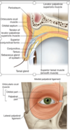
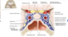
Decribe the four Layers of Fascia of the eye orbit

A
  1. periorbita & orbital septum & common tendonous ring
  2. bulbar sheath
  3. Fascia surrounding the extraocular muscles and recti to form check ligaments
  4. meningeal dura
2
Q

Why does one see papilledema with increased ICP

A

the meningeal dura surrounds the optic nerve, arachnoid and pia and is continuous with the sclera. Consequently, when there is increased intracranial pressure, axoplasmic flow from the axons of the optic nerve is restricted, and they become swollen at the optic disc (papilledema)

3
Q

define chalazion

A

Blockage and inflammation of a tarsal gland

Tarsal plates are associated with glands (tarsal glands).

4
Q

what are the three muscles of the eyelid

A
  1. Levator palpebrae superioris
  2. Superior tarsal muscle
  3. Orbicularis oculi
5
Q

what is the innervation of the eye muscles

A
  1. Levator palpebrae superioris-

CNIII

  1. Superior tarsal muscle-

sympathetic fibers

  1. Orbicularis oculi-

CNVII

6
Q

what provides special and general sensory information to the eye

A

CN II optic- special sensory

CN V1- general sensory

7
Q

What are the branches of V1

A

NFL

  • Nasociliary
  • Frontal
  • Lacrimal
8
Q

name the branches of the frontal nerve and what it innervates

A

supraorbital and supratrochlear nerves

Supraorbital=>brow, forehead, parts of scalp

Supratrochlear=> medial upper lid and forehead

the frontal nerve is a branch of the trigemmenal

9
Q

what does the lacriminal nerve supply

A

lacrimal gland, Conjunctiva, lateral superior eyelid

Supplies innervation to the lacrimal gland—parasympathetics via greater petrosal n (CNVII) hitch a ride on this nerve

The lacrimal nerve runs along the lateral wall above the lateral rectus, supplies the lacrimal gland* and lateral upper lid

10
Q

what are the branches of the nasocilary n and what does it supply

A

PLICA: posterior ethmoid, long ciliary, infratrochlear,communicating branch to ciliary ganglion, anterior ethmoid

  • Anterior and posterior ethmoidal nerves: to sinuses and nasal cavity
  • Long ciliary nerve: cornea, conjunctiva; sympathetic fibers to dilator pupillae, afferent limb of corneal blink reflex.
  • Infra trochlear nerve: skin of medial eyelids, tip of nose, conjunctiva, lacrimal sac
  • communicatingbranchto ciliary ganglion- sensory root
11
Q

what innervates the corneal blink reflex

A

long cilliary branch of the nasocilliary nerve

12
Q

wht provides parasympathetic innervation to the eye orbit

A

CN III and VII provide parasympathetic innervation to the orbit; these parasympatheticsalways travel on a branch of CN V.

13
Q

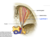
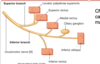
review the anatomy of the cavernous sinus and the neurovasculature commonly impacted by a cavernous sinus thrombosis

A

O - oculomotor nerve
T - trochlear nerve
O - ophthalmic branch of trigeminal nerve
M - maxillary branch of trigeminal nerve
C - internal carotid artery
A - abducensnerve*
(T - trochlear nerve again)

O TOM CAT

14
Q

ID the 2 branches of CN III and the muscles they innervate and which one carries PNS and SNS fibers

A

Superior branch

  • levator palpebrae superioris
  • superior rectus
  • carries SNS fibers that innervate a smooth muscle(the superior tarsalm)

Inferior branch

medial and inferior rectus & inferior oblique

preganglionic PSNS to ciliary ganglion (visceromotor)

15
Q

what can cause ptosis

A

Loss of function of either the levator palpebrae superioris (PNS) or superior tarsal muscle (SNS) results in a ptosis of the upper eyelid. Thus, ptosis may be caused by either CNIII or sympathetic damage.

16
Q

Why is the position of PSNS fibers relative to CNV fibers clinically relevant? Third nerve palsies are often caused by aneurysms. This aneurysm will compress the parasympathetics first, and you will see a blown pupil before you see mm effects.

and aneurysm at junction of Pcomm(posterior communicating) & ICA

what are third nerve palsies typically caused by

A

aneurysm that will compress the parasympathetics first, and you will see a blown pupil before you see mm effects. Because of PSNS fibers relative to CNV fibers CNV is also impacted

17
Q

what are the 3 sympathetics targets in the eye how to they reach their target

A
  1. Pupillary dilator
  2. Superior tarsal muscle
  3. Lacrimal gland

All sympathetics originate in the lateral horn of the spinal cord and synapse in the superior cervical ganglia. Post-ganglionics travel in a plexus surrounding the internal carotid (the carotid plexus), then take a convoluted route

18
Q

describe the sympathetic path to the lacrimal gland

A

lateral horn of the spinal cord –> synapse in the superior cervical ganglia –> travel in the carotid plexus –> leave as the deep petrosal nerve + join the greater petrosal nerve–> nerve of the pterygoid canal, and follow the same path as the parasympathetics to the lacrimal gland

19
Q

Inferior oblique

innervation

action

A

CN III

elevation, lateral movement and abduction

20
Q

superior oblique

innervation

action

A

CN IV

Depression, Abduction, medial eye rotation

21
Q

lateral rectus

innervation

action

A

CN VI

abduction

22
Q

medial rectus

innervation

action

A

CN III inferior branch

adduction

23
Q

superior rectus

innervation

action

A

CN III superior branch

elevation, adduction
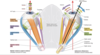
24
Q

inferior rectus

innervation

action

A

CN III superior branch

depression, adduction

25
Q

levator palpebrae superioris

A

CN III superior branch

26
Q

What cranial nerves carry preganglionic parasympathetic (general visceral efferent) fibers

A

CNIII, VII, IX, X; CNIII and CNVII carry these into the eye or orbit

27
Q

what characterizes trochlear nerve injury

A

inability to focus on near objects

28
Q

what would a lesion of the facial nerve result in

A

Loss of taste from the anterior two-thirds of the tongue

Partial sensory denervation of the auricle

Increase in loudness of sound

Loss of tear production

29
Q

what CN is damaged

A

left LR is unable to move the eye left.

30
Q

why do you need to conduct an eye exam before a LP

A

papilledema would indicate increased CSF pressure

31
Q

Following a partial thyroidectomy a patient presents with signs and symptoms that you believe indicate his cervical sympathetic trunk was inadvertently transected during the procedure. Which of the following would NOT be consistent with your suspicion?

A

Absence of sweating on forehead

Ptosis

Pupillary constriction

Redness and increased temperature on the forehead

32
Q

A 10-year-old boy fractures the floor of the right middle cranial fossa in an automobile accident/ Physical examination reveals loss of emotional tearing on the ipsilateral side. What nerve is most likely damaged

A

1.Greater petrosal. The lacrimal nerve conveys autonomics to the lacrimal gland, but would not be damaged by a fracture of the middle cranial fossa

33
Q

What nerves enter the orbit through the intraconal space

A

CNII, IIIi, IIIs, VI and nasociliary

34
Q

where is CN VI

what does it innervate

why is it suseptible to damage

A

Abducens nucleusis located in caudal pons, in the facial colliculus. Exits the inferior pontine sulcus, i.e. near midline at ponto-medullary junction. Can be damaged in nucelus or at pontine base due to its track

•contains lower motor neuron cell bodies whose axons (CN VI) innervate lateral rectus

can be damaged because its long and thin

35
Q

where is the CN IV located

what does it innervate

A

Nucleus= located in caudal midbrain (caudal to oculomotor nucleus) –> decussates in tectum of midbrain –> exits dorsal midbrain caudal to inferior colliculus –> passes through lateral wall of cavernous sinus & superior orbital fissure

Innervates superior oblique: depresses (infraducts) adducted eye; intortsabducted eye

Nerve encircles midbrain in subarachnoid space

36
Q

what would you see clinically if CN IV was injured

A

Eye elevated, extorted

Patient cannot look down & in

vertical diplopia

37
Q

where is CN III located

what does it innervate

what would you see clinically if it was injured

A

nucleus is in rostral midbrain

  • Parasympathetic nucleus = Edinger-Westphal, located just dorsal
  • CN III emerges medially between midbrain and pons (between PCA & SCA) –> lateral wall of cavernous sinus & superior orbital fissure

innervates all extraocular muscles except lateral rectus & superior oblique

eye “down & out”, severe ptosis, dilated pupil… can be caused by aneurysm in pcomma

38
Q

what are the 3 gaze centers and their location within the brain

A

vertical gaze (up and down- midbrain- CN III

vergence (converse, diverge)- midbrain- CN III

lateral gaze (horizontal)- midbrain (medial rectus) and pons (lateral rectus)- II and VI

CN III midbrain, CN VI- pons

39
Q

what occurs when you move the eye to the left

A

activation of abducens (CN VI) in left pons to move left lateral rectus –> simultaneously a second population of neurons send fiber bundles called medial longitudenal fasiculus (MLF) across the midline and to the left pons sends LMN to midbrain to activate right medial rectus- oculomotor (CN III)

40
Q

whata are the two bones that form the roof of the orbit

A

lesser wing of sphenoid, orbital plate of frontal bone

41
Q

what are the bones that form the lateral wall of the orbit

A

frontal and orbital surface of the zygomatic bone

greater wing of the sphenoid

42
Q

what is the strongest wall of the orbit

A

lateral

Fractures of the lateral orbital wall are usually accompanied by severe facial trauma

most common site for a fx of the lateral wall is the sphenozygomatic suture line

43
Q

what forms the apex of the orbit and what openings does it have

A

Sphenoid

  1. medially: optic canal for optic n./ophthalmic a.
  2. laterally: superior orbital fissure for a number of nerves (III, IV, V1, VI) & superior ophthalmic v.; it separates the greater and lesser wings of this bone

3.inferior orbital fissure between sphenoid and maxilla: through here brs. of maxillary nerve and artery pass; also veins from deep face region pass through here connecting with veins within orbit

44
Q

what bones form the floor of the orbit

A

maxilla

zygomatic bone

palatine bone

45
Q

what bones form the medial wall of the orbit

A

lacrimal

ethmoid

lesser wing of sphenoid

46
Q

what does the lacrimal fossa contain

A

The lacrimal fossa of the lacrimal boneholds the lacrimal sac NOT the lacrimal gland

47
Q

ID the nerves passing through the superior orbital fissure

A

“Live Frankly To See Absolutely No Insult”

48
Q

Mnemonic for intraconal space

A

With the exception of the obliques (and levator palpebrae superioris), all extraocular muscles attach to a common tendinous ring. The area defined by the common tendinousring is called the intraconalspace

Mnemonic for intraconal space: NASO2: nasociliary,abducens,sympathetic nn, O2=2 divisions of oculomotor n, superior and inferior

49
Q

Orbital rim fractures are a relatively common fracture of the orbit. These fractures involves the three bones that form the outer rim of the orbit. What are these bones?

A

Maxilla, zygomaand frontal

50
Q

The superior orbital fissure connects the orbit with what cranial fossa

A

middle

51
Q

The superior orbital fissure connects the orbit with what cranial fossa

A

The superior orbital fissure connects the orbit with what cranial fossa

52
Q

Your patient has a lesion of a nerve outside the intraconalspace. What eye movements will help you determine the integrity of this nerve

A

.Three nerves are located outside the intraconalspace—frontal, lacrimal and trochlear. Of these, only one is a motor nerve (trochlear). The trochlear nerve innervates the superior oblique muscle. To check the integrity of the SO, ask the patient to look inward (toward the nose) and down.