Week 3 - A - Anatomy 3 - Anatomy of Anaesthesia for labour Flashcards
(32 cards)
The reproductive system of the female has both motor and sensory function Reproductive motor system function
- * - Uterine “cramping”
- * - Uterine contraction
- * - Pelvic floor muscle contraction
When do each of these happen usually?
- * Uterine cramping - menstruation
- * Uterine contraction - during labour
- * Pelvic floor muscle contraction - during sneezing or coughing
The reproductive system sensory function is largely for pain
- * pain from the adnexae
- * pain from the uterus
- * pain from the vagina
- * pain from the perineum
What is the adnexae? What type of axonal fibres transmit pain?
The adnexae are the appendages of an organ - in this case the uterus - the adnexae are the ovaries and the fallopian tubes
Tye Aδ or type C nerve fibres transmit pain
Type C are unmyelinated and thin
It is fundamental to know whether the body structure is in the pelvis or perineum for knowing which nerve fibre types are carrying out which function IF the structure is in the pelvis, which nerve fibre modalityies carry out the motor and sensory function? If the structure is in the perineum, which nerve fibre modalities carry out the motor and sensory function?
Pelvis
* Body cavity structure and therefore autonomic (parasympaethtic and sympathetic) for motor and visceral afferents for sensory
Perineum
* Body wall structure and therefore somatic motor and somatic sensory nerves
For anaesthesia therefore have to decide between numbing the visceral afferents or somatic sensory fibres What is said to be the dividing line between pelvis and perineum?
The dividing line is meant to be the levator ani muscle - the pelvic floor

State whether the nerve supply is hormonal (sympathetic or parasympathetic), visceral afferent, somatic motor or somatic sensory
- * Uterine “cramping” (e.g. menstruation)
- * Uterine contraction (e.g. during labour)
- * Pelvic floor muscle contraction (during sneezing)
- * pain from the adnexae (ovaries + fallopian tubes)
- * pain from the uterus
- * pain from the vagina
- * pain from the perineum
- * Uterine “cramping” (e.g. menstruation) - hormonal
- * Uterine contraction (e.g. during labour) - hormonal
- * Pelvic floor muscle contraction (during sneezing) - somatic motor
- * pain from the adnexae (ovaries + fallopian tubes) - visceral afferent
- * pain from the uterus - visceral afferent
- * pain from the vagina -
- superior = pelvis (visceral afferent),
- inferior = perineum (somatic sensory)
- * pain from the perineum - somatic sensory
Pain from vagina will change as superior vagina is in the pelvic cavity and therefore innervated by visceral afferents Vagina passes into the perineum through the pelvic floor and inferior vagina is therefore perineum so somatic sensory control The superior aspect of the pelvic organs ‘pain’ sensation enters the spinal cord different to the inferior aspect What is it that the organs touch that differentiate this?
Superior aspect of pelvic organ - in contact with peritoneum Inferior aspect of pelvic organ - not in contact with peritoneum

The pelvic organs both use visceral afferents to relay sensation to the spinal cord however * Superior aspect of pelvic organs / touching the peritoneum - Run alongside sympathetic fibres * Inferior aspect of pelvic organs / not touching peritoneum - Run alongside parasympathetic fibres What are the different spinal cord roots for these exact fibres?
Superior aspect of pelvic organs - run alongside the sympathetic fibres to enter the spinal cord at levels T11-L2 Inferior aspect of pelvic organs - run alongside parasympathetic fibres to enter the spinal cord at levels S2,3,4
Where is pain from the superior part of the pelvic organs therefore percieved? Where is pain from the inferior part of the pelvic organs therefore percieved?
Pain from superior part - perceived as suprapubic Pain from inferior part - percieved as dermatomal to S2,3,4 - perineum
Structures crossing from pelvis to perineum, e.g. urethra, vagina: above levator ani – in the pelvis What will the nerve modality be? What will it run alongisde? What level of the spinal cord will it enter? below levator ani – in the perineum What will the nerve modality be? What will it run inside? What level of the spinal cord will it enter?
Above levator ani - Visceral afferents running alongside parasympathetic nerve fibres entering the spinal cord at S2,3,4 level Below levator ani Somatic sensory running within the pudendal nerve entering the spinal cord at S2,3,4 level
Sympathetics travelling down to pelvic cavity – come from T11-L2 sympathetic chains either side of the vertebral column The sympathetic chains send fibres to the superior hypogastric plexus The parasympathetic coming from S2,3and4 come out of the sacral foraminae and these parasympathetic fibres are known as what?
The parasympathetic nerves coming from S2,3,4 are known as the pelvic splanchnic nerves

Where is it that the sympathetic fibres travelling down to the pelvic cavity and the pelvic splanchnic fibres mix?
The area where these mix is known as the inferior hypogastric plexus, this plexus therefore sens mixed fibres to the pelvic organs ie uterus

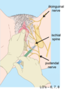
What nerve modalities does the pudendal nerve carry?
Carries somatic motor, somatic sensory and sympathetic fibres Parasympathetic fibres are already given off before the formation of the pudendal nerve and therefore pudendal nerve does not carry
T11-L2 and S2-S4 – two areas for pain sensation in the female
Which organs travel back to T11-L2 and which to S2,3,4
- * Uterus
- * Uterine tubes
- * Ovaries
- * Superior vagina
- * INferior vagina
- * Cervix
- * Perineal muscles and skin
Visceral afferents - T11-L2 (in contact with peritoneum)- run alongside sympathetic fibres
- * Uterus, uterine tubes and ovaries
Visceral afferents - S2,3,4 - run alongside parasympathetic fibres
- * Superior vagina, cervix
Somatic sensory fibres - S2,3,4 - run within pudendal nerve
- * INferior vagina, perineal muscles and skin
Everything below peritoneum (not in contact) but above levator ani is still pelvic cavity and visceral afferents but follows parasympathetic fibres which insert at the S2-4 Describe the pathway for pelvis or perineum pain sensation

Perineum - body wall - somatic sensation - pudendal nerve - S2,3,4 -
Pelvis - Body cavity - Autonomic/visceral afferent
Touching peritoneum - more superior - follows sympathetic back - T11-L2
Not touching peritoneum - more inferior - follows para back - S2,3,4

What are the three types of anaesthesia for a women in pregnancy?
Spinal anaesthetic Epidural anaesthetic Pudendal nerve block
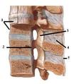
At what level does the spinal cord become the cauda equina? What level does the subarachnoid space end? What is the end of the spinal cord known as? What spinal cord level is anaesthetic put in for a spinal anaesthetic?
Cauda equina at L2 vertebral level (L1/L2) Subarachnoid space ends at S2 End of the spinal cord is known as the conus medullaris SPinal anaesthetic is at L3/4 vertebral level (can be done at L4/5)
How is the L3/4 intervertebral space found for the spinal anaesthetic?
A line is drawn between the most superior points on the anterior superior iliac spines This is known as the intercristal line and it is said to run through the L4 spinous process - therefore above this is the L3/4 intervertebral space

What structures does the needle pass through in a spinal anaesthetic?
Skin Subcutaneous fat Supraspinous ligament Interspinous ligament Ligamentum flavum Epidural space Dura and arachnoid mater (usually are joined)
What do these ligaments connect: Supraspinous ligament? Interspinous ligament? Ligamentum flavum?
- Supraspinous ligament - connects the tips of the spinous process
- Interspinous ligaent - runs between the spinous processes
- Ligamentum flavum - runs vertically from lamina to lamina
- Anterior and posterior longitduinal ligament = 1 and 2 respectively
When pushing through he dura (basically pushes through the dura and arachnoid mater at the same time and therefore you may here a little pop and in the subarachnoid space (may have heard a little pop previously when entering the epidural space) )Can aspirate a small amount of fluid (should be clear) to prove in the subarachnoid space Once injected, onset will take place within about 5 mins. If blood is aspirated, what may have happened?
If blood comes out of the spinal needle instead of CSF when the stylet is removed, it is likely that an epidural vein has been punctured. The needle should simply be advanced a little further.
Epidural means only going into the epidural space and not the subarachnoid What does the needles pass through for this? Which is quicker between spinal anaesthetic and epidural anaesthetic?
Needle passes through Skin, subcutaneous fat, supraspinous ligament, interspinous ligament, ligamentum flavum, epidural space Spinal anaesthetic has a quicker onset of action but epidural anaesthetic lasts longer
What is the benefit of the epidural anaesthetic over the spinal anaesthetic? Which is likely to cause a headache?
IN an epidural – The area where the needle will be inserted is numbed with a local anesthetic. Then the needle is inserted and removed after the catheter has passed through it. The catheter remains in place. The anesthetic medicine is injected into the catheter to numb the body above and below the point of injection as needed. The catheter is secured on the back so it can be used again if more medicine is needed. Spinal anaesthetic is more likely to cause a headache
Are you able to carry out a continous infusion with a spinal anaesthetic?
Spinal anaesthesia is a one time shot whereas epidural has a catheter for continuous infusion if need be
Sympathetic outflow originates from autonomic centres in the brain What level do sympathetics exit the spinal cord before travelling to sympathetic chains running the length of the vertebral column?
They exit the spinal cord in levels T1-L2 and pass into all spinal nerves (anterior and posterior rami / named nerves)