Workshop 9 - malignant epithelial tumors Flashcards
(56 cards)
What differences can we have between BT and MT?
( morphological caracteristics )
- differentiation and anaplasia
- rate of growth
- local invasion
- metastasis
Differentiation
of MTs and BTs
Refers to morphological and functional similarity of neoplastic cells with cell origin
MTs : showing various degrees of differentiantion
- WD - well differentiated forms
- ND - non differenciated forms
- anaplastic forms
BTs : well - differentiated tumors
***anaplasia or lack of differentiation is a characteristic feature of malignancy
characteristic feature of malignancy
( differentiation )
anaplasia
and
lack of differentiation
what rate of growth have the MTs and the BTs?
MTs : rapid rate
- WD MTs : grow more slowly
- ND Mts : grow rapidly
BTs : slow rate
Invasion of BTs
- grow as masses at the site of the origin of the tumor
- grow locally - causing compression of adjacent tissues
- usually have capsules that separate them from the tissue
- they are not all encapsuled but they are clearly defined
- not all are wel defined , except hemangioma
Invasion of MTs
- the growth is not limited at the site of the origin
- grow rapidly , invading adjacent tissues, cause damage
- not well defined
( exc renal nuclear cell carcinoma - has capsule )
MTs are not well defined
Name an exception
renal clear cell carcinoma
- grows slowly
- has capsule
Metastasis
definition
Metastasis are tumoral implants located at distance from primary tumor site
characteristic of MTs
4 ways of spreading
Metastasis
4 ways of spreading
- Local
- Lymphatic dissemination
- vascular dissemination
- Transcelomic
Local metastasis
tumor spreads by direct way
in adjacent tissues
along cleavage plans and nervous fibers
Lymphatic spread
Macro and micro
tumor cells disseminate along lympthatic vessels
causing secondary lymphatic metastasis
- Macro
lymph nodes : increase volume, loss of structure - Micro
tumor cells replace the normal lymph node
Vascular dissemination
Macro , Micro
tumor cells disseminate by hematogenous way
( tumor emboli)
in this way a primary tumor cause secondary visceral metastases in :
liver, lung, adrenal, brain etc
- Macro
Affected organ is increased in volume and presents many tumor nodules, well defined, uncapsuled - Micro
can be similar to primary tumor or not
transcelomic dissemination
primary abdominal and thoracic tumor disseminate alog the mesothelial surfaces
ex. pleural cavities
Histological features of BT and MT
Benign
-
Differentiation : Well - diff.
resembling with cell origin - Mitosis : few
- Nucleus / Cytoplasm Ratio : normal 1/4
- homogenous cell shape and size
Malignant
- Differentiation : Failure of cell diff.
- **Mitosis : **many
- **Nucleus / Cytoplasm Ratio : **high 1/1
- cell and nuclear pleomorphism
histological feature of BT
Benign
- **Differentiation **: Well - diff.
resembling with cell origin
- **Mitosis **: few
- Nucleus / Cytoplasm Ratio : normal 1/4
- homogenous cell shape and size
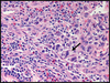
Histological features of MTs
Malignant
- Differentiation : Failure of cell diff.
- Mitosis : many
- Nucleus / Cytoplasm Ratio : high 1/1
- cell and nuclear pleomorphism
Characteristic features of Mts
- aplasia of lack of differentiation
- rapid rythm of growth
- invasion
- metastasis
Stages of development of neoplasia
and
progression of dysplasia to neoplasia
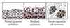
- normal epithelium
- dysplastic epithelium
- CIS - carcinoma in situ
- micro invasive carcinoma
- invasive carcinoma
Stages of development of neoplasia
and
progression of dysplasia to neoplasia
Dysplastic epithelium
mild , moderate and severe
- cytologically : defines morphological neoplastic features of cells characterized by incomplete maturation and increasing of mitosis number
- causes : ex. chronic inflammation
- may develop into neoplasia
Stages of development of neoplasia
and
progression of dysplasia to neoplasia
CIS- carcinoma in situ
- represents an early stage of neoplasia
previous to invasion - Cytologically : characterized by cell and nuclear pleomorphysm and increased mitotic activity
- Histologically : disruption of normal architecture , but BM remains intact
- may progress to neoplasia
Stages of development of neoplasia
and
progression of dysplasia to neoplasia
Micro invasive carcinoma
results by invasion of cancer cells
into subjacent stroma ( 5 mm in diameter )
Stages of development of neoplasia
and
progression of dysplasia to neoplasia
Invasive Carcinoma
corresponds to an advanced cancer
presenting clinical manifestation
Features of malignant epithelial tumors
- common in adults and elderly
- diagnosis established in stahe 0 (CIS) :
100% healing - usually present lymphatic metastasis
Histogenic classification of carcinomas
- Epidermoid / squamocellular carcinoma
- adenocarcinoma