Intro to Brain Anatomy-Medial View Flashcards
1
Q
Review Deck 1
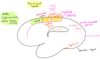
Name the parts pointed in this image

A

2
Q
Name the parts of the brain in the Frontal and Parietal lobes seen from the Medial View
A
Starting from the frontal and superior part of the brain going down:
- Cingulate Sulcus
-
At its end
- Marginal Ramus of cingulate sulcus
-
At its end
- CingulateGyrus
- Corpus Callosum
- Thalamus
At the anterior of the brain:
- Pre-cuneus
- Parieto-occipital Fissure
- Cuneus
- Calcarine Sulcus
- Lingual Gyrus
- called like this because tongue shaped

3
Q
Where is V1 found?
A
Within the banks of the calcarine sulcus
4
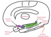
Q
Supplemental Motor Area (cortex)
SMA
A
- Location:
- Besides the paracentral sulcus and the cingulate sulcus
- Also known as M2
5
Q
Where is the Paracentral Sulcus?
A
- Only visible from the medial view
- Between the limit of the brain and the cingulate sulcus
6
Q
Cuneus
A
- Location:
- Between:
- Parieto-occipital fissure
- Calcarine sulcus
- Between:
- Is a region containing many sulci and gyri
7
Q
Paracentral Lobule
A
- Location:
- Between brain border and cingulate sulcus
- yellow part of the image
- Between brain border and cingulate sulcus
- Contains:
- continuation of the Central sulcus
- continuation of M1 of the foot
- continuation of S1 (somatosensory cortex) of the foot

8
Q
The Paracentral Lobule contains the ____ and _____ of the part of the body called _______
A
- M1
- S1
- foot
9
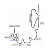
Q
Parahippocampal Gyrus
A
- Contains (From up to down):
- Thalamus
- Hippocampal Sulcus
- Pyriform Cortex
- Entorhinal Cortex
- Parahippocampal Cortex
- Rhinal Sulcus
- Collateral Sulcus
- Fusiform Gyrus
- Occipito-Temporal Sulcus
- Inferior Temporal Gyrus (ITG)
- L4 p.22:
- Used to be referred as Rhinencephalon (nose brain)
- Patient H.M.
- Important for Memory consolidation
- Pyriform cortex
- Entorhinal cortex
- Parahippocampal cortex
10
Q
Hippocampus

A
- Also known as archicortex
- First cortex
- Only one layer of neurons
*
11
Q
Limbic Cortex
A
- Connects the cingulate gyrus and the parahippocampal gyrus
- Phylogenetically old cortex
- Used to be called limbic lobe
- Less than 6 layers

12
Q
Cingulate Gyrus
A
- Part of the Limbic Cortex
- 5 layers of 5.5
13
Q
Insula
A
- Location:
- Within the Sylvian fissure, covered by the frontal, parietal and temporal opercula
- Has a central sulcus
- 3 short gyri:
- motor processing
- 2 long gyri:
- somatosensory processing
- Sits under the pre and post central gyri of the lateral surface

14
Q
Heschl’s Gyri
A
- Also known as Transverse Temporal Gyri
- On top of the temporal lobe
- Contains the Primary Auditory Cortex (A1)
15
Q
Corpus Callosum
A
- “The Great Cerebral Commissure”
- Connects the cortex of both hemispheres
- Divided into 4 parts:
- Rostrum
- Genu
- Body
- Splenium

16
Q
Anterior Commissure
A
- Communication of the cortex of the anterior temporal lobes
