11. Abdomen V Flashcards

(184 cards)

1
Q

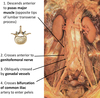
What is the abdomenal course of the ureters?

A

1) Crosses over PSOAS major
2) Crosses over genitofemoral nerves (part of lumbar plexus, easy to identify as it always crosses over PSOAS major muscle)
3) Gets crossed obliquely by gonadal vessels (e.g. ovararian or testicular arteries)
4) Crosses the bifurcation of common iliac artery (L4/L5) to enter pelvis

2
Q

What shape is the right suprarenal gland?

A

Pyramid

3
Q

What shape is the left suprarenal gland?

A

Crescent

4
Q

What is the nervous innervation of the kidneys?

A

Sympathetic:

Has vasomotor roles, regulating blood flow and renin secretion

It is from the renal plexus which follows the renal arteries

Post-ganglionic sympathetic from T10-L1

Parasympathetic:

This is contested as some texts state that parasympathetic fibres branch from vagus nerve however others say that there is no evidence of parasympathetic innervation of the kidneys.

Regardless, renal function is not dependent on innervation as is shown by renal transplants where the kidneys work just fine without being innervated.

Renal function is regulation ‘by’ hormones.

5
Q

What is the arterial ‘supply’ of the kidneys

A

Renal arteries, which divied into five segmental arteries (4 anterior and 1 posterior)

These then divide to lobar arteries, then interlobar arteries, arcuate artereis and then interlobular arteries

6
Q

What are some characteristics about suprarenal glands?

A
  • Endocrine glands
  • present on upper/superior pole of kidneys
  • the right suprarenal gland is pyramidal whereas the left suprarenal gland is crescent-shaped
  • Suprarenal glands are surrounded by renal fascia, hence even if kidney is disturbed the suprarenal glands are shielded
  • They contain a yellow cortex and a brown medulla
7
Q

What is the yellow cortex of the suprarenal glands derived from?

A

Mesoderm

8
Q

What is the brown medulla of suprarenal glands derived from?

A

Neural Crest

9
Q

What is the function of the yellow cortex of the suprarenal glands?

A

Secretes androgens and corticosterioids

10
Q

What is the function of the brown medulla of the suprarenal glands?

A

Secrete catecholamines (adrenaline and noradrenaline)

11
Q

What are the three main arteries of the suprarenal glands and where do they branch from?

A
  1. Superior suprarenal artery - branch from the inferior phrenic artery
  2. Middle suprarenal artery - comes off aorta directly
  3. Inferior suprarenal artery - comes off renal artery
12
Q

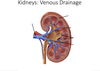
Where does the right suprarenal vein drain into?

A

IVC

13
Q

Where does the left suprarenal vein drain into?

A

Left renal vein (more efficient than traversing aorta to get to IVC)

14
Q

List some functions of the kidneys

A
  1. Acid-Base balance
  2. Excretes most waste products of metabolism
  3. Water and electrolyte balance of the body
  4. Secretes hormones and renin into the bloodstream

Kidneys filter around 113-114 pints of blood per hour

Filters all the blood in the body around 400 times a day

blood flow to the kidney is greater than to the heart

15
Q

Are kidenys primary or secondary retroperitoneal?

A

Primary retroperitoneal

16
Q

Which kidney is lower than the other and why?

A

Right is lower due to the presence of the liver above it (pushing it downwards)

17
Q

What is the vertebral position of the kidneys?

A

Located between T12 and L3

18
Q

What is the vertebral level of the hila of the kidneys?

A

L1

19
Q

Which ribs surround the kidneys?

A

Ribs 11 and 12

20
Q

Why do ribs move upon inspiration?

A

Because they are closely associated with the diaphragm

21
Q

What are the anterior relations of the right kidney

A
  • Superior pole - right suprarenal gland/adrenal gland (pyramid shape)
  • Liver - anterior surface of kidney ‘in contact’ with liver, but separated by peritoneum (hepatorenal pouch of morison)
  • Descending duodenum - close contact with hilum of right kidney
  • Right colic flexure - in contact with lower lateral part of kidney
  • Small intestine - lower medial side of kidney, separated by peritoneum

The surfaces of the kidney in contact with the liver and the small intestines are the surfaces which are ‘covered’ in periotoneum

22
Q

What are the anterior relations of the left kidney?

A
  1. Superior pole - left suprarenal gland/adranl gland (crescent shape)
  2. Stomach - separated by perionteum
  3. Spleen - separated by peritoneium (lienorenal/ splenorenal ligament)
  4. Pancreas - middle part of kidney
  5. Left colic flexure - lateral middle part of kidney
  6. descending colon - inferior middle part of the kidney
  7. Jejunum - lower medial part of kidney, separated by pertoneium

Thus, surfaces in contact with the stomach, spleen, and jejunum have periteonum ‘covering’ it

23
Q

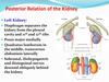
What are the posterior relations of the kidneys?

A

Posteriorly, kidneys are closely associated with the muscles of the posterior abdomenal wall:

  1. Superior pole is closely associated with the diaphragm
  2. Medially kidneys are closely associated with the PSOAS minor/major muscle
  3. ‘Middle laterally’ kidneys are closely associated with quadratus lumborum muscle
  4. Laterally the kidneys are closely associated with the trasversus abdominus muscle
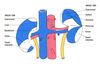
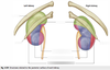
24
Q

Label this image

A
25
What are the functions of kidney/renal coverings and what actually are they?
Kidneys are enclosed in many fascial layers which hold the kidney up in place against the posterior abdominal wall, and 'support' them. 1. Renal Capsule - **closely adherant with surface of kideny** 2. Perinephric Fat - **within perineal space** (*perinephric fat is sandwiched between the renal capsule and the renal fascia)* 3. Renal Fascia - **condensation of areolar connective tissue.** Renal fascia is continuous with **-transversalis fascia (inferiorly)** and **-fascia over diaphragm (superiorly)** 4. Paranephric Fat - **external to the renal fascia**
26
What is the renal capsule?
Touch fibroconnective tissue surrounding the renal cortex
27
What is the renal cortex?
Outer layer surrounding the renal medulla, contains nephrons and renal tubules
28
What demarcates the renal pyramids?
_Renal cortex extends into the medulla as renal columns_, demarcating the renal pyramids of the medulla, with the apex of the pyramid being the **renal papilla** which drains into minor calyx
29
What is the minor calyx
Structures receiving urine from collecting ducts of renal pyramids via the renal papillae
30
Major calyx
site at which many minor calyces drain
31
Renal pelvis
Site at which major calyces unite, conveys urine to the proximal ureter
32
Hilum
medial aspect of each kidney, where renal pelvis emerges fro kidney. Hilum is site at which vessels, nerves and lymphatics emerge from or enter the kidney.
33
Label this diagram
34
What is another way of describing the renal pelvis?
The dilated superior portion of the ureter
35
Structures in the renal hilum:
Renal artery, Renal Vein, Ureter
36
What fills the renal sinus?
Perinephric fat (surrounds structures like renal pelvis and hilum)
37
What is the order in which vessels and ureters enter/leave the hilum of the kidneys?
VAU = Vein, Artery, Ureter
38
What other structures pass through hilum?
Sympathetic fibres (e.g. least splanchnic nerves) and lymphatic vessels
39
What are the five vascular segments of the kidney?
1) apical (at the very top of the kidney) 2) caudal (at the very bottom of the kidney) 3) anterior superior 4) anterior inferior 5) posterior Vascular segments arise as a result 'of' them being supplied 'by' one of the five different segmental arteries
40
What are the arteries to the kidney?
1. **renal arteries** (branch of aorta, L1/L2, right is longer than left) 2. **segmental arteries** (five of them, four of which are anterior to the renal pelvis and one of which is posterior to the renal pelvis) 3. **Lobar arteries** (going to lobe of kidney which is renal pyramid plus superior cortex) 4. Branches into **interlobar arteries** which run in between the lobes 5. These arch over the base of the pyramid as **arcuate arteries** 6. Arcuate arteries give off more branches known as **interlobular arteries** (these extend into the cortex and give afferent glomerular arterioles for Bowman's Capsule)
41
What course do the renal veins follow?
Follow track of renal arteries
42
Which veins receive blood from efferent glomerular veins from Bowman's capsule?
Interlobular veins
43
Which arteries 'give off' afferent glomerular arterioles to the Bowman's capsule?
Interlobular arteries
44
Label this image
45
How many times longer is the left renal vein than the right renal vein?
3x
46
What is relation of left renal vein to right renal vein when it enters the IVC?
Left renal vein enters IVC superiorly to right renal vein
47
Which side is preferred for live donor nephrectomy?
_left_ (due to **longer renal vein**, less associated with renal vein thrombosis like the right as well as right having shorter vessels)
48
What is the effect of an aneurysm of the superior mesenteric artery
Can compress the left renal vein (because the superior mesenteric artery passes over it)
49
Which other veins drain into the left renal vein?
1. left gonadal vein 2. left inferior phrenic vein 3. left suprarenal vein
50
What is the position of the right renal vein?
Posterior to the second part of the duodenum (descending duodenum) and head of pancreas
51
What is the route of the lymphatics of the kidney?
* Lymphatic vessels follow the route of the renal veins * They then drain into the para-aortic nodes such as the **right lumbar nodes** (which are the pre-caval nodes, post-caval nodes and lateral-caval nodes in front, behind and to the side of the IVC respectively) and the **left lumbar nodes** (lateral to the aorta) * These nodes then drain into the right and left lumbar trunks * Right and left lumbar trunks then drain into the cisterna chyli
52
Is renal function dependendent on nervous innervation?
No, it is more to do with hormonal regulation. In renal transplants there is complete transection of innervation and kidneys function just fine
53
Is there evidence of parasympathetic innervation of the kidneys?
Some texts suggest the vagus nerve has a role in parasympathetic innervation of the kidneys however more recent research shows there is no evidence of parasympathetic innervation to the kidneys
54
What is the function of the sympathetic innervation of the kidneys?
Vasomotor control, regulating blood flow and renin secretion From the renal plexus which follows the renal artery Pre-ganglionic fibres from T10 to L2
55
What causes staghorn caliculi?
Recurrent UTIs -\> increase ammonia production -\> increased pH -\> decreased phosphate solubility thus the phosphate accumulates into a calyx (renal stone)
56
How long is the ureter?
25-30cm
57
What type of contractions occur in the ureter?
peristaltic
58
What does the ureter continue from?
It begins as a continuation from the renal pelvis
59
What is the course of the ureter in the abdomen?
1. **descends anterior to the PSOAS major** muscle 2. **descends anterior to genitofemoral** nerve 3. gets **crossed over obliquely by the gonadal vessels** 4. **traverses the bifurcation of the common iliac artery (L4/L5)**, at the level of the _sacroiliac joint_, the ureters enter the pelvic brim
60
Ureter course in the pelvis
1. crosses anteriorly to internal iliac artery then reaches ischeal spine 2. At the level of the ischeal spine, the ureter turns **anteromedially** to enter upper lateral angle of bladder At the termination of the ureter, it gets crossed over by the **vas deferens/ductus deferens in males** or **uterine artery in females** When the ureter enters the bladder, it passes **obliquely through the bladder wall** for 3/4 of an inch before opening into bladder cavity. -this oblique angle of entrance of ureter into bladder is important to prevent backflow of urine.
61
What are bony landmarks which can be used to track course of the ureters?
**Sacroiliac joint** (where the ureters enter the pelvic brim) and **ischeal spine** (where ureteres turn anteromedially to enter bladder)
62
What are effects of ureteric calculi on the muscular wall?
May cause distention Also, can get **ureteric colic** which is severe pain due to **contraction of ureteric muscles to overcome an obstruction** usually due to stone or clot formation. Ureteric calculi can also cause intermittent/complete obstruction of urinary flow
63
Why does ureteric colic cause pain?
Because ureter contracts and distends to try to move renal stone along - that muscular spasm causes a lot of pain
64
What are the ureteric constrictions
Regions of the ureter which are narrower and hence calculi get stuck here more easily 1. ureteropelvic junction 2. crossing over common iliac artery 3. site of entrance to bladder
65
What arteries supply blood to the ureters?
1. Renal artery 2. Gonadal artery 3. Common iliac arteries 4. Internal iliac arteries
66
What is the maximum volume of urine the bladder can hold?
1L
67
What kind of organ is the bladder?
Extraperitoneal (sub-peritoneal)
68
What structure is the bladder posterior to?
Pubic symphysis
69
What is the location of the bladder?
When empty it lies in the lesser (true) pelvis, but when full it can extend into the abdominal cavity
70
What is the retro-pubic space of retzius?
'empty space' between pubic bone/symphysis and the bladder (so a space anterior to the bladder)
71
What is the vesico-uterine pouch?
Pouch between bladder and uterus in females
72
What is the position of the anterior uterus and vaginal wall compared to the bladder?
Posterior
73
What is the recto-uterine pouch?
Pouch which is between rectum and uterus
74
What is the rectovesical pouch?
pouch between bladder and rectum (only present in males)
75
what is the position of the prostate compared to bladder?
Directly inferior to bladder
76
What is the position of the seminal vesicle to bladder?
Postero-inferior
77
What attaches to and extends from the apex of the bladder?
Median umbilical ligament which extends all the way up to the umbilicus. The median umbilical ligament is a remnant of the urachus which is a foetal remnant of the allantois (this used to drain contents of the foetal urinary bladder into the yolk cac)
78
What structure does the apex of the bladder point towards?
The pubic symphysis
79
Label this image
80
Label this image
81
What structure is surrounded by the neck of the bladder?
urethra
82
What is the pubovesical ligament?
Fibromascular band (thickening of superior fascia of pelvic diaphragm) which attaches to pubic symphysis and wraps around neck of bladder and urethra to hold it above pelvic floor (important for continence)
83
What is stress incontinence?
In women who have 'given' birth, the pubovesical ligament can weaken as well the pelvic floor and levator ani muscles. Consequently, the neck of the bladder can prolapse below the pelvic floor when for example sneezing leading to incontinence (stress incontinence) due to increased intra-abdominal pressure
84
What is the puboprostatic ligament?
Fibromuscular band (thickening of superior fascia of pelvic diaphragm) connected to pubic symphysis, wrapping around the prostate. The neck of the bladder is surrounded by internal urethral sphincter (smooth muscle) has functions in maintenance of continence as well as widening neck of bladder for micturition
85
What kind of muscle are the wall of the bladder?
**Detrusor muscle (smooth muscle)** Inner layer = longitudinal muscle fibres Middle layer = circular muscle fibres Outer layer = longitudinal fibres
86
What kind of epithelium lines the inside of the bladder?
**Transitional epithelium** Type of squamous epithelium which is normally round in shape but becomes flattened as the bladder distends. Hence, it is important as it 'allows' stretching of bladder upon filling without damage to the lining.
87
What is the pattern of attachment of the vesical mucosa in the bladder?
Attached loosely to most parts of the detrusor muscle, except at the trigone where it is attached tightly. Trigone is a smooth triangular area
88
What is importance of vesical mucosa?
When the bladder is empty it forms these folds. When bladder is fills the vesical mucosal folds stretch out to become smooth
89
What is the inter-ureteric bar?
Extension of the smooth muscle of the ureters, connecting the ureteric orifices
90
What is the ureteric orifice?
**Slit-like opening** of ureters into the bladder ureters open into bladder **obliquely** to prevent backflow of urine the ureteric orifices are also **encircled by loops of detrusor muscle** to further prevent backflow
91
What does the urethra of a male of have that females don't?
**Internal urethral sphincter** which 'guards' the internal urethral orifice
92
What are the vesical arteries?
Arteries 'supplying' the bladder. In both males and females, arterial supply of bladder is via the **superior vesical artery** which is a branch of the anterior internal iliac artery (via umbilical arteries) However, in _males this is supplemented with the inferior vesical artery_ (also branch of anterior internal iliac) and in _females by vaginal artery_ (also branch of internal iliac)
93
Where does the vesical venous plexus drain into?
**Internal iliac vein** Can communicate inferiorly with: * inferior prostatic vein (males) * utero-vaginal plexus (females - plexus of veins draining the uterus and vagina)
94
Where do the pelvic splanchnic nerves travel through?
The inferior hypogastric plexus Then they synapse at ganglia on or near the bladder
95
What kind of innervation are the pelvic splanchnic nerves?
parasympathetic (S2-S4)
96
Where do the sympathetic nerves synapse?
synapse on pre-vertebral plexus e.g. coeliac or superior hypogastric plexus. Post-ganglionic fibres then travel to bladder Sympathetic nerves are T10 - L2 spinal nerves
97
Which nerve fibres are needed for micturition?
parasympathetic (pelvic splanchnic nerves) as they cause contraction of detrusor muscle and relation of any sphincters/ pelvic floor muscle around neck of bladder
98
Which fibres are needed for urinary retention?
Sympathetic (T10-L2) as it causes relaxation of detrusor muscle and contraction of sphincters activated during ejaculation to close **internal urethral sphincter** to inhibit backflow of urine to bladder and to inhibit urination into female during copulation
99
What are the visceral nerve afferents for?
Sensation and pain Stimulated by stretching Travel with sympathetic fibres (superior bladder) and parasympathetic fibres (inferior bladder) In babies, when bladder is full, the vesical afferents are stimulated. This activates the pelvic splanchnic nerve fibres which travel through vesical plexus to 'innervate' the bladder (contract it) and inhibit the sphincter muscle (relax it) - micturition. As we get older we learn to overcome this reflex.
100
What structures lie between the internal urethral orifice/meatus and the external urethral orifice/meatus in the female?
1. pelvic floor 2. deep perineal space/pouch (*potential space between deep perineal fascia of pelvic floor and perineal membrane)* 3. perineal membrane 4. external urethral sphincter 5. para-urethral glands
101
What kind of muscle is the external urethral sphincter?
**Striated muscle** under voluntary control (**innervation - pudendal nerve**)
102
What kind of epithelium lines the urethra?
proximal 2/3 - transitional epithelium distal 1/3 - stratified squamous epithelium
103
Label this image
104
What are the dimensions of the female urethra?
40cm long, 6mm wide (diameter)
105
What is the deep perineal pouch?
Potential space between the deep fascia of the pelvic floor (superiorly) and the perineal membrane (inferiorly)
106
What is the perineal membrane?
A layer of tough fascia which is perforated by the urethra
107
What kind of muscle is the internal uretral sphincter (males)
smooth muscle, involuntary control (autonomic innervation)
108
What are the four parts of the male urethra?
1. preprostatic 2. prostatic 3. membranous 4. spongy So-called as it passes through these different regions (e.g. just before the prostate, the prostate, the perineal membrane, the corpus spongeosum)
109
what is the position of the urethral opening in the female?
posterior to clitoris and anterior to vaginal opening external urethral orifice opens into the **vestibule** (cleft between labia minora)
110
Where is the internal urethral sphincter in males?
Present in the pre-prostatic part of the urethra smooth muscle (sympathetic innervation which constricts it during ejaculation to stop semen flowing into the bladder)
111
Where is the external urethral sphincter?
in the deep perineal space
112
Which part of the urethra is the ejaculatory duct?
Prostatic
113
What is the navicular fossa?
Dilated terminal portion of the urethra in the glans penis (the terminal part of spongy urethra is dilated)
114
Where is the bulbo-urethral gland located and where does it empty into?
It is **present in the deep perineal pouch** but **empties into the spongy urethra** (pre-ejaculate fluid called cowper's fluid)
115
Label this image
116
Label this diagram
117
What does the apex of the sacrum articulate with?
The coccyx
118
What are the boundaries of the posterior abdominal wall?
Superior: ribs 11 and 12 posteriorly: lumbar vertebrae (L1-L5) - they form a lordosis shape (concave curvature) lumbar vertebrae articulate inferiorly with the sacrum (which is made up of fused sacral vertebral bodies to form that pyramidal shape of the sacrum)
119
What is a feature of the inferior aspect of the ischium?
They are flared inferiorly as this is what we sit on (flared at the ischial tuberosity)
120
What are the muscles of the posterior abdominal wall?
Medial to lateral: * Psoas major * psoas minor * quadratus lomborum * iliacus
121
What is the origin and insertion of PSOAS major?
Wide origin: **T12 - L4** Insertion: fibres fuse with the iliacus muscle forming the iliopsoas muscle which inserts into the **lesser trochanter of the femur**
122
What is the innervation of PSOAS major?
L1-L3
123
What is the function of the PSOAS major muscle?
**Flex thigh at the hip joint** and **flex the trunk (make a prostration position)**
124
What is the origin and insertion of the iliacus muscle?
Origin: the **iliac fossa** Insertion: fibres fuse with fibres of PSOAS major to become iliopsoas muscle which inserts into the lesser trochanter of the femur
125
What is the innervation of the iliacus muscle?
L2-L4
126
What is the function of the iliacus muscle?
Flex thigh at hip joint and flex trunk
127
Does everybody have a psoas minor muscle?
Not present in everyone
128
What is the origin and insertion of psoas minor?
Origin: vertebral bodies of **T12-L1** Insertion: **iliopubic eminecne, pecten pubis**
129
What is the function of psoas minor?
assists with trunk flexion
130
What is innervation of psoas minor?
L1
131
What is the origin and insertion of quadratus lumborum?
Origin: **Iliac crest** Insertion: **inferior border of rib 12** and **vertebral bodies of L1-L5**
132
What is the function of quadratus lumborum?
Extends and laterally flexes the vertebral column
133
What is the innervation of quadratus lumborum?
**T12** and **L1-L4**
134
Which muscles cause flexion of the thigh at the hip joint and flex the trunk?
PSOAS major (psoas minor aids flexion of the trunk) and iliacus
135
Which muscle causes extension and lateral flexion of the vertebral column?
quadratus lumborum
136
What marks the superior boundary of the posterior abdominal region?
Diaphragm (this separates the thoracic cavity from the abdominal cavity)
137
What is the insertion of the right crus of diahragm?
Vertebral bodies of L1 - L3
138
Where is insertion of the left crus of the diaphragm?
Vertebral bodies of L1-L2
139
What is a way to describe the diaphragm?
Musculo-tendinous sheet
140
What passes posteriorly to the median arcuate ligament?
The aorta
141
What are the three arcuate ligaments of the diaphragm?
Median Medial Lateral
142
What passes posteriorly to medial arcuate ligament?
PSOAS major
143
What passes posterior to lateral arcuate ligament?
quadratus lumoborum
144
What structure does the central tendon of the diaphragm have a close relationship with?
Pericardium of the heart
145
What is interesting about the right dome of the diaphragm?
it is slightly higher than the left (perhaps due to presence of the liver)
146
What does the diaphragm attach to posteriorly?
Floating ribs (11 and 12)
147
What does the diaphragm articulate with in the midline?
The vertebral bodies Right crus articulates with bodies of L1-L3 Left crus articulates with bodies of L1-L2
148
How is the oesophageal opening of the diaphragm 'formed'?
The right crus goes upwards, diaphragm fibres form an oesophageal sphincter around the oesophagus where it enters diaphragm (T10)
149
At what level does the IVC pass through the diaphragm and what other structure passes with it?
T8, right phrenic nerve (left phrenic vein has its own opening on left side of the central tendon)
150
At what level does the oesophagus traverse the diaphragm and what other structures pass with it?
T10, anterior and posterior vagal trunks Hiatus hernia of the stomach can compress the vagal trunks
151
At what level does the aorta pass through the diaphragm and what other structures pass with it?
T12, Thoracic duct and azygous vein
152
What plexus does the posterior abdominal wall have?
Lumbar plexus (ventral rami of lumbar plexus of spinal nerves L1-L4/L5)
153
which spinal nerve/nerves become the iliohypogastric?
L1 (variable contribution from T12)
154
which nerve/nerves become the ilioinguinal?
L1 (variable contribution from T12)
155
Which nerves become the genitofemoral?
L1-L2
156
Which nerve/nerves become **lateral femoral cutaneous nerve of the thigh**
L2-L3
157
Which nerve/nerves become femoral?
L2-L4
158
Which nerve/nerves become the obtruator nerve?
L2-L4
159
Where do the nerves of the lumbar plexus run and from where do they arise?
They run in the neurovascular plane between transversus abdominis and internal oblique muscle All nerves of the lumbar plexus either come from the side of psoas major muscle or pass through it
160
Why is the genitofemoral nerve easy to find?
It always travels on top of PSOAS major muscle
161
What is the sensory function/cutaneous branch of the iliohypogastric nerve (L1)?
* posterolateral gluteal skin * skin over mons pubis
162
Motor functinon iliohypogastric (L1)
internal oblique and transversus abdominis
163
What is the sensory/cutaneous branch of iliohypogastric?
- skin over medial thigh - skin of root of penis and anterior scrotum (male) or mons pubis and labia majora (female)
164
What is motor branch of ilioinguinal nerve?
- transversus abdominis muscle - internal oblique muscle
165
What is the sensory/cutaneous branch of the genitofemoral nerve (L1-L2)?
Genital branch: skin of anterior scrotum (males), mons pubis and labia majora (females) Femoral branch: skin of anterior upper thigh
166
What is motor branch of genitofemoral nerve (L1-L2)?
Genital branch only has motor functions : motor innervation to cremaster muscle
167
What is the sensory/cutaneous branch of lateral cutaneous nerve of the thigh (L2-L3)?
Anterior and lateral skin of the thigh to the knee
168
What is the cutaneous branch of the obtruator nerve (L2-L4)?
Medial skin of the thigh
169
What is the motor branch of the obtruator nerve (L2-L4)?
* obtruator externus muscle * pectineus muscle * muscles of anterior compartment of thigh
170
What is the cutaneous/sensory branch of the femoral (L2-L4)?
Skin of anterior thigh and medial surface of the leg
171
what is the motor branch of the femoral nerve (L2-L4)?
* iliacus muscle * pectineus * muscle of anterior compartment of thigh
172
How long is the aorta?
13cm
173
Where is the aortic hiatus of the diaphragm?
T12
174
At what level does the aorta terminate?
L4
175
How do we describe the branches of the aorta?
As bein either paired or unpaired
176
What are paired arteries and which arteries are paired?
Arteries which come off the right and left sides of the aorta * **intercostal arteries** * R. and L. **superior phrenic arteries** * R. and L. **inferior phrenic arteries** * R. and L. **middle suprarenal arteries** * R. and L. **renal arteries** * R. and L. **gonadal arteries** (these are high up the abdominal wall because embryologically the gonads develop high up and then descend downwards into pelvis, gonadal artereis = ovarian arteries in females and testicular arteries in males) * **Lumbar arteries (four pairs on right and left side) -** run in the neurovascular plane between transversus abdominis and internal oblique muscle
177
At what level does the aorta bifurcate into the right and left common iliac arteries?
L4
178
What happens to the external iliac artery?
Travels under the inguinal ligament and becomes femoral artery
179
What do the internal iliac arteries do?
'supply' pelvic structures with blood and do so by dividing into anterior and posterior branches e.g. the anterior branch of the internal iliac artery gives rise to the superior vesical artery (which gives off branches to vas deferens and bladder)
180
What is an aortic aneurysm?
**Dilation of aorta due to weaking of the aortic wall** Risk is increased in patients with: * atherosclerosis * high blood pressure * diabetes
181
At what level is the IVC 'formed'
L5 as the right and left common iliac veins unite It pierces the diaphragm at T8 (caval opening) and right phrenic nerve runs with it
182
How do kidneys develop embryologically?
**Develop in pelvis then ascend up to posterior abdomenal wall** Sometimes an _ectopic/pelvic kidney_ can occur when this ascent does not happen as normal hence kidney is in pelvic cavity or inguinal position. However, _ectopic kidneys are usually structurally and functionally normal_ **Misposition of the ureters predisposes them to obstruction which can result in seconday hydrophreosis (swelling of kidney)**
183
What bony landamrks may be used to locate the constrictuions of the ureter in a radiograph?
1) ureteropelvic junction (t**ransverse processes of vertebra laong psoas major muscle**) 2) crossing over common iliac artery (**sacroiliac joint**) 3) entrance into bladder (**line from ischeal spine to bladder**)
184
Why do lumbar vertebrae have large vertebral bodies?
For pressure support, to support body weight