13. Special Studies Flashcards
(34 cards)
what must you discontinue before an angiogram?
glucophage
(patient may develop metabolic acidosis)
what are the phases of a bone scan?
4 phases
- immediate, early, flow, or angiogram (may be referred to by any of these names)
- pool
- delayed
- fourth phase
what is the fourth phase of a bone scan used for?
bone uptake for a patient with PVD
peripheral vascular disease
when are the phases of a bone scan imaged?
- immediate: 2-3 seconds
- pool: 2-3 minutes
- delayed: 2-3 hours
- fourth: 24 hours
what does each phase of the bone scan represent?
- immediate: blood flow
- pool: soft tissue
- delayed: bone activity
- fourth: bone uptake in PVD patient
what structures normally light up on a bone scan?
- epiphysis of growing child
- fracture
- tips of scapula
- bladder
- sternum
- intercostals (ribs)
what is the half-life of technetium-9?
6 hours
what is a likely diagnosis if bone scan lights up in the first and second stages, but not the third?
cellulitis
name a way to test between charcot disease and osteomyelitis?
- ceretec scan (tagged wbc)
- indium-111
what does gallium-67 test for?
acute inflammation and infection
how long does it take for gallium-67 to work?
2-3 days (it’s very expensive)
why would you use a technetium and gallium scan together?
what cell is tagged with indium-111 test?
white blood cells
(as does the Ceretec scan)
why is indium-111 used?
highly sensitive and specific
for acute soft tissue and osseous infection
what causes increased signal intensity on T1 MRI image?
fat
what causes increased signal intensity on T2 MRI image?
(FIIT)
- fluid
- infection
- inflammation
- tumor
for MRI, what are the main indications for STIR imaging?
- edema in high lipid regions (ie bone marrow)
- cartilage evaluation
what is fat-saturated MRI used for?
evaluation of fat
what is “gradient echo” also known as?
steady state magnetization
what is gradient echo used for?
joint imaging
what are two uses for gadolinium?
- intravenous: quickly distributed into ECF; distributed to places with increased vascularity, such as neoplasms and inflammation; cellulitis and walls of abscesses will enhance, pus will not
- intra-articular: tests cartilage integrity
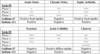
how will a stress fracture appear on each MRI image?
- T1: linear zone of decreased signal intensity surrounded by a less defined area of signal intensity
- T2: linear zone of decreased signal intensity surrounded by an increased SI due to edema
- STIR: increased SI because fatty bone marrow is suppressed
how will osteomyelitis show up on MRI?
- T1: break in cortex, decreased signal intensity in bone marrow
- T2: break in cortex, increased signal in bone marrow
how will avascular necrosis appear on MRI?
- decreased signal intensities on T1 and T2
- double rim sign on STIR and long T2 (inner margin will show increased SI, as granulation tissue; outer margin will show decreased SI, as mineralization)
