14 - Devo of Urinary System Flashcards
(48 cards)
What are the precursor tissues for the kidney and urinary tract?
Intermediate mesoderm: kidney, caylecesm pelvix, and ureter
Endoderm: epithelial lining of the urinary bladder and urethra.
- The smooth muscle and connective tissue in the walls of these organs are derived from splanchnic mesoderm.

How does the intermediate mesoderm develop? When it is seen?
Seen at days 18-20 and eventually separates from the paraxial mesoderm.
Forms the nephrogenic cord that will later be called the urogenital ridge.
What allows for the intermediate mesoderm’s kidney-forming ability? What directs kidney formation?
Kidney forming ability mediated by signals from the paraxial mesoderm
TFs Pax2, Pax8, and Lim1 expresed in intermediate mesoderm direct kidney formation
What are the urogenital derivatives from the intermediate mesoderm?
- Kidneys
- Ureters
- Gonads
- Genital ducts
This is why you can get defects that affet both the kidneys and the urogenital system
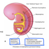
What forms within the nephrogenic cord in the thoracolumbar region of the embryo?
A mesonephric kidney: transient functional structure with two components:
- mesonephric duct
- mesonephric tubules

What is the function of the mesonephric duct in the mesonephric kidney? What is the cloaca?
Source of inductive signals for kidney structures
Solid cord which eventually canalizes and extends caudally through the nephrogenic cord to fuse with the cloaca (day 26)
Cloaca is the dilated caudal end of the primitive hindgut; a transient, common outlet for the UG and GI systems

What are mesonephric tubules? How are they formed?
Immature nephrons: inductive signals from the duct induces tubule formation; differential occurs cranial to caudal along nephrogenic cord.
All tubules induced are not present at the same time.

What does the metanephric (mature) kidney form from? Where does it form?
Forms in the pelvic region from the caudal aspect of the nephrogenic cord.
Forms from the metanephric diverticulum (ureteric bud) and the metanephrogenic mesenchyme (metanephric lastema)
Both of these are derived from intermediate mesoderm.
What occurs between the metanephric diverticulum and the metanephrogenic mesenchyme?
Reciprocal inductive interactions occur - the epithelial-mesenchyme interactins between them are mediated by GFs, secreted factors, patterning genes, and ECM changes.

They cause the metanephricdiverticulum to branch and the metanephrognic mesenchyme to differentiate into nephrons.
What is seen in Potters syndrome?
- Renal agenesis
- Severe urinary obstruction
- Features of facial compression, growth retardation, limb deformities (contracted limbs)
- Pulmonary hypoplasia because you need amniotic fluid to develop the lung
Fetal urine contributes to amniotic fluid volume, esp in the third trimester. Fetus swallows fluid and excretes urine; provides an environment of protection and mobility; growth factors present, lung development.
What is oligohydramnias and polyhydramnios?
Oligohydramnios: too little amniotic fluids, associated with renal agenesis, polycystic kidney disease and urethral obstruction.
Polyhydramnios: excessive amniotic fluid, associated with DB, multiple gestation, anencephaly (without part of brain and/or skull), esophageal atresia
Inductive interactions between the ___________ and the _______ result in nephron formation.
Metanephric diverticulum (MD) and the metanephrogenic mesenchyme (MM).
Signals frmo MM such as GDNH and RA induce formation of the MD, and the MD branches (cells express Ret, a GDNF receptor)
Expanded tip of the MD is called the ampulla, a key signaling center for nephron induction.
What is the function of the ampulla?
Signals from the ampulla direct the arrangement of nephrons and collecting ducts.
Amupllae begin to disappear at about 32 weeks and no new nephrons are formed after all ampullae diappear.

The metanephric diverticulum (MD) signals: ____ and ____ prevent the metanephrogenic mesenchyme cell apoptosis and induce a subset of MM cells to aggregate around the ampulla.
FGF2 and BMP7
What occurs in the early stages of nephron formation?
Signals from the apulla cause the metanephrognic mesenchyme to form an epithelial vesicle.

Formation of the mature nephron involves differentiation and differentual growth of what? What nephron derivatives are made from each part of this?
The S-shaped tubule:
- proximal part forms the DT and loop of henle
- middle part forms the proximal tubule
- distal part forms the renal corpuscle

How does the vasculature form in the kidney?
Vascular spouts from the intersegmental arteries are induced to grow toward the forming kidney.
Only induced mesenchyme secretes angiogenic growth factors (VEGF) that attracts the vascualr sprouts to the forming kidney.
What is seen in multicystic dysplatic kidney?
Most often unilateral (this differentiates it from polycystic kidney disease which is bilateral), multiple cysts of varying sizes making the kidney non-functioning.
Primitive ductules and cartilage seen; atretic ureter (uretal atresia)
Contralateral kidney hypertorphies
Bilateral disease is rare and fatal.

What is seen clinically with multicystic dysplastic kidney?
Potential for abnormalities of the contralateral kidney, such as vesicoureteral reflux, approximately 28%
HTN potentially
Failure of large MCKD to regress may be indication for nephrectomy (surgical removal)
Non-genetic (polycystic kidney disease is genetic)

Remodeling and differental growth of the branchting metanephric diverticulum results in the formation of what? Describe the growth.
Formation of collecting ducts, calyzes, pelvis and ureter.
There’s little growth of early generations of branches, and faster growth of of polar branches.
Exapnsions of the 3rd to 6th generations of branches form the calyces, pelvis, and ureters. Branches distal to the 5th and 6th generations form collecting ducts.

What is visible in the fetal kidney but not the adult kidney?
Renal lobes
Each kidney lobe ends in a pyramid shaped renal papilla which empties into a minor caylx.

What are some anomalies that result in variations in kidney size, histological organization or number?
Hypoplastic kidneys: may be small and normal or small because of abnormal development
Dysplastic kidneys
Duplications of ureter or kidney (partial or complete)
Horshoe kidney: fusion prevents complete ascent

How does the metanephric kidney get where it’s supposed to be in the body?
Develops in the pelvis and “ascends” into the abdomen because of differential growth of the embryonic body.
- pelvic and lumbar kidneys can result from failed or incomplete ascent
- extra renal vessels result from failure to atrophy during ascent
Gonads develop in the abdomen and descend.

How do people get polycystic kidney disease?
It’s inherited: either autosomal recessive or autosomal dominant


