14.1 Eye disease Flashcards

(76 cards)

1
Q

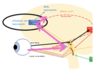
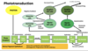
What is required for us to be able to see?

A
  • Eye
    • Clear cornea
    • Clear lens
    • Functioning retina and optic nerve
  • Brain
    • LGN (lateral geniculate nucleus)
    • Superior colliculus
    • Edinger-Westphal nucleus
    • III nucleus
    • IV nucleus
    • VI nucleus
    • Frontal lobe (voluntary eye movements)
      • Causes body movements
    • Vestibular nuclei
      • Gaze stabilisation
    • Occipital lobe
      • Pursuit eye movements & accomodation
    • Dorsal stream (where it is?)
    • Ventral stream (what it is?)
    • CN III, IV, VI
2
Q

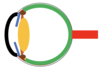
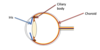
What do different areas of the eye do?

A
  • Cornea = refraction
  • Lens = focussing
  • Iris = controlling amount of light entering the eye
  • Retina = phototransduction
  • Optic nerve = carrying electrical signals to the brain
3
Q

What is the function of the cornea & the layers?

A
  • Main function is refraction
  • 5 layers
  • NO blood vessels but can grow but affect vision
  • O2 (oxygen) from the atmosphere
4
Q

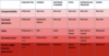
What is normal refraction called and give examples of refractive errors and how to correct them?

A
5
Q

What is astigmatism & explain it

A
  • Cornea is not spherical
  • Different radii of curvature in different axes
  • In this case the vertical radius is shorter than the horizontal radius therefore the curvature of the cornea is steeper in the vertical axis than the horizontal axis.
  • To correct this a cylindrical lens is needed with different powers in the different axes.
6
Q

List some corneal diseases

A
  • Corneal ulcer
  • Corneal dystrophy
  • Keratoconus
  • Corneal oedema
7
Q

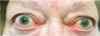
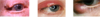
What is this & explain it

A

CORNEAL ULCER

  • Usually due to poor hygiene in contact lens wearers
  • Can be treated with antibiotics
8
Q

What is this?

A

Corneal dystrophy (rare)

9
Q

What is this?

A

Keratoconus

  • Treatment with corneal graft (given to allow good vision)
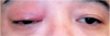
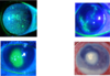
10
Q

What is this?

A

Corneal oedema

  • Due to failure of endothelial pumps
  • Treatment with corneal graft
11
Q

What is this?

A

Corneal graft

  • Given to allow good vision
12
Q

Explain what happens in the lens

A
  • Some refraction
  • Accommodation/fine focus:
  • Circumferential ciliary muscle contracts allowing lens capsule to relax, lens becomes more spherical
    • As we age this gets worse
13
Q

What is cataract & what are the risk factors & treatment?

A
  • Cataract: clouding of the lens
  • Risk factors:
    • Age
    • Diabetes
    • Corticosteroids
    • Congenital
    • Trauma
  • Treatment
    • Can be treated by surgery
14
Q

What is this condition?
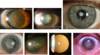
A

Cataracts

15
Q

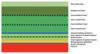
What are all the layers in the retina?

A

Inner at top

Outer at bottom (photoreceptors where transduction occurs)

16
Q

What is the blood supply to the retina?

A

Blood from:

  • Central retinal artery
    • Supplies innermost layers
  • Choroid artery
    • Supplies outermost layers
17
Q

What is the imaging of this picture?

A

Optical coherence tomography

18
Q

What is the foveal dip?

A
  • Where light entering the eye causes the sharpest image
    • Increase in visual acuity
    • Increase in determination of colour
  • Fovea appears slightly darker as it is thinnest here
    • Choroid is most visible
    • Hence, light can get straight to photoreceptors
19
Q

What is the concentration of rods & cones in the retina?

A
  • RODS
    • 6.5 million
    • Increase numbers at the fovea
  • CONES
    • 120 million
    • None at fovea
    • Widely spread throughout the retina
  • OPTIC DISC
    • NO photoreceptors at the optic disc
20
Q

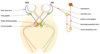
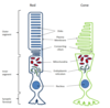
Explain photoreceptors & the structure of them

A
  • Photoreceptors is where phototransduction occurs
  • Rods = scotopic vision (vision under LOW light levels)
  • Cones = photopic vision (vision under WELL-lit conditions)
  • DISKS hold opsin molecules
    • Rhodopsin in rods
    • Iodopsin in cones
    • Light stimulates the isomerisation of retinal which activates opsin leading to hyperpolarisation of the photoreceptor
  • 3 different opsins present in all cones but one predominates in each of three different cones:
    • Bluecones containing mostly blue-sensitive opsin are excited chiefly by a wavelength of around 420 nm,
    • “Green” cones by a wavelength around 530 nm,
    • “Red” cones by a wavelength near 560 nm
21
Q

Explain phototransduction in detail

A
22
Q

Role of ON-OFF switches in phototransduction

A
  • Are to do with the modification of signal by transduction
23
Q

List some retinal diseases

A
  • Colour blindness
  • Retinal vascular occlusion
  • Diabetes
  • Macular degeneration
  • Retinal detachment
24
Q

Explain colour blindness & how it can be tested

A
  • ​There are many forms of colour blindness
    • ​Depends on which cones are affected (total loss/do not function properly)
  • Red-Green colour blindness is the most common type
  • Naming of colour blindness:
    • Prot- = Red (L) cones
    • Deuter- = Green (M) cones
    • Trit- = Blue (S) cones
    • -anomaly = malfunctioning
    • -anopia = absent
  • Tested
    • Using the Ishihara test plates
25
Explain **central retinal artery occlusion** (CRAO)
* Common in *diabetes* & *hypertension* * Causes the *inner retinal* cells to die * If retina loses blood supply then the retina thins as cells die * Vision is *_very poor_* * *_​_*Sudden or total visual loss
26
What is **diabetic retinopathy** & treatment
* Small blood vessels leak or get blocked (forming *micro-aneurysms*) * Leads to *oedema*, *exudation* and *haemorrhage* in inner retinal layers * Worse diabetes = INCREASE likely to have this * Treatment * With **anti-VEGF** injections/steroids
27
What is **wet age-related macular degeneration**?
* *Inflammation* & *hypoxia* due to aging eye (forms INCREASE VEGF) * New blood vessels grow from the *_choroid_* through the retinal pigment epithelium * These *leak* and *bleed* causing *oedema* and *haemorrhage* in the outer retinal layers * This causes *_distorted vision_* as **photoreceptors** are *displaced* *
28
Why do you get a *distorted image* with **wet ARMD** (age related macular degeneration)?
1. Pixels: Each cone is responsible for seeing one part of the face and that is mapped exactly in the *occipital cortex* 2. If *_oedema_* *separates* the cones their pixels will be different 3. The brain assembles the image as though cones are in correct place therefore image appears distorted
29
What is **retinal detachment** & *symptoms* & *treatment* for it?
1. **Posterior vitreous detachment** (PVD) 2. Vitreous remains attached to part of the retina, the traction causing a tear 3. Fluid breaks through the break in the retina between the *photoreceptor* layer and the *retinal pigment epithelium* 4. Fluid continues to leak thanks to gravity creating a **retinal detachment** **Symptoms** * *Flashing lights* * *Potential shadow* in vision **Treament** * Retina reattached ASAP * As photoreceptors _NOT_ recieving adequate blood supply
30
What is the **blood supply** to the *optic nerve*?
31
Explain **optic nerve defects**
* Nerve fibre defects in **superior part** --\> cause *inferior field* defects 1. As light from environment by refraction crosses over so from bottom of visual field in environment goes to the superior retina 2. Fibres from the superior part of the retina enter at the superior part of the *optic nerve* *SAME FOR INFERIOR PART*
32
What are **optic nerve** problems due to?
1. *Inflammation* (optic neuritis) 2. *Glaucoma* 3. *Ischaemia* 4. *Compression*
33
What can **optic nerve** problems cause?
* *Reduced* visual acuity * *Altered* colour vision * Visual field *defects*
34
What is **optic neuritis** & what can it be related to?
* *Inflammation* of optic nerve. Can result in variable visual loss including *colour vision problems* but usually recovers within a few months * Can be related to **Multiple Sclerosis** (50%) or be **post-viral** * Red objects do not appear bright red but more brown * Optic nerve can look normal if *optic neuritis* in *posterior part* * Optic nerve will be seen as *swelling* if *optic neuritis* in *anterior part*
35
What is **glaucoma** & the *treatment* for it?
* Progressive optic neuropathy causing the loss of the nerve fibre layer causing **CUPPED OPTIC DISC** appearance and corresponding **VISUAL FIELD DEFECTS** * Commonly associated with raised **INTRA-OCULAR PRESSURE** * Causes field loss depending on which part of the *optic* nerve is damaged the most * **TREATMENT** * Various *topical drops* * Surgery (more serious cases)
36
What is **anterior ischaemic optic neuropathy** & treatment?
* When *branches* of **posterior ciliary arteries** are affected * Most commonly due to *_microvascular occlusion_* related to *diabetes*, *hypertension*, *atherosclerotic disease* , but can be due to *inflammatory* causes including **Giant Cell Arteritis** (serious rheumatological disease where: losing appetite & feeling very unwell & pain in jaw & hard to comb hair as scalp tender) * _NOT_ caught early enough = VISUAL loss can occur --\> **IRREVERSIBLE** * Once 1 eye blind INCREASE likely without treatment other eye can become blind * **TREATMENT** * **​**HIGH dose of *steroids*
37
What is **optic nerve compression**?
* Optic nerve is surrounded by **fat** & **bone** for protection from injury * The eye is in an enclosed space –***_The Orbit_*** * Things that can cause *proptosis* (forward movement of the eye) & *compression* of the optic nerve: * *Inflammatory* conditions (*_thyroid eye disease_*) * *Infection* (*_orbital cellulitis_*) * *Haemorrhage* * *Tumours*
38
What is **thyroid eye disease** & treatment?
* Inflammation of the **extra-occular muscles** & **proptosis** (forward movement of eye) * **TREATMENT** * **​***Steroids* * *Radiotherapy* * *Surgery*
39
What is **orbital cellulitis** & treatment?
* **Inflammation** of eye tissues behind the *orbital septum*. It is most commonly caused by an *acute spread* of infection into the eye socket from either the adjacent ***_sinuses_*** or through the ***_blood_*** * Usually in **children** * Causes **protosis** & optic nerve **compression** * **TREATMENT** * **​**Imaging * Surgery
40
What *conditions* can stop us from seeing & the areas that they affect?
* Corneal opacity * Cataract * Optic neuritis * Glaucoma * AION * Optic nerve compression * CRAO * Retinal detachment * Wet ARMD * Diabetic maculopathy
41
What comprises the **visual pathway** and explain where in the brain (diagram)?
42
Where can **visual field defects** occur and explain what they cause?
**Pituitary lesions** --\> damage to *optic chiasm* = *_bitemporal hemianopia_*
43
Why is there **macular sparing**?
This is because the **macular** area is supplied by the *middle cerebral artery* & *posterior cerebral artery* so when visual cortex (dense hemianopia) is affected = preservation of central vision
44
What happens to the *electrical signals* from the optic nerve (draw this as a diagram)?
* ***Occipital lobe*** = allows us to focus on moving things * *Pursuit eye movements* & *accomodation* * Sends messages to **LGN** & **superior colliculus** & **Edinger-Westphal nucleus** * **​**These send messages to *III nucleus, IV nucleus, VI nucleus* which control *eye movements* * **Vestibular nuclei** * *Gaze stabilisation* so, allows our surroundings to stay _still_ whilst we move * When *drinking/ear infection* this can be affected * **Ventral stream** * **​**Pathway from *occipital lobe* to *temporal lobe* * Responsible for interpreting **WHAT** we see * *Recognition of objects, facial recognition, colour memory* * **Dorsal stream** * **WHERE** is it? * *Spatial awareness, looking for things* * From the *occipital lobe* to the *frontal lobe* * **Frontal lobe** * **​**Voluntary eye movements * Connects to *motor cortex* & enables us to respond appropriately * **WHAT** we gonna do about it? e.g. *run away* from fire
45
What are the **eye muscles** & what nerve are they innervated by?
**III nerve** = SR, IR, MR, IO **IV nerve** = SO **VI nerve** = LR
46
What is **pursuit** (movement) & what muscles does it involve? What is **accomodation** & what muscles does it involve?
* **Pursuit** movement = eyes move in *same* direction * *Medial rectus* & *contralateral lateral rectus* * **Accommodation** = eyes turn in (medially) * BOTH *medial recti*
47
What is the **accommodation reflex** & explain its pathway
* Causes eyes to *converge* thanks to III nerve (short ciliary nerve from ciliary ganglion) * Causes **pupil constriction** & **accommodation** of lens Pathway: 1. *Visual cortex* 2. to *EW ganglion* 3. to *PS fibres* 4. to *Ciliary ganglion* 5. to *Short ciliary nerve* Pupil **constriction**: * *Circumferential muscle fibres* in the *iris contract*. Pupil becomes smaller **Accommodation** of lens * Circumferential *ciliary muscles contract*, zonules relax and elastic capsule of lens relaxes. Lens becomes more spherical
48
Explain **pupil responses** and the different types of innervation & *drugs* that can allow these responses to happen
* Pupil responds to light --\> _NO_ involvement of *visual cortex* 1. Fibres from *optic tract* peal off at *Pre-tectal ganglion* then decussate into *Edinger-Westphal ganglion* 2. To the *ciliary ganglion* via the short ciliary nerve 3. **Constriction** of pupil (BILATERAL) 4. _Abscence_ of light = puppils dilate again * Active **DILATING** due to *sympathetic* innervation (NA - noradrenaline) * Useful for examining fundus * **Phenylephrine** –alpha 1 agonist (dilates) **Tropicamide**, **Cyclopentolate**, **Atropine** - cholinergic antagonists (dilate and reduce accommodation) * Active **CONSTRICTION** due to *parasympathetic* innervation (ACh - acetylcholine) * ***Pilocarpine***-cholinergic agonist
49
How can you check **pupil responses**?
**PERLA** - **_P**_upils _**E**_qual & _**R**_eactive to _**L**_ight & _**A_**ccomodation * However, usually only check patient's reactivity to **LIGHT** * ​This is because usually if react to **LIGHT** will react to **accommodation** * BUT, if _do not_ react to light then, test accommodation * Test for **LIGHT** * **​***Pupil constriction* * Test for **ACCOMMODATION** * ​*Pupil constriction* * *Convergence* * *Lens accommodation*
50
Where do the following take place (& through what systems): 1. Pursuit & accommodation 2. Pupil reflexes 3. Keeping the image still 4. What is it? 5. Where is it? 6. What next?
1. Occipital cortex 2. Pre-tectal and E-W nuclei, Parasympathetic and Sympathetic systems 3. Vestibular input 4. Ventral stream 5. Dorsal stream 6. Frontal lobe
51
What is **blindsight** & explain this
* As goes via this pathway we are _NOT_ consciously aware of it * Bilateral **occipital damage** demonstrates *blindsight*
52
What is **human echo location**?
The ability of the brain to turn **sound** into a **visual image**
53
What are the causes of pain in the eye?
* Foreign body * Trauma * Reduced tear film * Corneal epithelial disturbance (*including dry eyes)* * Inflammation * Raised IOP (*intra-ocular pressure)*
54
What **cranial nerves** are involved in the eye?
* II ***Optic*** nerve * III ***Oculomotor*** nerve * IV ***Trochlear*** nerve * V ***Trigeminal*** nerve * **Opthalamic** division * Upper Eye Lid including Palpebral Conjunctiva * **Maxillary** division * Bulbar Conjunctiva * Cornea * Ciliary Body * Iris * **Mandibular** division * Lower Eye Lid including Palpebral Conjunctiva * VI ***Abducens*** nerve * VII ***Facial*** nerve
55
Explain the **mechanism of pain** (types and types of pain receptors)
TYPES OF PAIN (in eye) * ***Nociceptive*** * ***​***Damage to non-neural tissue * ***Neuropathic*** * Damage to nerve * ***Inflammatory*** * ​Activation of nociceptors by inflammatory mediators * ***Sudden raised Intra-ocular pressure*** * ***​***Activation of nociceptors by ATP TYPES OF PAIN RECEPTORS (in eye) * Found on *surface* of *eye, conjunctiva, cornea*, anterior chamber & iris * *LENS & RETINA* = _NO_ nerve fibre endings for pain
56
What areas of the eye would have to be **examined** in order to find the **cause** of **pain**? (& what device would you use?)
* Eyelids * Conjunctiva * Cornea * Anterior Chamber * Iris Can examine these using a ***slit lamp***
57
Draw the anatomy of the **eyelid** & annotate it
58
What is **blepharitis** & the treatment for it?
* Inflammation of eyelids * Is **squamous** type * Very common **_TREATMENT_** * Warm compresses * Lid hygiene
59
What is **m****eibomianitis** & the treatment for it?
* Inflammation of *meibomian glands* (OR blocked = excess secretion formes and **DISTURBS** *tear film* * **Sebaceous** type * Very common **_TREATMENT_** * Warm compresses * Lid hygiene
60
What are **tears** made from?
1. **Aqueous** component * From *lacrimal gland* 2. **Oily** component * From *meibomian glands* * Prevents *dehydration*
61
What are the different types of **lumps** & **bumps** on the eye and briefly explain them & treatment for one
* **CYST OF MOLL** (left pic) * Blocked gland of Moll (modified sweat gland) * **STYE** (middle pic) * Blocked sebaceous gland related to hair follicle * **CHALAZION** (right pic) * Blocked meibomian gland * In *tarsal plate* thus, secretions formed and causes lump * *Treatment* * *​*Massaging eyelid (opens up *Meibomian glands*) * Warm compresses * Surgery (*severe)*
62
What is **entropian**, **ectropion** & **trichiasis**?
* **ENTROPION** (left pic) * Eyelid turned in (as *loose*), lashes rubbing on cornea (pain, treatment needed) * Usually when people get OLDER (as *less* orbital fat) and less tension in muscles acting on eyelid * **ECTROPION** (middle pic) * Eyelid turned out (less painful) * Usually when people get OLDER (as *less* orbital fat) and less tension in muscles acting on eyelid * **TRICHIASIS** (right pic) * Lashes turned in rubbing on cornea (eyelid in incorrect position)
63
What are some **lid diseases** & what happens to the **lashes**, **lid position** & what **glands are involved**?
64
What are some **conjunctiva** diseases?
* **BACTERIAL CONJUNCTIVITIS** (top left) * Sticky discharge * Lasts 3-4 days * **VIRAL CONJUNCTIVITIS** (bottom left) * Watery discharge * Lasts many weeks * **ALLERGIC CONJUNCTIVITIS** (right) * **​**Chemosis * Lid changes
65
Where can **foreign** **bodies** enter in the eye?
* **SUB-TARSAL FOREIGN BODY** (left) * **CORNEAL FOREIGN BODY** (right) * Usually due to high speed injuries *e.g. drilling*
66
What are the **layers** of the **cornea** and briefly describe them
Main function of the *cornea* is **refraction** * **Epithelium** * Several layers of cells * Barrier to water and bacteria entering the cornea * Maintains a smooth optical surface for refraction * **Bowman’s layer** * **Stroma** (keratocytes) * **Descemet’s** membrane * **Endothelium** * Single layer of hexagonal cells * Allows transport of nutrients including glucose into the cornea * Pumps water out to maintain clarity of stroma
67
What are some **common corneal problems**?
* **DRY EYES** (top left) * *Aqueous* part of tear production _NO_ longer working adequately as epithelial cells start dying off * **CORNEAL ABRASION** (bottom left) * Painful * e.g. *poked in the eye* * **DENDRITIC ULCER (HSK)** (top right) * **BACTERIAL ULCER** (bottom right) *Fluorescein* is used to show **epithelial defects**
68
Explain how the **anterior chamber** of the eye works & label (draw)
* *Ciliary body* * Produces *aqueous* component which circulates * When blocked = INCREASEin pain * *Trabecular meshwork* * *​*Aqueous component leaves via here into _venous system_
69
What are the symptoms of **acute angle closure glaucoma? (AACG)**
* **Severe pain** (ATP mediated) * **Vomiting** * **Reduced vision** * **​**As aqueous has _nowhere_ to go thus, through *cornea* and becomes water logged * **Rainbow haloes** (corneal oedema-altered refraction of light)
70
What is the **mechanism** of **AACG**?
* **HYPERMETROPIA** (usually occurs in these people) * Long sight-smaller eyes * Shallower AC * Enlarged lens (when older) * Pupil dilates in low light * Iris sticks to enlarged lens * Aqueous cannot flow in to AC pushes iris forward * Iris closes off already narrowed angle * IOP rises
71
How can you **treat** *AACG*?
* **Yag laser Iridotomy** * ​Prevents further attacks of AACG * Allows *aqueous* content to always get into **anterior** chamber
72
What is **uveitis**?
**Uveitis** is the inflammation of any or all of these structures (i*ris, ciliary body, choroids)*. Can be related to other systemic inflammatory diseases
73
What does the **uveal tract** consist of?
* Iris * Ciliary body * Choroid
74
What is **acute anterior uveitis**? & treatment?
* Is the **inflammation** of the **iris** which can cause the eye to become *red* * Can be idiopathic or related to auto-immune or other inflammatory conditions * ***Treatment*** includes *steroids* & dilating drops where *iris* has got stuck to the lens * *Iris stuck to lens* *(posterior synechiae)*
75
What conditions could cause a **red eye**?
When DECREASED *vision* &/OR *photophobia* = INCREASE serious condition
76
What is **ophthamology**?
**Ophthalmology** is a branch of medicine dealing with the *diagnosis*, *treatment* and *prevention* of *diseases* of the *eye* and *visual system*. It can be either a *surgical* or *medical specialty*. Most Ophthalmologists are *super-specialised*