14.1 The Neurobiology of Vision Flashcards

(43 cards)

1
Q

Label the diagram

A
2
Q

Label the diagram and the lobes

A
3
Q

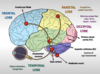
Where do the nerves from the retina go?

A
  1. The nerves leave the eye in the optic nerve and synapse in the lateral geniculate nucleus (LGN)
  2. Fibres then go to the visual cortex

Transmits all light information into electical information to the brain

4
Q

Label the diagram

A
5
Q

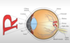
What is the purpose of the cornea?

A
  • Involved in focusing light into eye
  • Transparent covering over eye: is a barrier between outside world and inner eye
  • Refracts light entering the eye
  • NO blood vessels
  • VERY sensitive to light
6
Q

What is the purpose of the pupil?

A
  • Small opening in eye by which light passes
  • Size of pupil can change which can be due to light or emotion (e.g. like someone pupil dilates)
  • LOW light –> pupils dilate, more light into eye
    • better to see when dark
  • HIGH light –>pupils constrict
    • LESS light into eye
7
Q

What is the iris?

A
  • The coloured portion of the eye
  • Regulates the amount of light into the eye
8
Q

What is the purpose of the lens?

A
  • Serves by focusing light to back of eye (refracts)
  • Transparent structure that helps refract light onto retina
9
Q

What is the fovea?

A
  • Indentation (small) at the back of the eye where there is high level detail processing of light
  • Region where upper retinal layers are THINNED
10
Q

What flips the inverted image from the retina?

A

The vision cortex

11
Q

What is vision?

A

Transduction of light

12
Q

What produces tears?

A

Lacrimal gland produces tears and passes through the lacrimal ducts

13
Q

What does vision depend on?

A

Depends on light sensitive cells in the retina at the rear of the eye

14
Q

How does light reach the retina?

A

Light is focussed by the cornea and lens onto the retina

15
Q

Explain the structure of the retina (layers)

& label the diagram

A
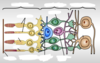
  • Photoreceptors (allow night vision)
    • ​Made of cones and rods
    • CONES
      • More in centre of retina
      • Only type in fovea of eye
    • RODS
      • More in outer areas of the eye
      • Used for peripheral vision
  • Outer plexiform layer
  • Horizontal layer
    • Form synapses with bipolar cells & photoreceptor cells
  • Bipolar cell
    • Connect photoreceptors to ganglion cells
  • Amacrine cell
    • Translate information
  • Inner plexiform layer
  • Ganglion cell
    • Gets info from retina & sends onto brain (via optic nerve)
    • Their axons leave the eye
16
Q

What do photoreceptors do & how?

A
  • Photoreceptors capture light
  • Have outer segments with stacks of membrane that increases their surface area greatly
  • Pigment molecules collect light
17
Q

What is a photon?

A

A single unit of light

18
Q

What is the wavelength of visual light and colours on the sides of the spectrum?

A

(BLUE) 380-750nm (RED)

Infra red Ultra-violet

19
Q

What are the kinds of photoreceptors?

A
  • 120 million RODS (more rods in nocturnal animals)
  • 6 million CONES
  • RODS
    • More sensitive to light and occur in the periphery (at low light)
  • CONES
    • Respond to different wavelengths
    • Mainly found in the fovea (day-light, when HIGH levels of light)
20
Q

What are these and label them and what is the difference between the two?

A

The outer segments are different

21
Q

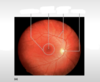
Label this fundoscopy image

22
Q

Label this diagram & briefly explain it

A
  • At the fovea (where information from the lens is focussed) there is a 1:1 ratio of photoreceptors : ganglion cells
  • At the retina there is a 50:1 ratio of photoreceptors : ganglion cells
23
Q

Explain receptor potentials in photoreceptors, bipolar cells, ganglion cells

A
  • Photoreceptors
    • HYPERPOLARISING membrane potential following stimulus
  • Bipolar cells
    • DEPOLARISING membrane potential
  • Ganglion cells
    • Recording of action potentials then sends it to the brain
24
Q

How many types of rods & cones are there?

A
  • Cones
    • Blue
    • Green
    • Red
  • Rods
    • Blue/green
25
Explain the anatomy of the visual system after the ganglion cell fibres
1. Ganglion cell fibres enter the **optic nerve** & then the **optic chiasm** 2. Fibres from the **nasal retina** cross over (**CONTRALATERAL** - opposite side) 3. Fibres from the **temporal retina** remain (**IPSILATERAL** - same side) **LGN** - Lateral geniculate nucleus 1. Nerve fibres from **LGN** go to the **visual cortex** 2. Synapse mainly in layer IV of cortex (**primary visual cortex** - at posterior of brain in *occipital lobe*)
26
What are the pathways from the **primary visual cortex**?
* **Dorsal pathway** --\> spatial vision e.g. WHERE something is (**FAST**) * To *parietal cortex* * **Ventral pathway** --\> colour and object vision e.g. WHAT (detail of what looking at) (**SLOW**) * To *inferatemporal cortex*
27
Explain **disorders of vision**
* Is rare in females * **More common** in *males* as on **X chromosome** (1 in 10) * Visual disorders can occur anywhere in the visual pathway * **Short** & **long** sightedness * **Neural deficits** * ***Colour blindness*** - caused by loss of one type of cone * ***Optic (retinal) neuropathy*** - specific to the retina, blood vessel damage & regrowth
28
What is **glaucoma** & the risk factors?
* When there is damage in the **connection** between the eye & the brain (usually in the optic nerve) * Up to **50%** of people with disease _don't_ know they have it * Causes **thinning** of the nerve fibre layer in the retina and gradually results in *_loss of vision_* and **_un_**treated glaucoma eventually leads to *_blindness_* * Risk factors: * Family history of *glaucoma* * Increasing age * Prolonged steroid use
29
What is **AMD** & the risk factors?
* **AMD** - age related macular degeneration * **Commonest** cause of blindness in western world * **Peripheral** vision remains _unaltered_ (causes blurry vision, wavy/distorted lines or a central blind spot * Risk factors: * Increase age * Female * Smoking
30
Explain *diabetic retinopathy*
Deterioration of the tight junctions of blood vessel wall causing **blockage**, **excudates** (plasma leakage), **haemorrhages**, **micro aneurysms**
31
Explain **retinal vein occlusion** & predisposing factors
* **Blockage** of a retinal vein. Predisposing factors are: * *Increasing age* * *Systemic hypertension* * *Raised Intra ocular pressure and long sighted spectacle corrections* * There is some visual loss depending on the severity of the occlusion but some recovery after 6 months
32
Explain **presbyopia**
* Means 'old eye' (can be particularly due to eye muscles) * Ages of 40-45 showing *difficulties* with reading. Other symptoms include holding reading material further from the eyes and blurry distance vision immediately after reading * Due to the lens becoming ***_inelastic_*** *making focusing on near objects difficult*. Rectified by the use of bifocals, varifocals, separate reading spectacles or contact lenses
33
Explain **cataracts** & how it can be treated
* Any **opacity** of the lens. May present as gradual *_blurring*_ of the vision, seeing _*double_* in one eye or increased sensitivity to glare * Cataracts are more common with *_increasing_* *_age_*. Excessive unprotected exposure to UV light, smoking, poor nutrition and prolonged steroid use also contribute * Cataracts can be **removed surgically** by replacing the *_lens*_ with a clear _*plastic lens implant_*
34
Explain **Balint's syndrome**
* Bilateral damage to parieto-occipital region * Region between parietal and occipital regions * Involved in spatial perception * Dorsal stream only is affected * Includes: * ***_Optic ataxia_*** * Difficulty reaching for objects under visual guidance * ***_Ocular apraxia_*** * Difficulty in visual scanning * ***_Simultanagnosia_*** * Difficulty perceiving more than one object at a time
35
What is **optic ataxia**?
Difficulty reaching for objects under visual guidance
36
What is **ocular apraxia**?
Difficulty in visual scanning
37
What is **simultanagnosia**?
Difficulty perceiving more than one object at a time
38
Explain what happens when there is **damage** to **V1**
* _No conscious awareness_ of being able to see * Known as ***_Blindsight_*** * ***_​_***Information goes *straight for processing* so information retained but without awareness * Patients report a ‘gut feeling’ but insist they cannot see
39
Explain what happens when there is **damage** to **V4**
* _No_ colour perception OR memory of colour * ***_Achromatopsia_***
40
Explain what happens when there is **damage** to **V5**
* *_Cannot process motion_* so objects moving become invisible * ***_Akinetopsia_***
41
Explain what happens when there is **widespread damage** to **all** but **V4**
* Severely impaired visual processing but colour information retained * ***_Chromatopsia_*** * Caused by *_carbon monoxide_* poisoning
42
What is **visual agnosia** & types?
*_Cannot_* perceive visual stimuli accurately * ***_Apperceptive_*** visual agnosia * Cannot perceive *_objects_* * ***_Prosopagnosia_*** * Failure to recognise *_faces_* * Damage to *fusiform* face area * Region of extrastriate cortex dedicated to face and complex object recognition * ***_Associative visual agnosia_*** * Inability to identify objects perceived visually * Form can be matched with similar objects or drawn from memory * Can still describe what objects are when named * Just can’t link pictures and words * Disruption to connections in ventral stream of the visual cortex * But no damage to those in the dorsal stream
43
What is **achromatopsia** & explain it
* _Inability_ to discriminate among different hues (a colour/shade) * Cannot see colour at all * Damage to visual association cortex * **Bilateral** * Affects both visual fields * _NO_ memory of colour * **Unilateral** * Only affects one visual field * Half the world is seen in grey-scale * Half the world is seen in colour * Also affects memory of colour * Patients cannot recall what colours objects were prior to brain injury