Lab4 Immunopathology Images Flashcards
What type of cells are these? What type of reaction?

Inflammatory allergic reaction
These cells are eosinophils
What type of cells? What type of reaction?

Acute inflammatory reaction
Neutrophils
What kind of reaction is this? What kind of cells do the arrows point to?

This is a chronic inflammatory reaction
Bottom arrow: lymphocytes
Top arrow: plasma cells
What kind of reaction is this? What types of cells present?
Granulomatous inflammation
Contains epithelioid histiocytes and Lymphocytes
What are the white spots in this bronchus?

Mucus. This is bronchial asthma
What type of disease does this bronchus wall show? How can you tell? What is the part around the arrows?
[Hint: lumen is top left]
Bronchial asthma
inflammatory infiltrate in bronchial wall
The black lines indicate hyperplastic submucosal glands
What does this show?
Hint: the dotted line is the epithelial surface of bronchial lumen, lumen is to left

Bronchial asthma
Mucus in bronchial Lumen
Inflammation in bronchial submucosa
What disease does this show? What do the arrows denote?

Bronchial asthma
The arrows denote hypertrophy and hyperplasia of bronchial smooth muscle
What condition in the broncus does this show? What are the cells?

Bronchial asthma
thickened epithelial basement membrane
Eosinophils
What type of cells are these?

Eosinophils by H&E stain
What type of cells are these?

Mast cells
With toluidine blue stain
What syndrome does this show [hint glomerulus]

Antibody mediated goodpasture syndrome
Glomerular necrosis
What does this show in the glomerulus? What are symptoms?

Goodpasture syndrome
glomerular necrosis and acute inflammation
Renal disfunction [high BP, bleeding from glomeruli, decreased glomerular filtration]
What does this show [hint goodpasture syndrome]
RBC casts in glomerular tubules
What does this show in goodpasture syndrome

RBC casts in urine
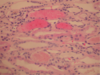
What does this show in goodpasture syndrome?

Pulmonary hemorrhage
What disease does this show in glomerulus? What path findings?

Post-streptococcal “Acute proliferative” Glomerulonephritis
Hypercellularity due to intrinsic and inflammatory cells
Obstructed capillary lumens
PAS stain
cells with brightest blue multiple nuclei next to the wall on left = neutrophils
What type of necrosis and cells? Possible disease?
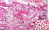
Fibrinoid necrosis
Neutrophils
Systemic vasculitis
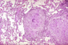
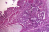
What does this show in pancreas? What type of hypersensitivity?

Diabetes Mellitus type I
cytotoxic T lymphocytes infiltrating islets to target beta cells
Type IV hypersensitivity
What do the arrows point to? What could this be in lung?

Granulomas
This could be Sarcoidosis
What is this?

Amyloid [with H&E]
What type of disease does this show? In kidney

Amyloids
What type of disease does this show? In heart

Amyloids
What does the wire loop lesion demonstrate?

thickened capillary wall due to immune-complex depositon