20. Genital disorders (Illustrated Textbook of Pediatrics) Flashcards
(26 cards)
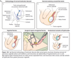
Inguinoscrotial conditions - Embryology
How are the testis formed?
Development of a testis from an early indeterminate
gonad is determined by genes associated with a Y chromosome.
For a testis to descend from its origin on the
posterior abdominal wall, it must produce testosterone
which acts on peripheral tissues. The testis, guided by
the mesenchymal gubernaculum, migrates down into
the inguinal canal (Fig. 20.1a). The structures that are
found in the scrotum in a boy (testis, vas and blood
vessels) or labium in a girl (attachment of the round
ligament of the uterus) pass through the abdominal
wall and pick up layers corresponding to those of the
abdominal wall. In a boy these make up the coverings
of the spermatic cord. In boys and girls there is
a remnant of peritoneal invagination, the processus
vaginalis (Fig. 20.1b), which, if it remains patent and
in continuity with the abdomen, explains why fluid or
abdominal contents can become a hydrocele or hernia,
respectively (Fig. 20.1c–e).

Inguinal hernia
Presentation and Complication
A hernia presents as a lump in the groin which may
extend into the scrotum (Fig. 20.2) or labium. They
are usually asymptomatic but may be intermittent,
visible during straining. On examination, sometimes
a lump or thickened cord structures can be palpated
in the groin.
The contents of the hernia may become irreducible
(incarcerated), causing pain and sometimes intestinal
obstruction or damage to the testis (strangulation). In
these circumstances the lump is tender and the infant
may be irritable and may vomit. The risk of incarceration
is much higher in infants than in older children.

Inguinal hernia
Management
Most hernias can be successfully reduced by ‘taxis’
(gentle compression in the line of the inguinal canal)
with good analgesia. Surgery can then be planned
for a suitable time when any oedema has settled and
the child is well. If reduction is impossible, emergency
surgery is required because of the risk of compromise
of the bowel or testis. In girls, sometimes the ovary can
become incarcerated within a hernia.
Surgery (see Fig. 20.1d) involves ligation and division
of the processus vaginalis, which has become the
hernial sac (herniotomy, removal of the hernia sac – as
opposed to herniorrhaphy in adults, when the inguinal
abdominal wall is also reinforced, usually with a mesh).
Beyond the first three months of age, this can be safely
performed as a day case.
Hydrocele
Signs/Symptoms
How to differentiate hydrocele from hernia
Management
A hydrocele has the same underlying anatomy as a
hernia, but the processus vaginalis, although patent,
is not sufficiently wide to form an inguinal hernia.
Hydroceles are usually asymptomatic and sometimes
appear blue. It is usually possible to feel the
testis, however tense the hydrocele. Sometimes the
hydrocele is separate from the testis (see Fig. 20.1e)
in the cord. The key to differentiating a hernia from
a hydrocele is the ability to ‘get above’ a hydrocele.
Hydroceles usually transilluminate (Fig. 20.3).
Although the processus vaginalis is often patent
at birth it usually closes within months. Hydroceles
therefore usually resolve spontaneously, and can be
managed expectantly. Surgery may be considered if it
persists beyond the first two years of life, but resolution
may take longer than this. In a girl, a hydrocele
(of the ‘canal of Nuck’) is much less common than
in boys.

Variocele
Cause(s)
Which side is more common?
Symptom(s)
Physical examination
Management
This is a scrotal swelling comprising dilated (varicose)
testicular veins and occurs in up to 15% of boys, usually
at puberty (Fig. 20.4).
Its cause is multifactorial; valvular
incompetence plays a role.
It is commoner on the left
side because of drainage of the gonadal vein into the
left renal vein, which also receives blood containing
catecholamines from the left adrenal vein.
It is usually
asymptomatic, but may cause a dull ache.
On examination
it may have a bluish colour and feel like a ‘bag of worms’. Sometimes the testis is smaller or softer than
normal.
Management is conservative if asymptomatic.
Occlusion of the gonadal veins can be achieved by
surgical ligation – through the groin laparoscopically
or by radiological embolization.

Undescended testis
Examination
Palpable/Impalpable
Retractile
Most undescended testes become arrested along their
normal pathway of descent (see Fig. 20.1a). Undescended
testes are present in up to 5% of newborn
term infants but are more common in premature
infants. By three months of age, only 1% are still undescended.
The diagnosis should ideally be made at the
routine examination of the newborn (Ch. 10. Perinatal
medicine) but since there is still a small spontaneous
rate of descent after this time the decision to operate
for undescended testis should be delayed.
Examination of the testes in babies must be made in
a warm environment and with warm hands. The testes
may be felt in the scrotum or may need to be delivered
by gentle pressure along the line of the inguinal canal
to the scrotum.
An undescended testis may be palpable or impalpable.
A palpable undescended testis is usually seen
or felt in the groin, but cannot be manipulated into
the scrotum. Occasionally it can be palpated below the external inguinal ring but outside the scrotum – the
so-called ‘ectopic’ testis.
If the testis is impalpable, it may be in the inguinal
canal but cannot be identified or it may be intraabdominal
or absent. If there are bilateral impalpable
testes, the karyotype must be established to exclude
disorders of sex development. This should be regarded
as a medical emergency.
A testis may also be retractile. The crucial difference
between a retractile and undescended testis is that a
retractile testis can be manipulated into the scrotum
with ease and without tension. Action of the cremaster
muscle (as seen in eliciting the cremasteric reflex by
light touch on the abdominal wall) pulls up the testis.
Parents of boys with a retractile testis often report that
the testis is sometimes obvious, particularly when the
boy is warm and relaxed, and sometimes not. This is
why a boy with a suspected undescended testis should
be examined in a warm environment and when warm
and relaxed.
Undescended testis - Investigations and management
Imaging is not helpful in the assessment of an undescended
testis.
Orchidopexy, the surgical placement of the testis
in the scrotum, is performed for the following reasons:
• Cosmetic – to achieve the same, symmetrical
appearance as other boys. This may be of
psychological benefit. If the testis is absent, a
prosthesis can be inserted when older.
• Reduced risk of torsion and trauma compared to
groin location
• Fertility – the testis needs to be in the scrotum,
below body temperature, in order to allow
spermatogenesis. The effect is probably marginal
in unilateral undescended testis but is more
important if bilateral. There is some evidence that
delaying orchidopexy beyond the first two years of
life adversely affects testicular development.
• Malignancy – increased risk in an undescended
testis, which is greater if bilateral or intraabdominal.
Placing the testis in the scrotum
facilitates self-examination but may not influence
the risk of malignancy.
The timing of orchidopexy depends on local
surgical and anaesthetic facilities, but should be performed
before or around one year of age. Thereafter,
spontaneous descent is unlikely, and there is evidence
that testicular growth, hormonal function and spermatogenesis
is improved by operating at this early age
rather than waiting until older.
Groin approach orchidopexy involves opening the
inguinal canal in a similar manner to herniotomy, mobilizing
the testis whilst preserving the vas and blood
vessels and placing it within the scrotum. It is usually
performed as a day case. An intra-abdominal testis is
usually managed laparoscopically; it may be amenable
to placement in the scrotum in a single operation or
may require a staged approach.
Regarding impalpable testes, about 10% have
regressed in development and are, in fact, absent.
Laparoscopy allows both diagnosis and treatment.
For a retractile testis, follow up is recommended
because some high testes require surgery to place
them in the scrotum. Whether or not this is true ascent
of the testis is controversial.
Torsion of the testis
Presents as…
Pain localised…
Must be distinguished from…
This is commonest in post-pubertal boys (Fig. 20.5a),
but may occur at any age, including the newborn
when it usually presents at birth and is believed to be
perinatal. It is usually very painful, with redness and
oedema of the scrotal skin. However, the pain may be
localised to the groin or lower abdomen, highlighting
the need to always examine the testes in a boy presenting
with sudden-onset pain in the groin, abdomen or
scrotum. It must be distinguished from an incarcerated
hernia. An undescended testis is at increased risk of
torsion, as is a testis lying transversely on its attachment
to the spermatic cord (the so-called ‘clapper
bell’ testis).

Torsion of the testis
Treatment
Outcome
Torsion of the testis must be treated within hours
of the onset of symptoms to lower the risk of testicular
loss. In fact, surgical exploration in any acute scrotal
presentation is mandatory (Fig. 20.6) unless torsion can be excluded with certainty (see below). Fixation
of the contralateral testis is essential because of the
increased risk of a contralateral torsion, especially if an
anatomical abnormality is present in the torted testis.
Outcome is variable, depending on time to correction.
If delayed, testicular loss is likely. In perinatal testicular
torsion, testicular loss is almost inevitable.

Torsion of appendix testis
More common than…
Pain…
Diagnosis
Management(?)
A testicular appendage (Hydatid of Morgagni) is a Mullerian
(paramesonephric) remnant usually located on
the upper pole of the testis. Torsion of the appendix
testis (Fig. 20.5b) tends to affect prepubertal boys and
is more common than torsion of the testis. Pain evolves
over days, but is not as dramatic as in testicular torsion.
Scrotal exploration and excision of the appendage is
often necessary because it cannot be differentiated
reliably from torsion of the testis. If a ‘blue dot’ can be
seen through the scrotal skin and pain is controlled
with analgesia, surgery may not be necessary.

Other acute inguinoscrotal conditions (1)
Acute scrotum - Infection
Infection may cause an acute scrotum. Epididymoorchitis
(Fig. 20.5c) is commoner in infants and small
children, and more likely with a pre-existing urological
or anorectal malformation. As it may be indistinguishable
from torsion, scrotal exploration may be necessary.
Doppler ultrasound of flow pattern in the testicular
blood vessels may allow differentiation of epididymitis
from torsion of the testis, but must not delay surgical
exploration if torsion remains a possibility. A urine
sample should be obtained to identify an associated
urinary tract infection. Pus should be sent at operation
for microbiology to characterize the nature of the infection,
but infection may be bacterial or viral. Antibiotics
are started empirically.

Other acute inguinoscrotal conditions (2)
Idiopathic scrotal oedema
Incarcerated hernia
In idiopathic scrotal oedema there is redness and
swelling extending beyond the scrotum into the thigh,
perineum and suprapubic area, but the testis is normal
and non-tender. It requires analgesia and review. It may
recur. An incarcerated hernia may also cause an acute
scrotum, although symptoms usually affect the groin.
Other acute inguinoscrotal conditions (3)
Trauma to the scrotum
Trauma to the scrotum is an uncommon cause of
testicular damage, but may need exploration, debridement
and surgical repair. Sexual abuse needs to be
considered in all genital injuries.
Other acute inguinoscrotal conditions (4)
Recurrent scrotal pain in boys
Recurrent scrotal pain in boys can be difficult to
manage. Any associated symptoms or signs such as
swelling or redness should be regarded as intermittent
testicular torsion and the testes fixed. Sometimes prophylactic
fixation is required to exclude intermittent
torsion as a cause for recurrent pain. In adulthood,
chronic scrotal pain can follow scrotal surgery.
Abnormalities of the penis - The foreskin (Prepuce)
Retraction
Protection
Differentiate from
Treatment
A normal foreskin does not retract in infancy, and
retraction should not be attempted. At 1 year of age,
about half of uncircumcised boys have a non-retractile
(normal) foreskin. Only 1% of boys over 16 years old
have a non-retractile foreskin. The prepuce develops
adherent to the underlying glans, and acts as protection
to the non-keratinised glanular and meatal squamous
epithelium in an environment where astringent urine
can cause inflammation or even ulceration. This can
be manifest as ammoniacal dermatitis (napkin rash) in
infants and young children, where the preputial opening
can be reddened and sore. It usually only needs reassurance
and attention to routine hygiene. This needs to be
differentiated from infection, or balanoposthitis, where
the redness is more extensive, and, crucially, there is a
purulent discharge. It occurs in about 3% of boys, reaches
a peak incidence around the third year of life, and recurs
in about a third. The infection is usually bacterial and
needs antibiotic treatment, either topical or systemic. As
it is rarely fungal, antifungal agents are not indicated.
Topical corticosteroids may sometimes be beneficial.
Ballooning of foreskin
Results from…
May also occur on…
Consequence
Ballooning of the foreskin on urination is a common
cause of parental concern. It can look dramatic but
seldom causes any trouble. It results from lysis of
preputial adhesions around the glans before those at
the preputial opening. Ballooning may also occur on
the shaft of the penis, arising from the attachment of
shaft skin below the coronal sulcus of the glans. Ballooning
stops when preputial adhesions have lysed
completely. It has no functional consequence, does not
represent obstruction, and does not need intervention.
Smegma
Appears as…
Intervention
Another cause of parental concern is sub-preputial
smegma. It appears as a lump which grows briefly,
seemingly under the non-retractile or partially retractile
foreskin. It is yellowish and malleable, and simply
comprises desquamated skin and secretions. There is
no need to intervene – it will discharge in due course
(with typical appearance of smegma – ‘cottage cheese’;
Fig. 20.7) when the preputial adhesions break down.

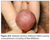
Non-retractile foreskin and phimosis
Commonest condition to give rise to a true phimosis…
When traction is applied (gently) to a normal foreskin,
the skin at the preputial opening is seen to evert, even
if it does not necessarily open up (Fig. 20.8). A foreskin
that is pathologically non-retractile will not do this,
and will truly render the glans ‘muzzled’, (Greek word
‘phimos’). This differentiates a foreskin that is simply
non-retractile (i.e. normal, physiological) from one
which is problematic. The commonest condition that
gives rise to a true phimosis is balanitis xerotica obliterans,
or BXO, which gives rise to progressive scarring which can extend onto the glans, into the meatus and
ultimately into the urethra. Typically this affects older
boys and young adults, and there is often a history
that the foreskin was normally retractile in earlier childhood.
Figure 20.9 shows the typical appearance of BXO.
BXO is the index indication for circumcision,
although there is some evidence that potent topical
steroids, closely monitored, can cause it to regress.
Paraphimosis
What is it?
Treatment
This is a condition, usually in post-pubertal boys, of
a retracted foreskin that cannot be reduced easily.
There is a ring of narrower skin. The glans swells,
and if the prepuce is not reduced it may result in
compromise of the blood supply to the glans. Treatment
(by reduction) is an emergency, which may
require general anaesthesia. Paraphimosis has been
regarded as an indication for circumcision, but this
is no longer considered to be the case unless the
foreskin is abnormal (as with BXO).
Circumcision
Medical reasons for circumcision
Other indications for circumcision
Complications
Circumcision remains a tradition in Jewish and Muslim
religions.
Medical reasons for circumcision include:
• BXO causing a true phimosis
• recurrent balanoposthitis causing refractory
symptoms
• prophylaxis of recurrent urinary infection,
especially in the presence of a congenital uropathy
(such as posterior urethral valves or vesicoureteric
reflux) or if renal reserve is limited
• if access to the urethra is required reliably for
intermittent catheterization, e.g. spina bifida.
There are inevitably other indications for circumcision,
some of which are highly dependent on the
individual family and surgeon. There is some evidence
that circumcision affords protection against transmission
of HIV and HPV (human papillomavirus), and there
are programmes promoting circumcision in newborn
infants and young adult males in some countries with
high prevalence of HIV infection.
There have been many techniques described for
circumcision, and complications are uncommon. Up to one boy in fifty has post-operative bleeding requiring
a return to the operating theatre. Infection in the skin
margin or ulceration of exposed granular skin may
occur. Meatal stenosis can also occur, more often after
circumcisions done for BXO, and this may require subsequent
surgery. Rarer complications include urethral
fistula.
Hypospadias
Arises from...
Three features (Check image)
Management
Complications
This is a common condition, with an incidence of up
to 1 in 200 boys. It is thought to arise from failure
of development of ventral tissues of the penis – in
particular failure of ventral urethral closure. For that
reason it is really a constellation of ‘ventral hypoplasia’
of the penis.
Typically there are three features, although their
occurrence is variable:
• a ventral urethral meatus – the urethral meatus is
variable in position (Fig. 20.10), but in most (80%)
is on the distal shaft or glans penis (Fig. 20.11a)
• ventral curvature of the shaft of the penis (formerly
called ‘chordee’) (Fig. 20.11b), more apparent on
erection
• hooded appearance of the foreskin – characteristic
in appearance because of ventral foreskin
deficiency but of no functional significance.
There is rarely an associated or underlying disorder
of sex development, and only very rarely another congenital
urinary tract abnormality. Investigation of the
urinary tract with imaging is not routinely indicated.
Surgery is not mandatory, especially in a distal
hypospadias when the penis and urinary stream are
straight. However, it may be performed on functional
or cosmetic grounds. The ultimate functional aim of
hypospadias surgery is to allow a boy to pass urine in
a straight line whilst standing, and to have a straight
erection. Surgery, if needed, is usually performed in the
first two to three years of life. The commonest surgical
complications are breakdown of the repair or meatal
narrowing. The prepuce may be preserved and reconstructed,
although for more proximal hypospadias
it is sometimes required for the repair itself. For this
reason it is important that a boy with hypospadias is
not circumcised before the repair.

Obesity’s effect on the penis
Variations in penoscrotal skin attachment and in the
infant or child’s body habitus may make the penis look
buried. This is common with obesity, when the only
treatment is weight loss, but improves with growth of
the penis after puberty. However, it may persist if there
is marked obesity.
Vulvovaginitis
The commonest problem is redness of the vulva.
In infants, this is often due to a nappy rash due to
ammoniacal dermatitis. Less often, the vulvovaginitis is
infective, occasionally with Candida infection (More common when waring diapers or being immunodeficient).
Vaginal discharge
&
Foreign bodies
Vaginal
discharge is common, and is usually innocuous unless
it is green or offensive when it may indicate infection.
Foreign bodies are more often suspected than
found; they are actually rare. The ‘red flag’ symptom
is a bloody vaginal discharge, and needs referral to a
specialist as vaginal rhabdomyosarcoma is a rare but
important cause in preschool girls.
